Scientists have achieved the restorative benefits of deep sleep in awake mice, without them actually sleeping. In a groundbreaking study published in Nature Neuroscience, researchers at the University of Wisconsin-Madison used optogenetics, a technique that uses light to control genetically modified neurons, to induce slow-wave “on/off” patterns characteristic of non-REM sleep in localized regions of the animals’ brains.
Sleep-deprived but active mice that received this artificial stimulation performed as well on memory and learning tasks as fully rested mice. The results support the idea that specific neural activity patterns, rather than unconsciousness itself, drive synaptic restoration and cognitive recovery.
This discovery suggests it may one day be possible to develop targeted therapies that counteract the effects of sleep deprivation and help treat cognitive decline in humans.
[Driessen, K., Squarcio, F., Tononi, G., & Cirelli, C. (2026). Induction of cortical on/off periods in awake mice fulfills sleep functions. Nature Neuroscience]
🧠 YOUR BRAIN KNOWS THE DIFFERENCE
What if typing and handwriting aren't the same after all?
Research shows that writing by hand activates more areas of the brain linked to memory, focus, and learning. That's why handwritten notes often help people remember information better than typed ones.
A simple pen and paper may be giving your brain a workout that a keyboard can't.
Source: van der Meer, A. Research on handwriting and brain activity, NTNU.
🫀Heart attacks may actually be caused by bacterial infections.
A groundbreaking study from Tampere University and the University of Oxford is reshaping our understanding of what causes heart attacks.
Long blamed primarily on cholesterol and lifestyle factors, new research now points to a hidden culprit: bacterial infections.
Scientists discovered that within the fatty plaques of coronary arteries, bacterial biofilms—gel-like communities of bacteria—can lie dormant and undetected for years. These microbial invaders, particularly strains like viridans streptococci commonly found in the mouth, evade immune detection and traditional antibiotics by embedding themselves deep within plaque tissue.
The danger arises when the body is hit with a viral infection, which ramps up immune activity and disturbs the biofilms. That disturbance can reactivate the bacteria, triggering a sudden surge of inflammation. In turn, this can weaken arterial plaques, causing them to rupture and form clots—leading to heart attacks. Researchers were able to map these biofilms in tissue from patients who died from cardiac arrest and found that antibodies could unmask their full structure. This discovery could pave the way for new diagnostics or even vaccines to prevent infection-triggered heart attacks, signaling a major shift in cardiovascular medicine.
Source: Viridans Streptococcal Biofilm Evades Immune Detection and Contributes to Inflammation and Rupture of Atherosclerotic Plaques. Journal of the American Heart Association, 2025.
Researchers induce the memory-boosting benefits of sleep in parts of the awake brain | Eric W. Dolan, PsyPost
A recent study suggests that triggering specific, sleep-like brain wave patterns in awake mice can provide the brain with the restorative benefits usually only gained by actually falling asleep. The findings indicate that the physical need for sleep, as well as the memory-boosting effects of a good night’s rest, might be replicated without the animal ever losing consciousness. This research was recently published in the journal Nature Neuroscience.
Sleep is a biological necessity for all mammals. It serves to reset the brain and body after a long period of wakefulness. When an animal is awake, it learns, moves, and experiences new things in its environment. All of this waking activity causes the microscopic connections between brain cells, known as synapses, to grow stronger and more numerous.
If synapses constantly grow stronger day after day without ever resetting, the brain would become physically overloaded, consume too much energy, and lose its ability to process new information. Deep sleep provides evidence of a massive resetting process across the brain. During non-rapid eye movement sleep, which makes up about eighty percent of total sleep in adults, the junctions between neurons that make memories are evaluated.
During this sleep phase, the brain protects important connections for long-term storage, prunes those that are less necessary, and makes space for new ones. The brain also experiences highly synchronized electrical activity. Millions of neurons will fire electrical signals all at once, creating what scientists call an “on” period. Immediately following this burst, the cells will collectively go silent, which is known as an “off” period.
This rhythmic switching back and forth creates slow brain waves that can be recorded by sensors. Scientists track this slow-wave activity to measure how badly an animal needs sleep. The longer an animal stays awake, the more intense the slow-wave activity will be once it finally falls asleep. As the animal rests over several hours, this activity gradually decreases, indicating that the biological need for sleep has been satisfied.
A research team from the University of Wisconsin-Madison, including Kort Driessen, Fabio Squarcio, Giulio Tononi, and Chiara Cirelli, wanted to test a specific question about these brain waves. The researchers previously showed that both rats and humans can exhibit sporadic, local slow-wave brain activity while awake if they are sleep-deprived. While those brief dips into sleep-like activity might not be enough to provide benefits, the team reasoned that a longer, more systematic version of this activity might allow part of the brain to rest while the animal remains active.
“What we’re essentially doing is forcing sleep in a local region of the brain. While that part is solidifying memories and restoring learning capacity, other parts stay aware/vigilant and connected to the environment,” said Chiara Cirelli, a professor of psychiatry at the University of Wisconsin-Madison. “Dolphins do something similar, sleeping with only one brain hemisphere at a time.”
To test this idea, the researchers used a technique called optogenetics. This method involves genetically modifying specific brain cells so they can be controlled by flashes of light. The scientists implanted tiny light-emitting devices, alongside electrical recording sensors, into the brains of adult mice. These implants were placed on both the left and right sides of the brain.
This design allowed the team to manipulate the neural networks on one side while using the opposite, untouched side as a natural control for comparison. In the first set of experiments, the researchers worked with nineteen genetically modified mice. They kept the animals awake for five hours by continually introducing new objects into their cages.
During the final thirty minutes of this sleep deprivation period, the scientists used light pulses to force the neurons on one side of the brain into rhythmic on and off periods. They tailored the light flashes to mimic the exact timing and duration of natural deep sleep waves. During this entire process, the mice remained awake and behaved normally, moving around their cages without interruption.
After the thirty minutes of light stimulation, the sleep deprivation ended, and the mice were allowed to fall asleep naturally. The researchers closely monitored the brain activity during the first hour of this recovery sleep. The side of the brain that received the artificial on and off periods showed significantly less slow-wave activity than the untreated side. Additionally, the neurons on the treated side fired with much less synchronization.
In sleep science, less synchronization provides evidence that the biological pressure to sleep has been successfully relieved in that specific area. The authors then asked if simply lowering the overall activity of the brain without a rhythm would have the same effect. Some scientists had suggested that an overall reduction in neuronal firing might be the mechanism needed to recover from the cellular fatigue caused by staying awake.
The researchers ran a second experiment with seven different genetically modified mice. Instead of creating a rhythmic on and off pattern, the scientists used a continuous beam of light to quiet the brain cells, broadly reducing their overall firing rates. When these mice were allowed to sleep, both sides of their brains showed the same high need for rest. This finding suggests that the specific rhythm of turning neurons on and off, rather than a general reduction in brain activity, is required to fulfill the restorative functions of sleep.
Next, the team looked at the physical connections between brain cells. They analyzed molecular markers of synaptic strength in twenty-four mice. These mice were split into three groups of eight based on their specific genetic modifications, including a control group. After keeping the mice awake and applying the rhythmic light stimulation to one side of the brain, the scientists immediately collected brain tissue without letting the animals sleep.
In the brain tissue, the researchers measured the levels of specific proteins that help transmit signals between neurons. They found significantly fewer of these receptors on the synapses of the light-stimulated side. This reduction mirrors the natural weakening of cellular connections that occurs during normal deep sleep. This specific weakening process tends to prevent the brain’s networks from becoming overloaded with information.
Finally, the researchers tested whether this artificial brain rhythm could rescue memory after a period of sleep deprivation. They used a behavioral test of tactile memory, an ability that relies heavily on rest. The team used thirty mice for a memory test involving floor textures. On the first day, the mice explored an enclosed chamber with two identical floor textures for fifteen minutes.
Afterward, the animals were divided into three testing groups. Nine mice were allowed to return to their cages and sleep normally. Thirteen mice were kept awake for an hour. Eight mice were kept awake for an hour but received the artificial on and off brain stimulation during that time.
The following day, the mice were placed back into the testing chamber. This time, the chamber featured one familiar floor texture and one entirely new texture. Because mice naturally prefer to explore novel environments, a well-rested mouse will spend more time investigating the new floor.
The mice that slept normally recognized the old floor and spent most of their time investigating the new one. The mice that were simply kept awake failed to recognize the familiar floor, spending equal time on both sides. However, the mice that received the rhythmic light stimulation while awake performed just as well as the well-rested mice.
While these findings are deeply informative, they require proper contextualization to avoid broad misunderstandings. Casual readers might misinterpret the study to mean that humans or animals could entirely replace a full night of sleep with localized brain stimulation. The authors note that completely disconnecting from the environment, as happens during natural sleep, is likely still necessary for the brain to process memories on a large, system-wide scale. The localized stimulation in this study only affected specific, targeted regions of the sensory and motor cortex, not the entire brain.
Another limitation is that the methods used in this study are highly invasive. Optogenetics requires the genetic modification of brain cells and the surgical implantation of hardware into the skull. Because of this, this exact technique cannot be tested on human subjects. The researchers also pointed out that artificial brain waves, depending on the specific type of cells targeted, can sometimes exhibit reversed electrical polarities compared to naturally occurring sleep waves.
Future research will likely focus on how these local rest periods affect the overall health of the brain over much longer stretches of time. Cirelli aims to learn whether similar effects could be replicated in humans using less invasive technologies, like transcranial stimulation. Understanding the exact mechanics of these on and off periods could eventually guide new treatments for severe sleep disorders or age-related memory issues.
“This research further decodes why we sleep and how we learn, which brings us a step closer to understanding how to better prevent and treat cognitive decline,” said Amy Bany Adams, acting director of the National Institute of Neurological Disorders and Stroke, which funded the research.
Read more:
https://t.co/XFlu5jukLn
We're learning a lot about cellular senescence, how to track it, and its role in aging and disease. Cover and commentary @CellCellPress
https://t.co/bFma1UocnS
A scientist spent 30 years studying an organ every textbook said was irrelevant. In 2026, two papers in Nature proved she had been right all along. The papers were not written by her.
Her name is Noel Rose Mackay. She is a thymic biologist who has studied the thymus since the 1990s, at a time when the field was considered a professional dead end.
The thymus is a small immune organ behind the breastbone. By the 1980s, medical consensus had settled: the thymus trains immune cells in childhood, shrinks at puberty, and stops functioning meaningfully in adults. Researching adult thymic function was considered a waste of time and grant funding.
Mackay and a small number of colleagues disagreed. They published research throughout the 1990s and 2000s arguing the thymus remained active in adults and that its ongoing T cell production mattered for immune health. The papers were published in smaller journals, cited rarely, and largely ignored by mainstream medicine.
For 30 years, clinical practice did not change. Radiologists reading millions of CT scans did not measure thymic health. Oncologists designing immunotherapy did not account for it. No clinical guideline mentioned it.
In March 2026, researchers at Mass General Brigham used artificial intelligence to analyse CT scans from over 25,000 adults. The AI found exactly what Mackay had argued for three decades.
Adults with healthier thymuses lived longer. 50% lower risk of death from any cause. 63% lower risk of cardiovascular death. 36% lower risk of lung cancer. In cancer patients receiving immunotherapy, stronger thymic health predicted a 37% lower risk of cancer progression and a 44% lower risk of death.
Two papers. Published simultaneously in Nature. Covered by Harvard Medical School, Mass General Brigham, and dozens of international outlets.
The researchers who wrote them work in artificial intelligence and cancer imaging. They were not thymic biologists. They were not looking for the thymus. The AI found it for them.
The science that spent 30 years being ignored was correct.
It took a machine looking at 25,000 scans without any prior assumptions to confirm what a small group of scientists had been saying for three decades.
Sometimes the reason a field is underfunded is not that the question is unimportant.
It is that the answer is inconvenient.
No, the peptide craze is not backed by science.
"What people are shooting up with out there I would give to mice."
“The influencer crowd has sort of created this perception that these are miracle drugs."— @mkaeberlein
New feature @nature
https://t.co/o0CHojoFNj
Silencing One Brain Gene May Reverse Autism Deficits | Neuroscience News
Summary: Researchers uncovered a precise molecular mechanism to restore NMDA receptor (NMDAR) hypofunction, a core pathology in autism spectrum disorder (ASD). The research shifts focus away from traditional, highly toxic systemic targets toward Slc6a20a/SLC6A20, a glycine transporter heavily localized within cognition-related brain regions like the cortex and hippocampus.
Utilizing antisense oligonucleotides (ASOs) in adult mouse models with SHANK2 and SHANK3 mutations, as well as human cortical organoids, the team successfully normalized NMDAR activity, corrected synaptic phosphorylation signaling cascades, and reversed deep-seated behavioral deficits without triggering standard brainstem respiratory side effects.
Key Facts
- The NMDAR Activation Dilemma: The NMDA receptor requires both glutamate and glycine to fully trigger. While NMDAR hypofunction drives multiple brain disorders—including ASD, schizophrenia, and intellectual disabilities, previous clinical trials failed because they targeted GlyT1, a glycine transporter dense in the brainstem, causing dangerous respiratory and motor side effects.
- Localization-Driven Safety Precision: To solve this, the IBS team targeted Slc6a20a, a distinct glycine transporter highly concentrated within higher-order cognitive zones (cortex and hippocampus) while remaining safely sparse in brainstem motor centers.
- Adult Behavior and Social Rescue: Administered to adult mice carrying mutations in major autism-risk genes SHANK2 and SHANK3 (models for Phelan-McDermid syndrome), a localized Slc6a20a-ASO successfully boosted NMDAR activity. The intervention reversed entrenched adult behavioral phenotypes, including deficits in social interaction, communication impairments, and repetitive motor loops.
- The Phospho-Proteomic Mechanism: Large-scale phospho-proteomic analysis revealed that the ASO does not simply change total protein numbers. Instead, it systematically normalizes abnormal phosphorylation patterns across critical synaptic signaling proteins and NMDAR regulatory networks.
- Human Cortical Organoid Validation: To confirm human translational viability, the team used CRISPR gene editing to build human cortical organoids with SHANK2 and SHANK3 mutations. Applying a human-targeted SLC6A20-ASO successfully restored NMDAR function back to near-normal baseline parameters.
- Extended Therapeutic Window Durability: A single dosing regimen of the engineered ASO sustained its neuroprotective efficacy for at least 8 weeks in vivo, demonstrating long-term operational stability with zero detectable adverse effects or toxicity trends.
- Broader Neuropsychiatric Utility: Because it targets endogenous signaling pathways rather than complex gene re-expression, this SLC6A20 paradigm offers a scalable template to treat an array of neuropsychiatric conditions anchored by NMDAR hypofunction, including schizophrenia.
---
Researchers have identified a promising new therapeutic strategy for autism spectrum disorder (ASD). A research team led by Director KIM Eunjoon of the IBS Center for Synaptic Brain Dysfunctions has now identified a promising new strategy for restoring NMDA receptor (NMDAR) function by targeting a glycine transporter called Slc6a20a/SLC6A20.
Impaired NMDAR function has long been implicated in a range of brain disorders, including autism spectrum disorder (ASD), schizophrenia, intellectual disability, and NMDAR encephalitis. Despite decades of research, attempts to restore NMDAR activity have produced mixed clinical results, highlighting the need for more precise therapeutic approaches.
The NMDA receptor requires not only glutamate but also glycine to become fully activated. Previous therapeutic approaches attempted to increase glycine levels by inhibiting GlyT1, another glycine transporter. However, because GlyT1 is widely expressed in brainstem regions involved in breathing and motor control, such treatments often produced limited benefits and undesirable side effects.
The researchers instead focused on Slc6a20a, a glycine transporter predominantly expressed in cognition-related brain regions such as the cortex and hippocampus.
Using antisense oligonucleotides (ASOs) to suppress Slc6a20a expression, the team investigated whether NMDAR function could be restored in mouse models carrying mutations in SHANK2 and SHANK3, two major autism-risk genes that are also associated with Phelan-McDermid syndrome and other neurodevelopmental disorders.
The results showed that Slc6a20a-ASO successfully restored NMDAR activity in multiple autism-related mouse models. The treatment also improved several behavioral abnormalities, including impairments in social interaction, social communication, and repetitive behaviors. Importantly, these therapeutic effects were observed in adult animals, suggesting that correction of NMDAR dysfunction may remain possible even after key stages of brain development have passed.
To understand the underlying mechanism, the researchers performed large-scale phospho-proteomic analyses. Surprisingly, the treatment had relatively little effect on overall protein abundance. Instead, it restored abnormal phosphorylation patterns in proteins involved in synaptic signaling and NMDA receptor regulation, suggesting that the therapy works by normalizing protein function rather than simply changing protein levels.
To evaluate its translational potential, the team extended the study to human brain models.
Using CRISPR gene editing, the researchers generated human cortical organoids carrying SHANK2 or SHANK3 mutations. These organoids exhibited reduced NMDAR activity similar to that observed in the mouse models. Treatment with an ASO targeting the human SLC6A20 gene restored NMDAR function to near-normal levels.
“Unlike gene re-expression strategies, SLC6A20 inhibition works by modulating endogenous signaling pathways and may offer a more practical therapeutic route,” said Director KIM Eunjoon. “The fact that the effect was reproduced not only in mice but also in human cortical organoids suggests that this approach may represent a promising therapeutic strategy for neurodevelopmental disorders characterized by NMDA receptor hypofunction.”
The researchers also found that a single administration of the ASO remained effective for at least 8 weeks without detectable adverse effects in the treated mice.
Beyond autism spectrum disorder, the findings may have broader implications for other neurological and psychiatric conditions associated with reduced NMDAR activity, including schizophrenia and certain forms of intellectual disability.
The findings establish SLC6A20 as a promising therapeutic target for restoring NMDAR function and provide a potential framework for treating a broader range of neurodevelopmental and neuropsychiatric disorders linked to NMDAR hypofunction.
Read more:
https://t.co/ijTqQsipeT
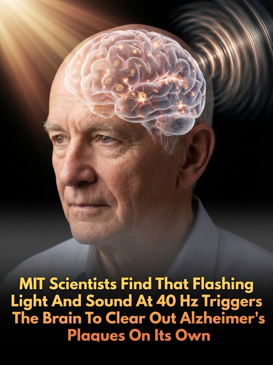
A noninvasive treatment using light and sound shown to trigger the brain’s natural waste-clearance system to combat Alzheimer's disease.
More than 7 million Americans are currently living with Alzheimer’s disease, a crisis projected to cost the U.S. healthcare system an estimated $409 billion in 2026. This treatment could change that.
In a groundbreaking preclinical study funded by the National Institute on Aging (NIA), researchers from MIT, Boston University, and Westlake University in China have discovered that noninvasive light and sound stimulation can significantly reduce levels of toxic amyloid proteins in the brain.
By exposing mice to flashing lights and auditory tones engineered to generate 40-hertz electrical gamma waves, scientists triggered a dramatic boost in the flow of cerebrospinal fluid.
This sensory stimulation effectively tapped into the brain’s glymphatic system—its internal waste-disposal network—prompting cells called astrocytes to expand, flush out debris, and clear the destructive plaques historically associated with Alzheimer’s disease.
The therapeutic mechanism relies on a delicate chain reaction in the brain. Researchers found that gamma wave stimulation coaxes specific inhibitory interneurons to release a vital hormone called vasoactive intestinal peptide (VIP).
This hormone signals the glymphatic system to increase fluid circulation, accelerating the elimination of toxic waste. When scientists experimentally blocked either the astrocytes' expansion or the interneurons' ability to produce VIP, the plaque-clearing benefits vanished.
These promising results suggest that simple, noninvasive sensory therapy could act as a potent tool to keep our brain's plumbing running smoothly, offering a highly accessible avenue for treating Alzheimer’s and other protein-accumulating neurological disorders.
source: Murdock, M. H., et al. Multisensory gamma stimulation promotes glymphatic clearance of amyloid. Nature, 627(8002), 149-156.
Scientists pinpoint an overlooked stretch of DNA linked to the main features of autism | Eric W. Dolan, PsyPost
A recent study published in Nature suggests that a specific genetic sequence, which does not produce proteins, plays a significant role in the core behavioral features of autism in males. By examining human genetics alongside genetically altered mice, scientists found that missing sections of this genetic material lead to social difficulties and repetitive behaviors without affecting general intelligence. These findings provide evidence that targeting specific brain circuits might eventually help support individuals with autism.
Autism spectrum disorder presents with primary characteristics like differences in social communication and repetitive behaviors. Roughly one in 50 children and youth in Canada have the condition. Despite the diverse ways people experience autism, changes in social interaction and repetitive actions are common across the spectrum. Many individuals with autism also experience additional conditions, such as intellectual disability or attention issues.
Separating the biology of core autism traits from these other conditions remains a significant challenge in genetics. Most known genetic variations linked to autism encode proteins and tend to influence broad brain development, making it hard to pinpoint what drives specific social and repetitive behaviors. A large international research team led by scientists at The Hospital for Sick Children, or SickKids, in Toronto initiated this project to study a specific genetic region on the X chromosome known as PTCHD1-AS.
This genetic region produces long non-coding ribonucleic acid, commonly known as RNA. Unlike typical genes that provide instructions for making proteins, long non-coding RNA molecules act as functional regulators within the cell. By interacting with other genes and cellular machinery, they help control how and when other genetic instructions are turned on or off. Researchers targeted PTCHD1-AS because it sits in a region close to other protein-coding genes that together have been linked to autism and intellectual disability.
“PTCHD1-AS gives us a new entry point to study the biology of ASD, sharpening our understanding of how specific biological pathways relate to key autism traits,” says Stephen Scherer, senior scientist of genetics and genome biology and chief of research at SickKids, and director of the McLaughlin Centre at the University of Toronto. “This is essential, because no new therapeutics in clinical trials are designed to modulate the main features of ASD.”
To begin their research, the scientists analyzed whole-genome sequencing data from more than 9,300 individuals in global databases. They identified 27 male individuals with autism from 23 unrelated families who were missing small pieces of DNA in the PTCHD1-AS gene. The scientists focused on males because the gene is located on the X chromosome, and females possess a backup X chromosome that provides protection. Statistical analysis indicated that these missing segments increased the odds of an autism diagnosis by more than two and a half times.
Notably, clinical records for these specific individuals showed fewer instances of intellectual disability or attention issues compared to the general autism population. When the researchers expanded their view to a broader group of 118 individuals with neurodevelopmental disorders, they found that those with PTCHD1-AS deletions predominantly exhibited core autism traits. This clinical profile provided evidence that the PTCHD1-AS region might specifically govern the core social and repetitive traits of autism.
To understand how this genetic deletion works in the brain, the scientists created two distinct genetically modified mouse models. In both models, they used gene-editing tools to delete a specific segment of the mouse equivalent of the PTCHD1-AS gene. They then subjected these male mice, along with genetically typical control mice, to a variety of behavioral and physiological tests.
The behavioral assessments revealed that the genetically modified mice engaged in significantly less social interaction. In a standard test using a three-chambered enclosure, these mice showed equal interest in a lifeless object as they did in another live mouse. They also spent significantly more time engaged in repetitive self-grooming compared to control mice.
The scientists also tested how the mice reacted to social odors, which rodents rely on heavily for communication. Typical mice will intensely sniff a new scent, like the urine of another mouse, and then lose interest over time. The genetically modified mice showed very little interest in new social odors and failed to adapt to them, indicating limited social responsiveness.
To assess communication, the researchers recorded the high-frequency sounds that mice make to each other. The mice missing the genetic segment produced fewer distinct vocalizations and communicated less intensely. At the same time, the researchers tested the mice on memory and complex learning tasks. The genetically modified mice performed just as well as the control mice on tasks involving navigating a puzzle box and remembering spatial cues.
“Our findings suggest there is a different biology involved with our PTCHD1-AS model compared to other ASD protein-coding models,” says Lisa Bradley, first author and research associate in The Centre for Applied Genomics at SickKids. This behavioral profile in mice perfectly mirrored the human data. It suggests that the PTCHD1-AS gene influences social and repetitive behaviors independently of learning and memory.
To see if the brains of these mice developed differently, the researchers scanned 50 mice repeatedly from their early postnatal period into adulthood. They observed subtle developmental differences in specific brain structures, such as the anterior cingulate cortex, and in nerve fiber tracts associated with sensory processing. To find out what was happening inside the cells, the research team examined brain tissue, focusing on an area called the striatum. The striatum is a deep brain structure involved in processing rewards, controlling movement, and regulating habits.
“When we examined gene and protein expression in this area, we saw changes in genes and proteins involved in regulating synaptic plasticity as well as myelination, the process that allows electrical signals to travel faster between neurons,” Bradley says. “This gives us a molecular pattern we can use for future studies into the biological effect of this non-coding gene in the brain.”
The scientists used advanced sequencing techniques to look at the RNA from individual brain cells, allowing them to see exactly which cellular pathways were altered. They discovered that the absence of PTCHD1-AS disrupted the production of molecules responsible for creating myelin. The scientists also found alterations in support cells called astrocytes, suggesting a mild level of brain inflammation specific to the striatum.
The scientists also analyzed thousands of proteins in the brain tissue using mass spectrometry. They found alterations in proteins involved in synaptic plasticity. Synapses are the tiny gaps where nerve cells communicate with one another, and synaptic plasticity refers to the brain’s ability to adapt and fine-tune signals in response to activity. This process is how the brain learns and adapts at a microscopic level.
The researchers measured the electrical activity in slices of the striatum and the hippocampus. In the hippocampus, which handles memory, the electrical activity and plasticity were entirely normal. However, in the striatum, a specific type of synaptic depression, a process that weakens connections between neurons, was significantly enhanced in the genetically modified mice.
“Through a multi-disciplinary approach combining human genetics, mouse models, multi-omics and electrophysiology, we’ve connected a non-coding gene to measurable changes in brain function,” says study co-author Graham Collingridge, senior researcher at the Lunenfeld-Tanenbaum Research Institute at Sinai Health, director of the Tanz Centre for Research in Neurodegenerative Diseases, and professor in the Department of Physiology at the Temerty Faculty of Medicine at the University of Toronto.
“Together, our research helps clarify how unique alterations in synaptic plasticity relate to the core features of autism,” Collingridge adds.
A particular family of enzymes known as conventional protein kinase C was notably reduced in these brain regions. The researchers traced these changes to reduced enzyme activity in a specific brain circuit connecting the cortex to the striatum. When the researchers chemically blocked these enzymes in normal mice, their brain tissue behaved exactly like the tissue from the genetically modified mice. This confirmed that the genetic deletion was actively changing how striatal neurons communicate.
While these findings are highly detailed, it is important to avoid misinterpreting the results as a universal explanation for autism. The PTCHD1-AS deletion accounts for only a small fraction of autism cases globally. Animal models also cannot perfectly replicate the complexities of human neurodevelopment or human social experiences. The study focuses exclusively on male individuals and male mice, meaning the exact role of this genetic region in females remains unaddressed.
The research team notes that by linking a specific gene and biological pathway to social and repetitive behaviors, these findings may be relevant across all autism diagnoses, regardless of clinical complexity. Future research will need to explore how these striatal circuits interact with other brain regions during early development. The next steps for the researchers include deeper investigation of the molecular, cellular, and circuit-level pathways influenced by PTCHD1-AS. By identifying potential targets driving those core features of autism, the scientists hope to inform future precision therapeutics for those who seek them.
Scherer, who is also a professor in the Department of Molecular Genetics at the Temerty Faculty of Medicine at the University of Toronto, notes the broader implications of the findings. “Beyond significantly advancing our understanding of autism as a human condition, the study shows how small changes in DNA can influence complex human behavior,” Scherer says. “It’s amazing to me how much of our disposition is genetically ‘hardwired,’ even in the traits that shape how we connect and interact.”
Scientific efforts of this scale require extensive backing from public and private institutions. The research received funding from multiple organizations, including Autism Speaks, the Autism Science Foundation, the Canada Foundation for Innovation, the Canadian Institutes of Health Research, Genome Canada, Ontario Genomics, the Government of Ontario, the Ontario Brain Institute, the Province of Ontario Neurodevelopment Disorders Network, the Simons Foundation Autism Research Initiative, the University of Toronto McLaughlin Centre, and the SickKids Foundation.
Read more:
https://t.co/6k8Z7OnjOi
The first participant to receive partial cellular reprogramming for eye disease (advanced glaucoma) in a pilot study of 12 patients was treated. Using 3 of the 4 Yamanaka stem cell factors to potentially achieve cellular rejuvenation @Nature
https://t.co/R4oJlrPhnS
Remote work is increasing isolation and harming mental health for millions of Americans, according to a major new study.
When the pandemic pushed millions of workers into home offices, Harvard Ph.D. student Emma Harrington was initially impressed by how productive she could be. But living alone, she soon experienced the downside: long stretches of profound isolation with almost no human contact.
That personal experience led Harrington and her colleagues to launch a large-scale investigation. Their study, analyzing data from more than 588,000 American workers, was published in the journal Science.
The findings are striking: remote workers now spend roughly one extra hour alone each day compared to those in non-remote jobs. This increased isolation is linked to significantly higher rates of mental distress, greater use of therapy, and more antidepressant prescriptions. The effects are especially severe for people living alone, who face an 83% higher likelihood of spending entire days with zero human interaction.
As remote and hybrid work becomes a permanent feature of modern life, the authors stress that companies and policymakers must actively address this hidden cost — ensuring that flexibility doesn’t come at the expense of human connection.
[Emanuel, N., Harrington, E., & Pallais, A. (2026). Home alone: Remote work, isolation, and mental health. Science, 392(6802). DOI: 10.1126/science.aec7671]
In today's @TheLancet there are 3 papers on cardiometabolic disease: biology, epidemiology, prevention/treatment.
The sobering and all to common story from womb to tomb conveyed in this graphic
https://t.co/rVs2Yz97NC
https://t.co/OpuJTia0bC
https://t.co/i714onXoCG
Why is basic research so important?
Listen to 2025 medicine laureate Mary Brunkow on why we never should stop investing in basic research.
Watch the full interview with Brunkow: https://t.co/9UYSR1rVJU
Middle age appears to be a pivotal window for brain health. Emerging neuroscience suggests that changes in neural connectivity begin years before noticeable cognitive decline. Protecting metabolic health, sleep quality, physical activity, and cardiovascular function during this period may help preserve brain resilience and extend cognitive performance well into later life.
This trial was an early phase one, as I understand it,
on patients who'd had extensive chemo already.
What will happen if used at time of diagnosis?
Will it work better with or without chemo?
Communication between the cells in our bodies is managed by substances called hormones. Each cell has a small receiver known as a receptor, which is able to receive hormones.
In the 1980s, Brian Kobilka successfully identified the gene that regulates the formation of the receptor for the hormone adrenaline. He and Robert Lefkowitz also discovered that the receptor was similar to receptors located in the eye that capture light. It was later discovered that there is an entire family of receptors that look and act in similar ways - "G-protein-coupled receptors". Approximately half of all medications used today make use of this kind of receptor.
Image 1) a three-dimensional model of the beta 2 adrenergic receptor-Gs protein complex etched in a glass cube. The glass cube was donated to the @NobelPrizeMuseum from 2012 chemistry laureate Brian Kobilka when he visited Stockholm to receive his Nobel Prize. Kobilka and his colleagues created the glass cube after revealing the structure of the molecules in 2011.
Image 2) Brian Kobilka in his office posing with a model of the active-state beta 2 adrenergic receptor bound to adrenaline. The model was made by Kolbilka's friend and colleague Dr. Roger Sunahara. Photo credit: Christopher Michel


![Rainmaker1973's tweet photo. Scientists have achieved the restorative benefits of deep sleep in awake mice, without them actually sleeping. In a groundbreaking study published in Nature Neuroscience, researchers at the University of Wisconsin-Madison used optogenetics, a technique that uses light to control genetically modified neurons, to induce slow-wave “on/off” patterns characteristic of non-REM sleep in localized regions of the animals’ brains.
Sleep-deprived but active mice that received this artificial stimulation performed as well on memory and learning tasks as fully rested mice. The results support the idea that specific neural activity patterns, rather than unconsciousness itself, drive synaptic restoration and cognitive recovery.
This discovery suggests it may one day be possible to develop targeted therapies that counteract the effects of sleep deprivation and help treat cognitive decline in humans.
[Driessen, K., Squarcio, F., Tononi, G., & Cirelli, C. (2026). Induction of cortical on/off periods in awake mice fulfills sleep functions. Nature Neuroscience]](https://pbs.twimg.com/media/HLgovTFa4AAAWsP.jpg)





![Rainmaker1973's tweet photo. Remote work is increasing isolation and harming mental health for millions of Americans, according to a major new study.
When the pandemic pushed millions of workers into home offices, Harvard Ph.D. student Emma Harrington was initially impressed by how productive she could be. But living alone, she soon experienced the downside: long stretches of profound isolation with almost no human contact.
That personal experience led Harrington and her colleagues to launch a large-scale investigation. Their study, analyzing data from more than 588,000 American workers, was published in the journal Science.
The findings are striking: remote workers now spend roughly one extra hour alone each day compared to those in non-remote jobs. This increased isolation is linked to significantly higher rates of mental distress, greater use of therapy, and more antidepressant prescriptions. The effects are especially severe for people living alone, who face an 83% higher likelihood of spending entire days with zero human interaction.
As remote and hybrid work becomes a permanent feature of modern life, the authors stress that companies and policymakers must actively address this hidden cost — ensuring that flexibility doesn’t come at the expense of human connection.
[Emanuel, N., Harrington, E., & Pallais, A. (2026). Home alone: Remote work, isolation, and mental health. Science, 392(6802). DOI: 10.1126/science.aec7671]](https://pbs.twimg.com/media/HKYkQK1WcAADqvv.jpg)


