We’re hiring at @DeepPiction 🧠🔬
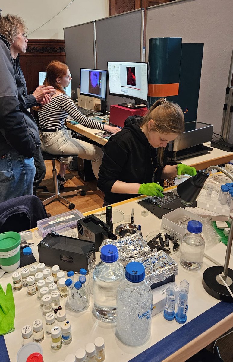
We are looking for a Scientist – Tissue Clearing & Image Analysis (m/f/d) to join our team in Munich.
You will help drive tissue-clearing, light-sheet microscopy, and quantitative 3D imaging projects across internal R&D programs and collaborations with biotechnology and pharmaceutical partners. The role combines hands-on experimental work, project ownership, workflow development, and close collaboration with our AI and image-analysis teams.
We’re looking for someone with strong experience in tissue clearing, fluorescence microscopy, light-sheet imaging, and large-scale 3D image analysis.
Please apply with one single PDF containing your CV and cover letter to:
[email protected]
Subject: Scientist – Tissue Clearing & Image Analysis (DP-TC-2026-01)
Applications will be reviewed on a rolling basis until the position is filled.
https://t.co/2PM9tWIzBZ
Today in @Nature, we report MouseMapper: foundation-model AI to map disease perturbations across the entire mouse body cell-by-cell.
In obesity, it revealed body-wide inflammation & unexpected facial nerve damage. 🧵👇🔉
https://t.co/BERf5GQ10Z led by @Dorie00 & @yingchen733
An AI foundation whole-body 3D model that assesses perturbations (such as obesity) across multiple systems (such as immune, neural) at the cell level. This is MouseMapper. Imagine HumanMapper someday @Nature@erturklab https://t.co/jsg3VMzOqg
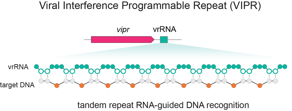
Excited to share our discovery of a new programmable RNA-guided DNA-targeting system hiding inside bacteriophages that predates CRISPR.
We call it VIPR (Viral Interference Programmable Repeat), and it uses an entirely new logic to find its targets.
Thread + link below.
🔬 This image by PhD student Jake Elton shows how lungs react to Aspergillus fumigatus. Yellow marks narrowed airways; red/blue are immune cells fighting fungal spores. MACSima imaging reveals how mould exposure may worsen asthma.
@UoEXCC@miltenyibiotec@drpetercook
🚨 Come join us @sheffielduni for a PhD on the evolution and development of feathers and flight! 🐣🔬
This project is supervised by @Mattowers1@AlexGFletcher, and I'm excited to be involved as a project advisor. Please share!
https://t.co/NHZLR3hXG6
I’ll be speaking about the mechanical development of crocodile and tortoise head scales next Wednesday at 4pm for the @TLM_Cambridge seminar series. Subscribe to the mailing list to hear more about my work, and that of Malte Mederacke!
https://t.co/dpuRUVpPHN
Delighted that my light sheet image of an embryonic chicken wing was recognised as an Image of Distinction in the @NikonSmallWorld 2025 Photomicrography Competition 🐤🔬
https://t.co/hxagA8mXcy
Spatial-DMT is now published in @Nature ! For the first time, we can simultaneously map DNA methylation and RNA transcription in tissue context. Great collab with @zhouwanding lab. Big congrats to Chin @Chin72155594 , Hongxiang @HongxiangFu and team! https://t.co/9K1vtltSqG
This is the skull of a tortoise embryo imaged with light sheet microscopy. Check out our new article in @iScience_CP to learn how both chemical signalling and mechanical forces sculpt their intricate head scales 🐢🔬
https://t.co/23ZfOYGx0q
We are thrilled to see that our recent Nature Biotechnology paper just became the most accessed publication in the journal within only three months (>225K accesses 🤩)—a fantastic achievement reflecting the power of teamwork!
This paper has 7 first authors: two computer scientists, two biologists, and three chemists working together. It perfectly illustrates something I’ve learned deeply over the past 10 years leading my group: great science is mostly the synergy of diverse minds and skills.
Building a successful team is like assembling Lego bricks—each person complements the others, fits precisely, and collectively forms something much greater than any single piece. Over the years, we’ve made mistakes and learned valuable lessons, particularly in finding team members whose values and work styles align with our team culture. Now, we’re more intentional about ensuring a mutual fit, making the collaboration enjoyable, reciprocal, and highly productive.
This paper is a perfect example: by combining high-resolution imaging and AI, our interdisciplinary team enabled unprecedented visualization and therapeutic development at the single-cell level throughout entire organisms.
Proud of the team effort behind this impactful research and deeply grateful to each member who made it possible!
What are your experiences working in a highly collaborative team vs. better focusing on your own project? The latter is totally fine and works perfectly for many academic labs.
https://t.co/2V6QZ2GdAm
ICYMI, our new work in @RSocPublishing's Open Biology shows how mechanical forces can drive patterning. By altering growth & material properties of the chicken embryo's skin in vivo, we induce a shift from molecular to mechanical patterning 🐣 🔬
https://t.co/dL8InW8nW7
Great to see our chicken wing image featured on the cover of @PLOSBiology 🐣🔬! To learn more about how these feathers develop, check out the full article here ⬇️
https://t.co/TszLAYdq8p
@LANEVOL@genevunige
Congrats to Rios Coronado et al. from @Stanford on their beautiful Cell cover using the UltraMicroscope Blaze🫀
📸 : Posterior aspect of a co-dominant human fetal heart. Coronary arteries labeled in black.
Multiomic Capabilities.
Many allow subsequent RNA detection (e.g., RNAScope) or H&E after protein imaging.
A few (e.g., STOmics, MACSima) offer integrated RNA + protein co-detection.
(Attribute 6/6) Multiomics capabilities
Akoya's PhenoCycler and PhenoImager preserve samples for sequential RNA analysis. Bruker's systems, Hyperion XTi, and InSituPlex enable co-detection with RNAScope® assays. Miltenyi's MACSima leads with 200+ proteins plus 27 RNAs via proprietary MICS and RNAsky™ technologies in the same workflow.