Check our Editor’s choice for the month ‘’Treatment Response Assessment Maps to Differentiate Tumor Recurrence from Radionecrosis: A Systematic Review and Diagnostic Meta-Analysis’’
This meta-analysis evaluated the diagnostic performance of treatment response assessment maps (TRAMs) MRI in differentiating tumor recurrence (TR) from radionecrosis (RN) after radiotherapy in CNS tumors. Across 7 studies (286 patients), TRAM demonstrated high sensitivity (89%) and moderate specificity (75%), with an overall AUC of 0.884, indicating strong diagnostic accuracy. TRAM appears particularly useful for ruling out tumor recurrence by leveraging delayed contrast enhancement patterns that distinguish contrast clearance from retention. Despite methodologic heterogeneity and limited study numbers, these findings support TRAM as a promising complementary tool in neuro-oncologic imaging.
https://t.co/nI1A4DFD6L
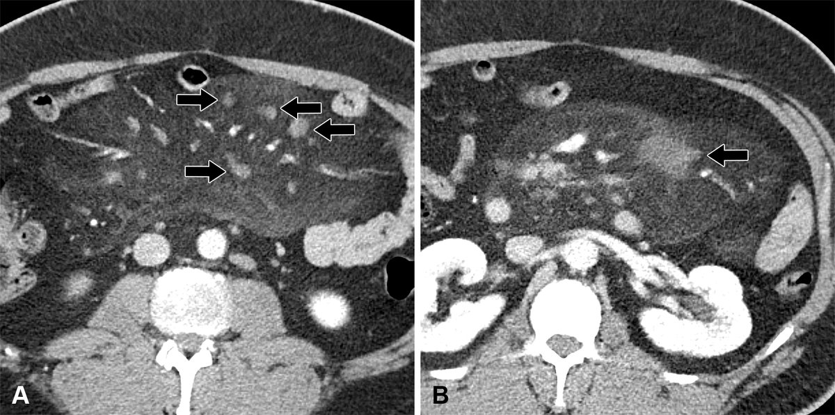
Incidental findings: common, often benign--but not always. This review updates ACR guidance, highlights gaps, and challenges prior recommendations across organs. A must-read to refine management and avoid unnecessary workup. @UCDRadiology https://t.co/SFsxFZA1LA
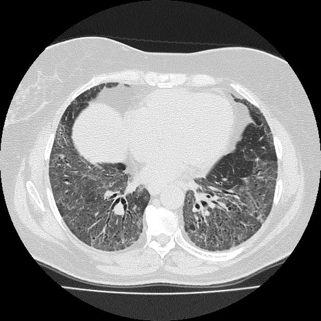
🩺 Case Answer | 51-Year-Old Male HRCT
The pattern demonstrates fibrotic consolidation with traction bronchiectasis and irregular reticulation, showing a central-to-peripheral continuity with upper lobe predominance, along the bronchovascular bundles, involving the hilar regions and extending to the periphery reaching the costal margins, along with cicatricial emphysematous changes.
This constellation is characteristic of fibrotic sarcoidosis.
While complicated silicosis remains a consideration, it typically demonstrates calcified conglomerate masses, background nodules, and frequently calcified mediastinal/hilar lymph nodes, often with relevant occupational exposure—which is not evident here. In contrast, no significant nodal enlargement or calcification is seen in this case, and sarcoidosis can present with predominant parenchymal fibrotic changes even in the absence of lymphadenopathy.
💡 Recognizing the emphysema as cicatricial is important, as it helps avoid misinterpretation as smoking-related emphysema (centrilobular or paraseptal).
📘 For more pattern-based HRCT interpretation of sarcoidosis, see my book Challenging Cases in Interstitial Lung Diseases: A High-Resolution CT Approach.
Important review of a common abnormality in clinical workflow!
Congenital Posterior Fossa Malformations: Imaging Patterns and Diagnostic Approach | RadioGraphics @RadioGraphics@RadG_Editor
https://t.co/oB9q82oUcI
TEACHING FILE.‼️‼️☣️
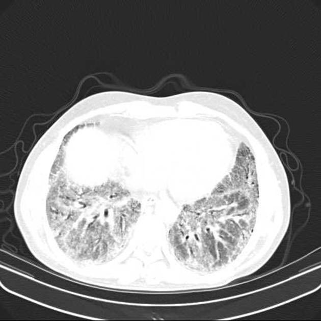
Non specific interstitial pneumonia (NSIP);
NSIP) is the second most common morphological and pathological pattern of interstitial lung diseases after UIP. NSIP is commonly associated with connective tissue disease (CTD), and a multidisciplinary team best decides the underlying diagnosis and management. Seen in adults aged 40-50yrs.
HRCT FINDINGS;
1. Ground glass opacities
2. Reticular opacities
3. Traction bronchiectasis
4.Lung volume loss
5. Consolidations in advanced cases.
Note ; the most prominent feature is ground glass opacities.
See attached images.
#Radres #Radiology #Radtwitter
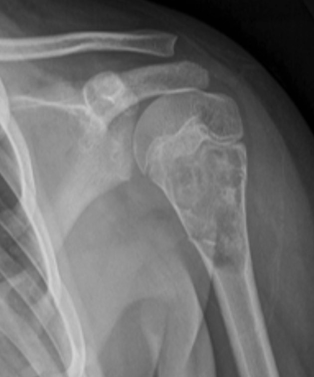
📖 #RadExam Study Guide 📖
A 13-year-old male presents with left upper arm pain. What is the most likely diagnosis?
A) Unicameral bone cyst
B) Aneurysmal bone cyst
C) Chondroblastoma
D) Enchondroma