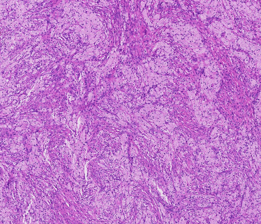
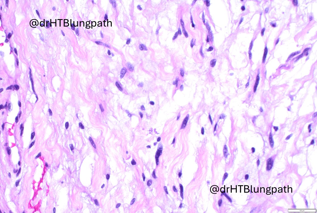
Interesting (histologically not challenging but rare) case of the day: young male presenting with a slowly growing posterior mediastinal mass (see pictures). Immunostains showed the target cells to be SOX10 positive and S100 essentially negative, while CD34 showed lattice-like staining in the background collagenous and myxoid stroma. STAT6 and keratins are negative. Beta-catenin showed no nuclear staining. What is your differential diagnosis? While not diagnostically challenging, making the right diagnosis here is important for the clinical implications.
#lung #pathology #PathTwitter #moffitt @USFpathology #pulmpath #thoracicpath
I want to recognize my colleague Dr. Yu Sun (@YuSun_YS@MoffittNews) for helping me on this case. Collaboration among colleagues is one of the greatest strengths of modern pathology (one of the main reasons why I became a pathologist), and it aligns deeply with the Moffitt's mission to contribute to the prevention and cure of cancer through transformative care, cutting‑edge science, and innovation. At Moffitt, teamwork is not just a cultural ideal but a core institutional value—reflected in its commitment to multidisciplinary excellence, shared expertise, and a collaborative environment supporting high‑quality, patient‑centered decision‑making.
PS: I am sorry for the watermarks. I have been alerted that my pictures have been published by others without my consent (I appreciate all of you who have asked for permission and acknowledge them).
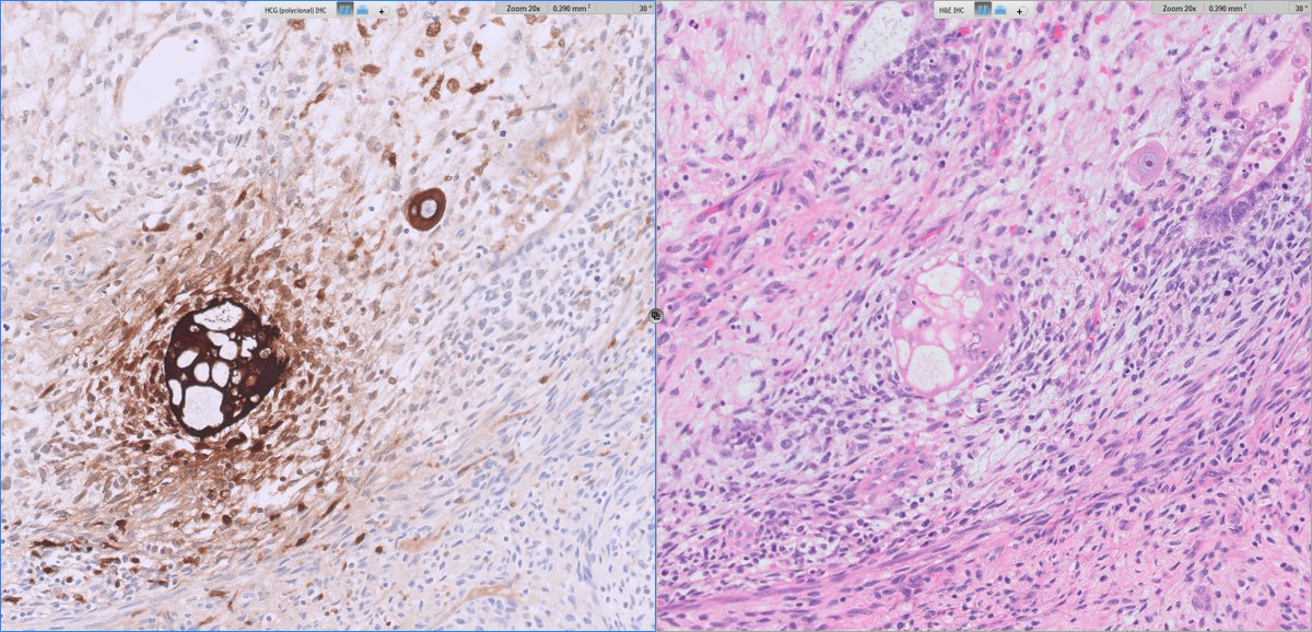
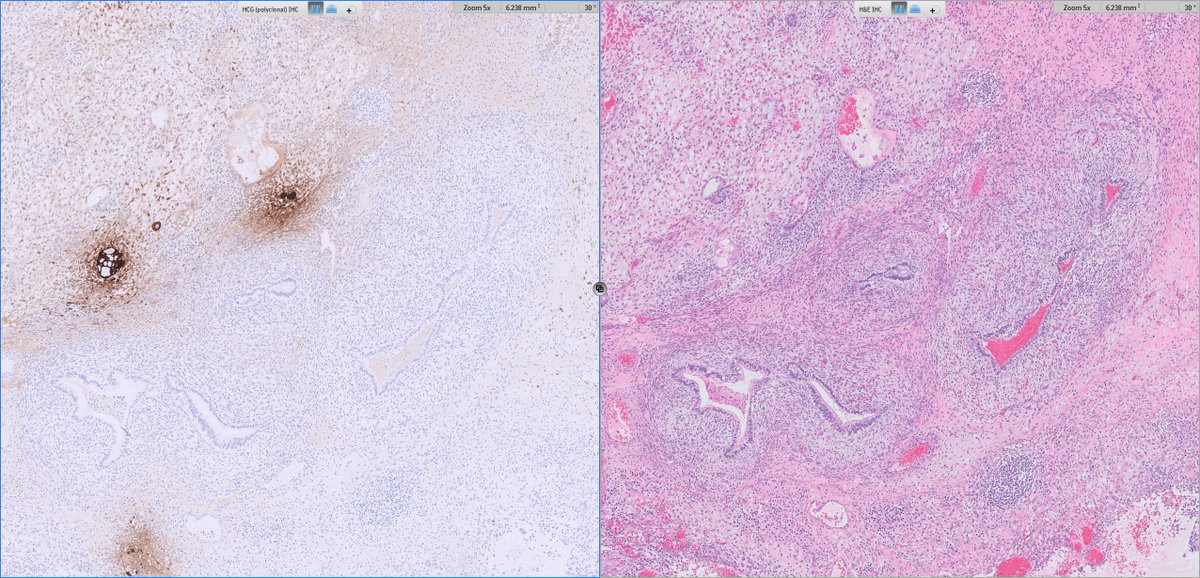
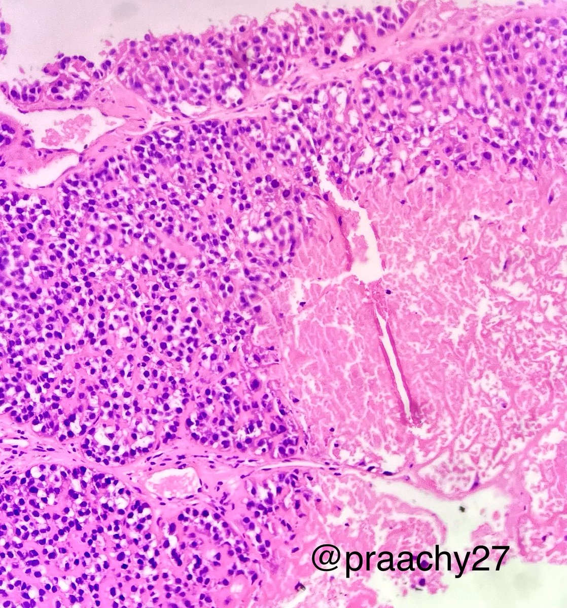
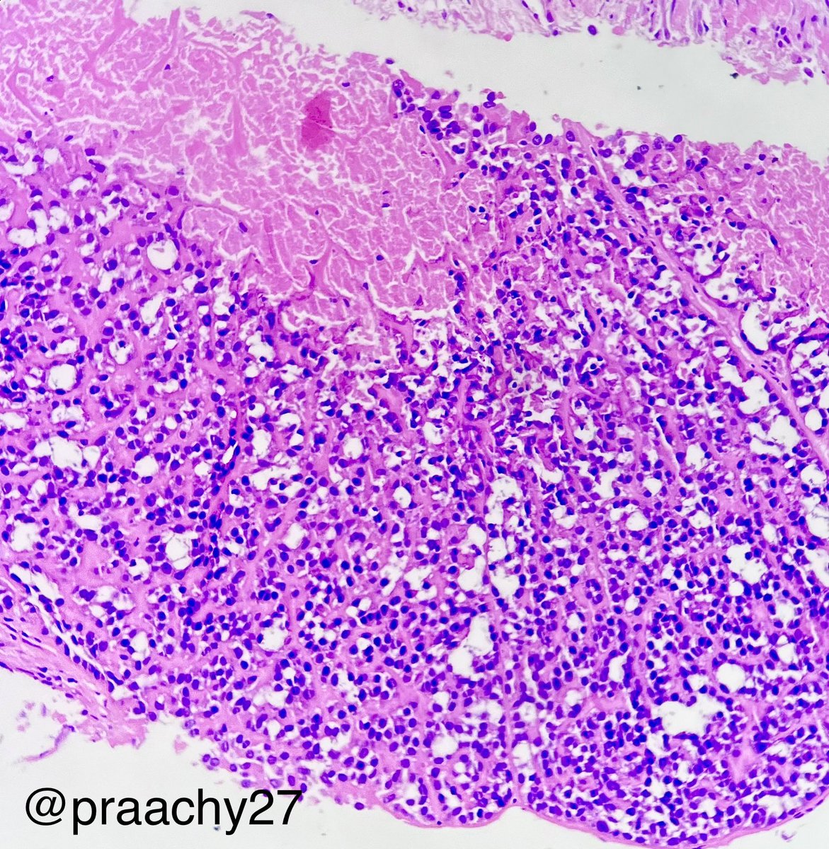
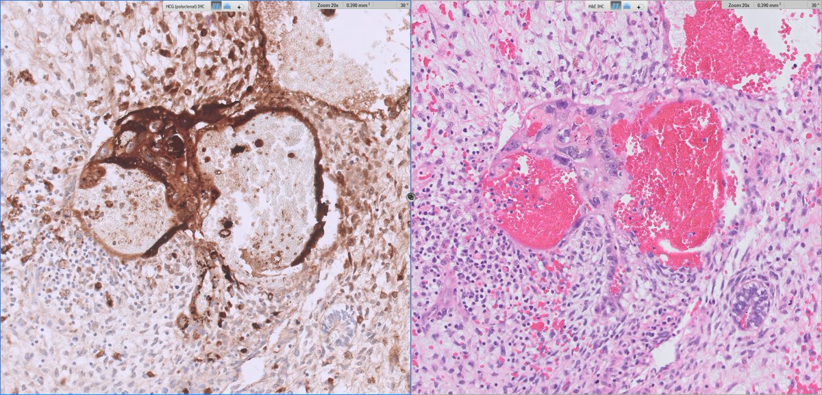
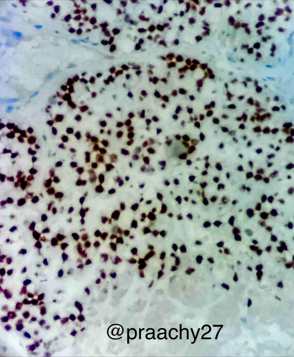
#GUpath orchiectomy:
mixed germ cell tumor (GCT) with admixed syncytiotrophoblasts
✔️may account for low serum elevations of HCG but NOT diagnostic of choriocarcinoma elements
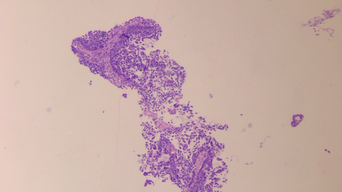
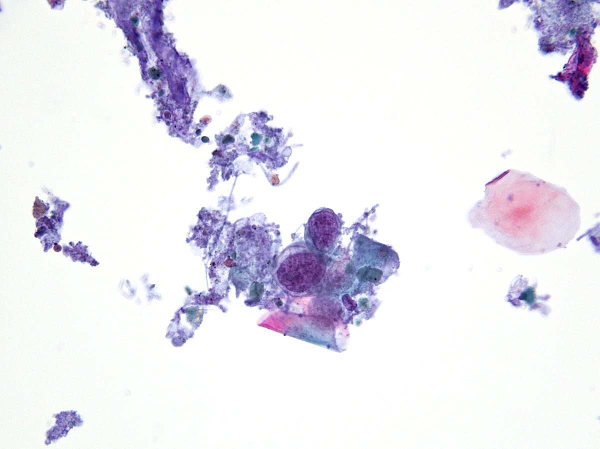
🫁 #CytoQuiz 🔬
A) Small cell lung carcinoma
B) SMARCA4-deficient undifferentiated tumor
C) NUT carcinoma
D) Large cell neuroendocrine carcinoma
#Pathology#Cytology#LungPath Clue in caption 🧐
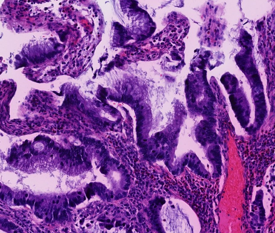
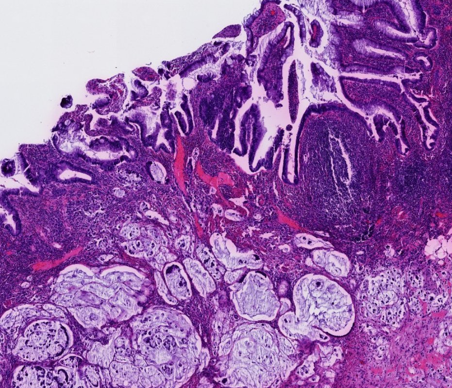
Patients with longstanding IBD (especially ulcerative colitis) are at risk for developing dysplasia and cancer.
Non-conventional dysplasia has been described in IBD:
- Hypermucinous (pic 1 and 2)
- Goblet cell deficient
- Dysplasia with increased Paneth cell differentiation
- Serrated dysplasia (including SSP/SSL-like, traditional serrated adenoma-like, and NOS)
- Dysplasia with terminal epithelial differentiation
P53 mutant pattern and loss of SATB2 expression can aid in the diagnosis of IBD-associated dysplasia.
The majority of non-conventional dysplasias were associated with ulcerative colitis and mucinous adenocarcinoma (pic 1).
Dr. Krasinskas - Tips and Hints in Surgical Pathology #USCAP #pathology #PathX
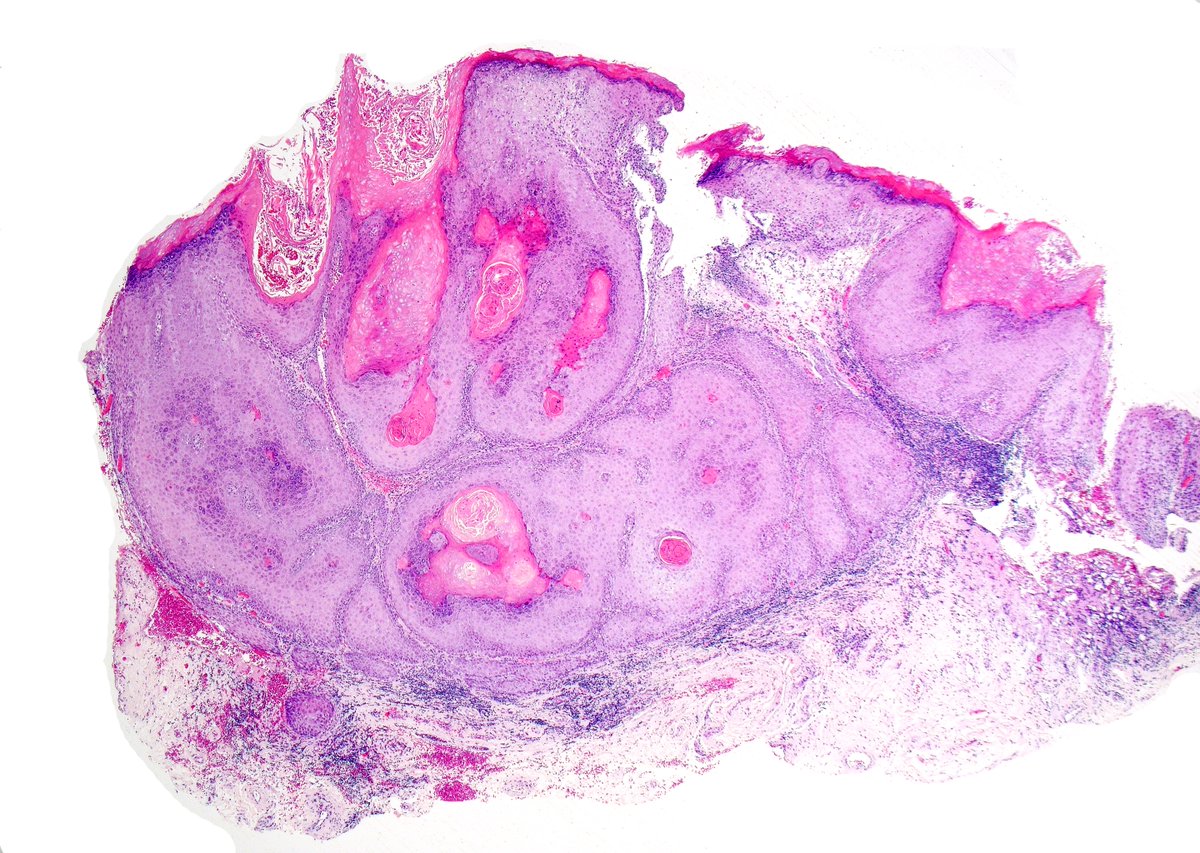
Verrucous carcinoma of the penis 🔬
Broad-based, exophytic lesion with pushing borders. Note the bulbous rete ridges and keratin-filled sinuses, classic for this low-grade variant. Despite its bland cytology, local invasion can be extensive. Early recognition is key.
#GUPath
Paneth-cell-like prostate adenocarcinoma: nests of tumor cells with abundant eosinophilic granular cytoplasm, reminiscent of intestinal Paneth cells—an uncommon morphologic variant. Recognition is key to ensuring accurate grading. #Pathology#GUPath#ProstateCancer#MedEd
Non-ossifying fibroma (NOF), see pics 1 and 2
Benign fibrohistiocytic tumor arising in the metaphysis of long bones in skeletally immature individuals (children)
Adult epiphyseal "NOF-like" lesions were previously called benign fibrous histiocytoma of bone but are now considered giant cell tumor of bone with degenerative changes.
H3.3 G34W IHC positivity supports GCTB (negative in NOF).
Dr. Lauer - Tips and Hints in Surgical Pathology #USCAP #pathology #PathX #PathTwitter
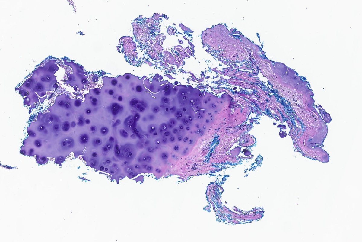
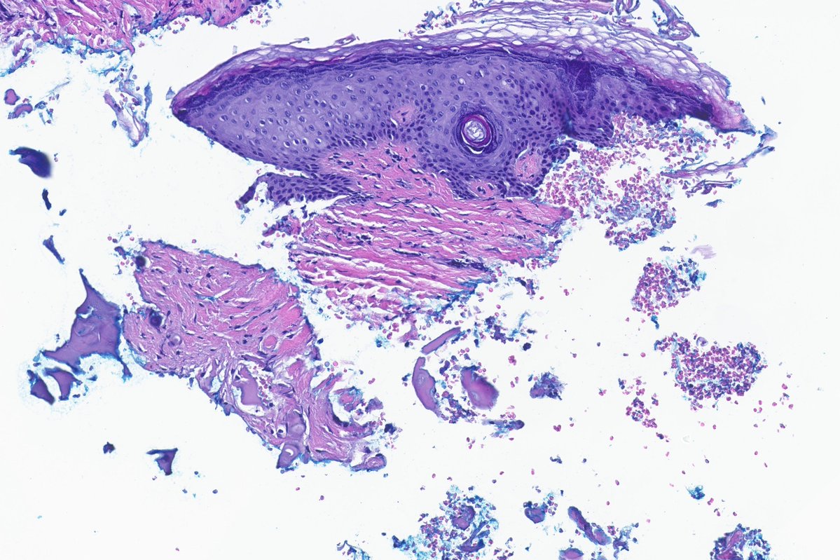
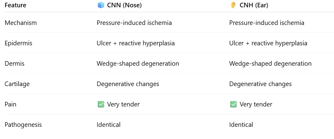
Let’s start the morning with pressure lesions! This one is rare but worth knowing—it's much more common on the ear.
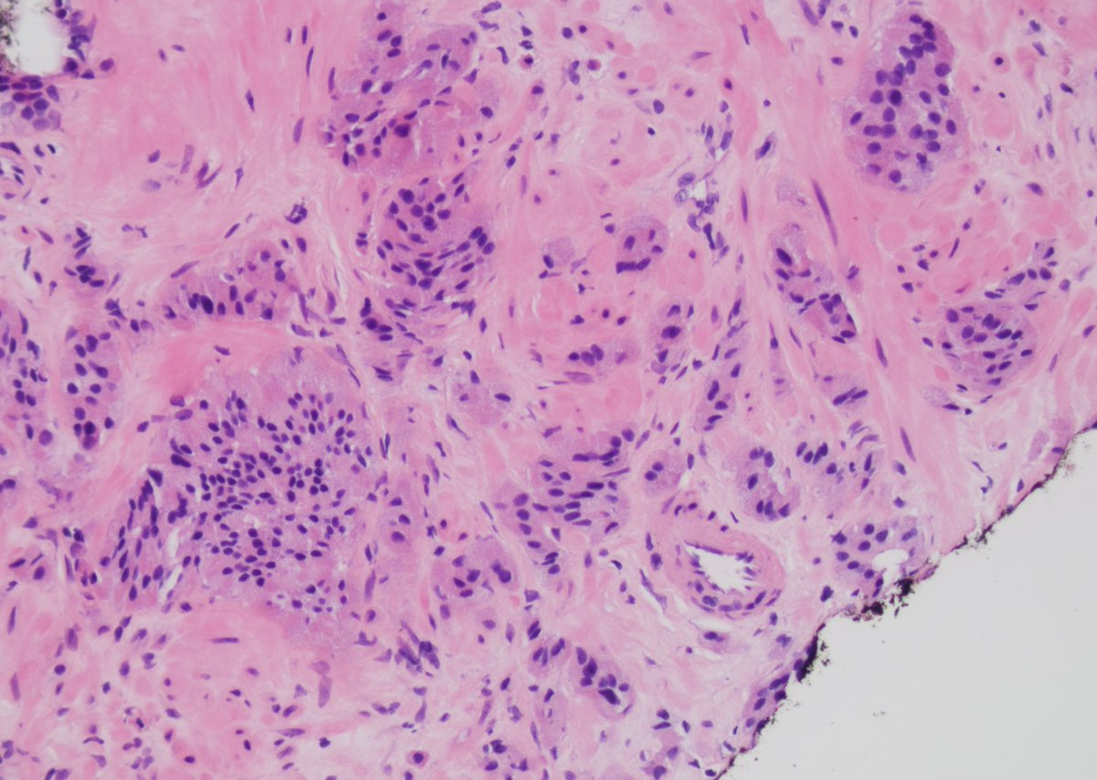
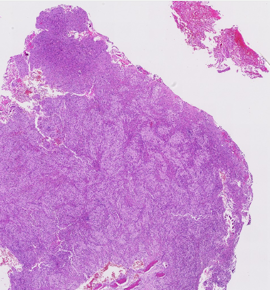
🛌Chondrodermatitis Nodularis Nasi (CNN):
👃 Painful, tender small nodule on the nasal tip/ala
⚠️ Central ulcer with wedge-shaped dermal ischemic degeneration
🧩 Underlying degenerative cartilage-chondrocyte dropout/fissuring
🔄 Pressure-induced ischemic repair pattern with granulation tissue — histologically identical to Chondrodermatitis nodularis helicis
#dermpath #dermatology #dermatologia #dermtwitter #pathology #pathologists #PathTwitter