🚨🔖Don't miss our new publication in @LIjournal!
🔷Comprehensive Spatial Transcriptomic Profiling of Cribriform, Intraductal, Atypical Intraductal Proliferation, and Ductal Adenocarcinoma of the Prostate
➡️Read here: https://t.co/ACUXXt4g5h
@MGBpathology@IntSocUropath@GU_Path_Society
#GUPath #MolPath #SpatialTranscriptomics #ProstateCancer
New from Laboratory Investigation 🔬 | Using spatial transcriptomics to characterize cribriform, intraductal, atypical intraductal proliferation, and ductal adenocarcinoma of the prostate. @TingZhaoPathdoc
🔗 Zhao et al. Read the article: https://t.co/Y9WBK8I1U1
🔖Check out the #AMP2026 abstracts published in The Journal of Molecular Diagnostics, highlighting the latest advances in molecular pathology!
💠https://t.co/YMISIdb3mx
@AMPath@JMDiagn#MolPath
The #AMP2026 abstract compendium is now available on the @JMDiagn website.
📖 Download it to see what's being presented this week in Seattle: https://t.co/CqkBlzjLId
📢 Mark your calendars!
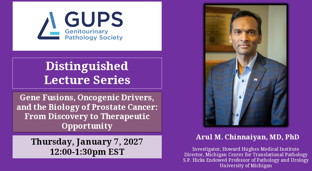
Join #GUPS for our Distinguished Lecture Series featuring Arul M. Chinnaiyan, MD, PhD
Topic: 🧬 Gene Fusions, Oncogenic Drivers, and the Biology of Prostate Cancer: From Discovery to Therapeutic Opportunity
📅 January 7, 2027
⏰ 12:00–1:30 PM EST
Already in Seattle for #AMP2026? Join us today for educational corporate workshops highlighting the latest technologies and innovations in molecular diagnostics!
@AMPath#MolPath
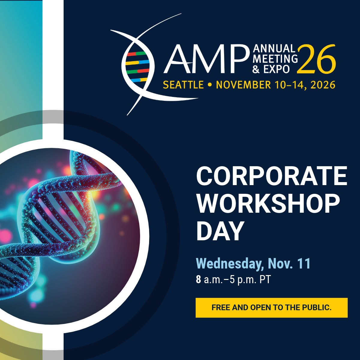
Tomorrow at #AMP2026: Corporate Workshop Day! Join us Wednesday for FREE educational workshops exploring emerging technologies and innovations in molecular diagnostics. Open to all attendees and the public. https://t.co/YT2FRatdnm
The #AMP2026 abstract compendium is now available on the @JMDiagn website.
📖 Download it to see what's being presented this week in Seattle: https://t.co/CqkBlzjLId
Thank you for this insightful comment, Riham! Argani et al. demonstrated that several "oncocytoid RCCs" arising in survivors of childhood neuroblastoma or medulloblastoma represent ESC RCCs harboring somatic TSC1/TSC2 alterations (PMID: 37522346). The patients with available treatment histories had received chemotherapy, although the specific agents were not detailed. The authors suggested a possible, incompletely characterized tumor-predisposition syndrome. However, investigating whether specific chemotherapeutic agents contribute to the acquisition of somatic TSC1/TSC2 alterations through treatment-associated mutational processes would also be an interesting area.
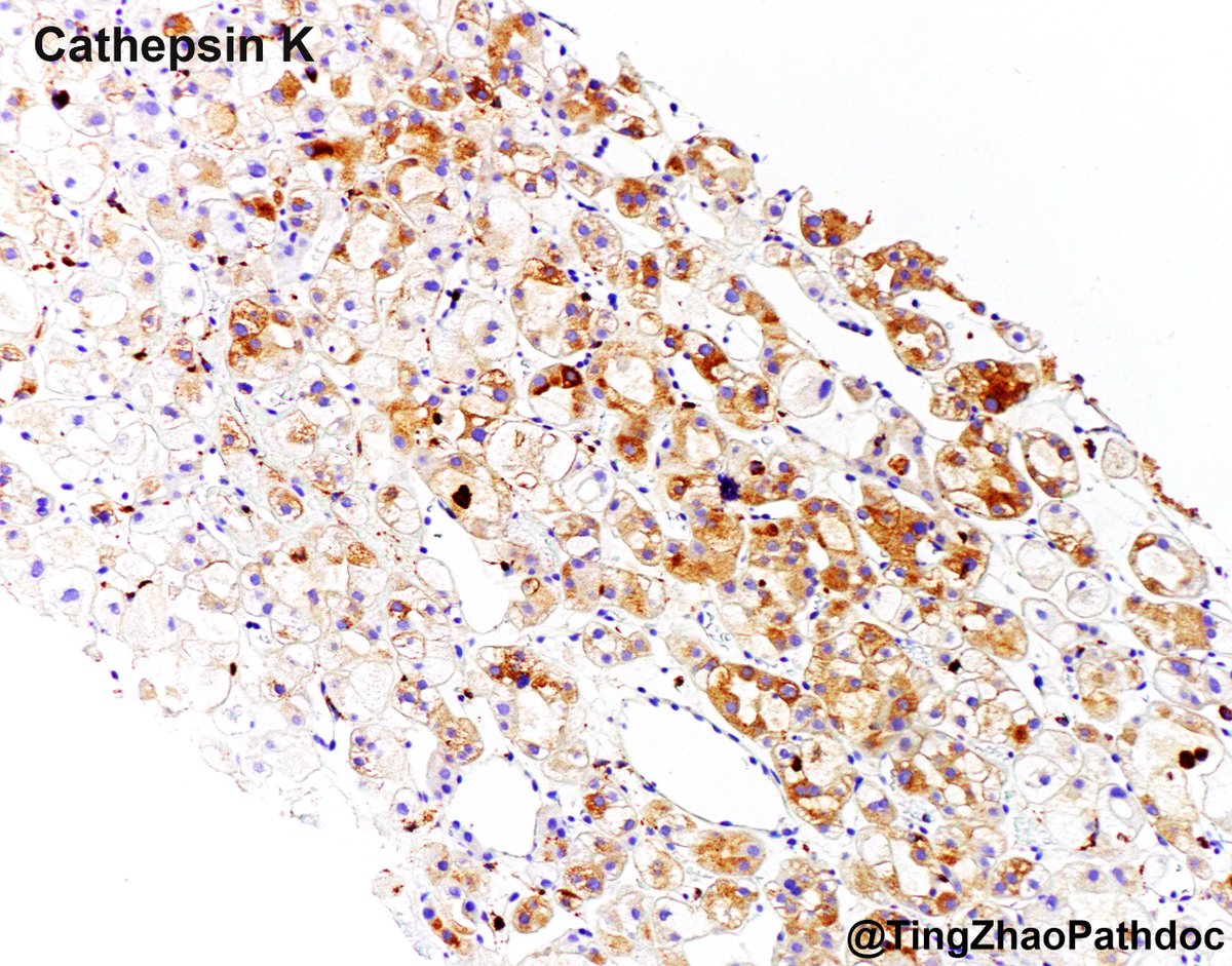
Great differentials! EVT and ESC RCC have overlapping morphologic, immunohistochemical, and molecular features. The tumor is CD117+ and CK20���. Taken together with the morphologic features, including intracytoplasmic vacuoles and prominent nucleoli, the tumor is best classified as EVT.
Great differential! EVT and ESC RCC have overlapping morphologic, immunohistochemical, and molecular features. The tumor is CD117+ and CK20−. Taken together with the morphologic features, including intracytoplasmic vacuoles and prominent nucleoli, the tumor is best classified as EVT.
@Trantor__ Nice catch! One mitosis was observed in the tumor. Rare mitoses are compatible with the diagnosis and have no proven prognostic meaning to date.
Great thoughts, Sergei! Both are important differential diagnoses, with overlapping morphologic, immunohistochemical, and molecular features. The tumor is CD117+ and CK20−, with retained FH expression. Taken together with the morphologic features, including eosinophilic cytoplasm with intracytoplasmic vacuoles and prominent nucleoli, as well as the additional IHC findings, the tumor is best classified as an EVT.