Broken kneecap: the deciding test is a straight leg raise.
Intact extensor mechanism, minimal displacement, brace it. Displaced, step off, or no straight leg raise, fix it. Bone heals. Quadriceps strength is the long game.
Aaron J. Krych, MD, demonstrates meniscal root repair using the SutureLoc™ implant and a FiberStitch™ 1.5 implant to augment the repair.
Watch the full video: https://t.co/8K45hDdULX
#Arthrex
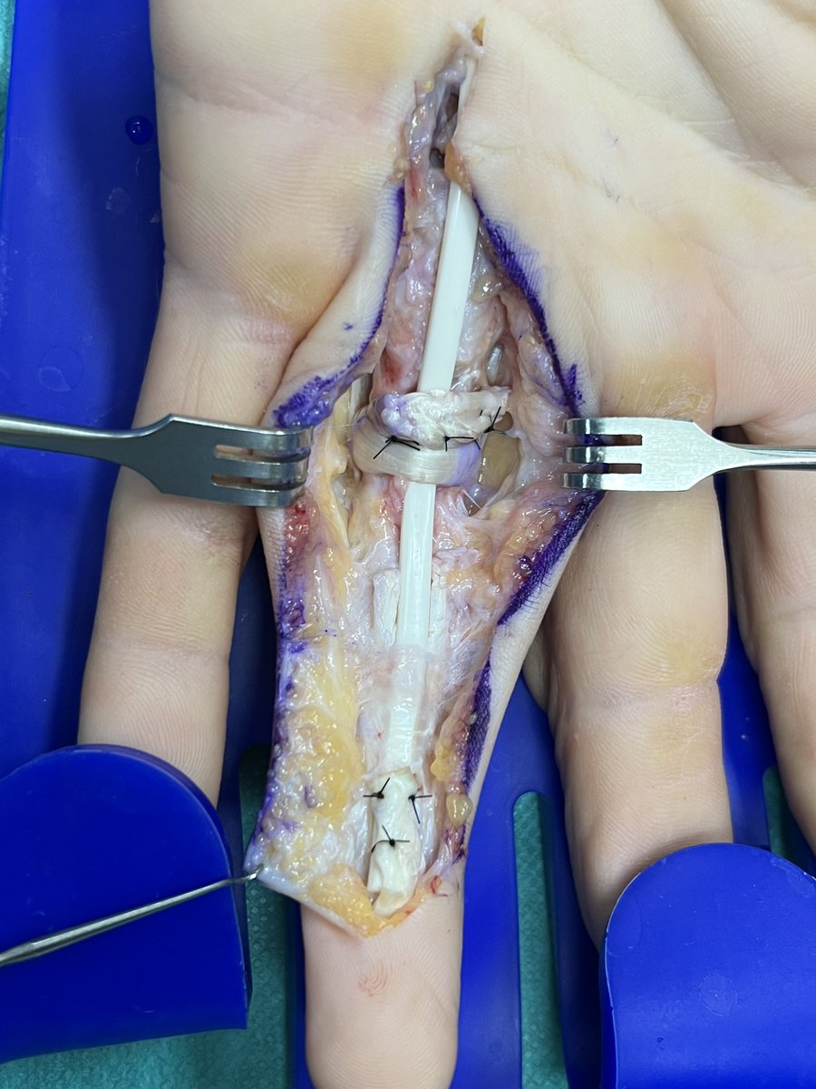
Que hacer si fracasa la sutura de un tendón?Reparación secundaria :
1* tiempo: reparación de poleas, nervios y varilla de silastic.
Movilización precoz y con esto crearemos una neocanal para el tendón definitivo en segundo tiempo.
Deep gluteal syndrome: role of ultrasound in its diagnosis
A 54-year-old patient with left paravertebral low back pain with gluteal and lower limb radiation of 16 months' duration. In the MRI of the hemipelvis, a fibrous band can be seen that is attached to the anterior face of the sciatic nerve, extending laterally to the greater trochanter and medially to the ischial tuberosity and the quadratus femoris muscle, corresponding to type 3 of the Hernando classification.
Dynamic ultrasound is performed, which shows reduced mobility of the sciatic nerve during knee and hip extension (comparative video of normal neural mobility: https://t.co/NiqvUemcdL).
Ultrasound has little value for the primary diagnosis of visualization of perineural fibrous bands, although it can often be appreciated a posteriori, guided by the findings in MRI. This could be useful for realizing fenestrations of the fibrosis after perineural infiltration using ultrasound control.
The great utility of ultrasound is the assessment of the mobility of the sciatic nerve during neurodynamic exercises, which in cases where it is decreased compared to the contralateral nerve, can be used as a secondary sign for the diagnosis of DGS.
young ,distal clavicle fracture for 30 days, non-synthesizable fragment your surgical conduct(note RX done with weight lifted from both upper limbs) @InvictaOrtho all twitters
Pes Anserinus/ Goose foot tendon/ pata de ganso, todos nombres que se usan para describir a la inserción tendinosa de los músculos Sartorio, Gracilis (Recto interno) y Semitendinoso en la cara anteromedial de la tibia proximal.
Clasificación Cierny Mader de #osteomielitis en adultos (1981):
Tipo I: Osteomielitis medular.
Tipo II: Osteomielitis superficial.
Tipo III: Infección localizada y estable.
Tipo IV: Osteomielitis difusa e inestable (articulaciones sépticas)