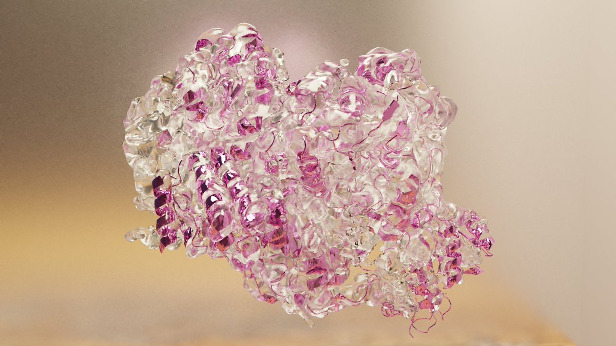
#cryoEM structure of ex vivo fibrils associated with extreme AA #amyloidosis prevalence in a #cat shelter, published now in @NatureComms https://t.co/tiVgRZmfQD
@James__Dunlea Hi James, thanks for the post. After a PhD and few postdocs I am now exploring the job market for industry, and this is an interesting layout. May I have your opinion on this: several ads I see require previous experience in industry. But without a 1st chance, how to get it?
The Lewy fold differs from the multiple system atrophy fold. Parkinson’s disease, Parkinson’s disease dementia and dementia with Lewy bodies share the same fold. Multiple conformers of assembled alpha-synuclein. As always with @SjorsScheres. https://t.co/4L2qSydgtB
Our structural study was headed by twitterless Stefano Ricagno @LaStatale@grupposandonato. @ChavesSanjuan collected beautiful cryo-EM data on a Talos Arctica 200kV, supervised reconstruction using #Relion and supported analysis. Negative stain prep by @valesper1.
Looking at amyloidogenic fibrils from heart tissue (ex vivo) on CryoEM and found this "bead repeat" contaminant I cannot identify... Anyone as any input? @ImagingArtifact ?
@SaelicesL@culkat5@Shumyla44@ImagingArtifact@SjorsScheres I have already thought about nucleic acids, but I'm not sure dimensions would fit. And what would "decorate" with such a regular spacing? I worked with nucleosomes and polymerases before (though not in microscopy) so of course it was my first thought
@culkat5@Shumyla44@ImagingArtifact@SaelicesL They are in at least 85% of our screen micrographs. Interestingly, they seem to be present in 2 different forms, single or clustered, though I'm not sure they are the same thing. And at times they seem attached to the amyloids, but it could be just co-exsistence @SjorsScheres
@culkat5@subletterdave@Shumyla44@ImagingArtifact@SaelicesL Glass pestles are used in the extraction process. I'm looking into the protocol to also identify possible non-biological contaminants. Though this is the same protocol used for other extractions, and it is interesting that we never saw this filament before.
@bwfalcon @ImagingArtifact Indeed the spacing seems that. I am looking into all possible protein coils that might be in the tissue. Interestingly, we have analyzed this type of tissue before, and never saw this contaminant. Thank you for your idea!
@SjorsScheres@ImagingArtifact@SjorsScheres it looks cool indeed, that is why I couldn't resist sharing and asking. Previous samples of the same type didn't have this one particular addition. Procollagen? Fibrinogen?
This human integrator complex, now released in #EMDB, resolved by #Singleparticle#cryoEM, gives new insight into how this protein works as a modular unit and identifies previously unknown sub-complexes.
@MolecularCell
DOI: https://t.co/J3ygcmE4q0
EMDB: https://t.co/BGilhXP3wG