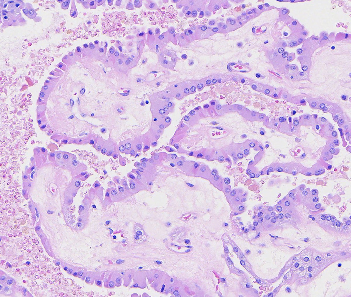
Papillary renal neoplasm with reverse polarity:
- Papillae lined by bland cuboidal cells with nuclei oriented AWAY from the basement membrane
- GATA3+
See the whole slide and more info:
https://t.co/3rAlL7a2Yu
Cortical microabscess formation is a clue to the presence of an infectious organism. Various bacteria, viruses, fungi, and even parasites may cause similar appearing lesions. This biopsy is taken from a renal transplant patient with acute kidney injury and microscopic hematuria. The low-power image shows a dense mixed cellular infiltrate involving the tubulointerstitium (Fig. 1). Higher magnification shows tubulitis with intratubular leukocytes and rare structures suspicious for fungal elements (Fig. 2). Gomori methenamine silver staining confirms the presence of budding yeast forms (Fig. 3 arrow) with hyphae and pseudohyphae. Fungal isolates from urine culture showed colony morphology characteristics consistent with Candida species.
#TeachingPoints #kidneypath #renal #pathology
A tumor with relatively bland nuclei can still be high-grade if it exhibits Pattern 4 or 5 architecture. This is the concept that many trainees initially miss.
#path_clicks#pathology#trending#progress#Histopathology
https://t.co/aMEIRGozAc
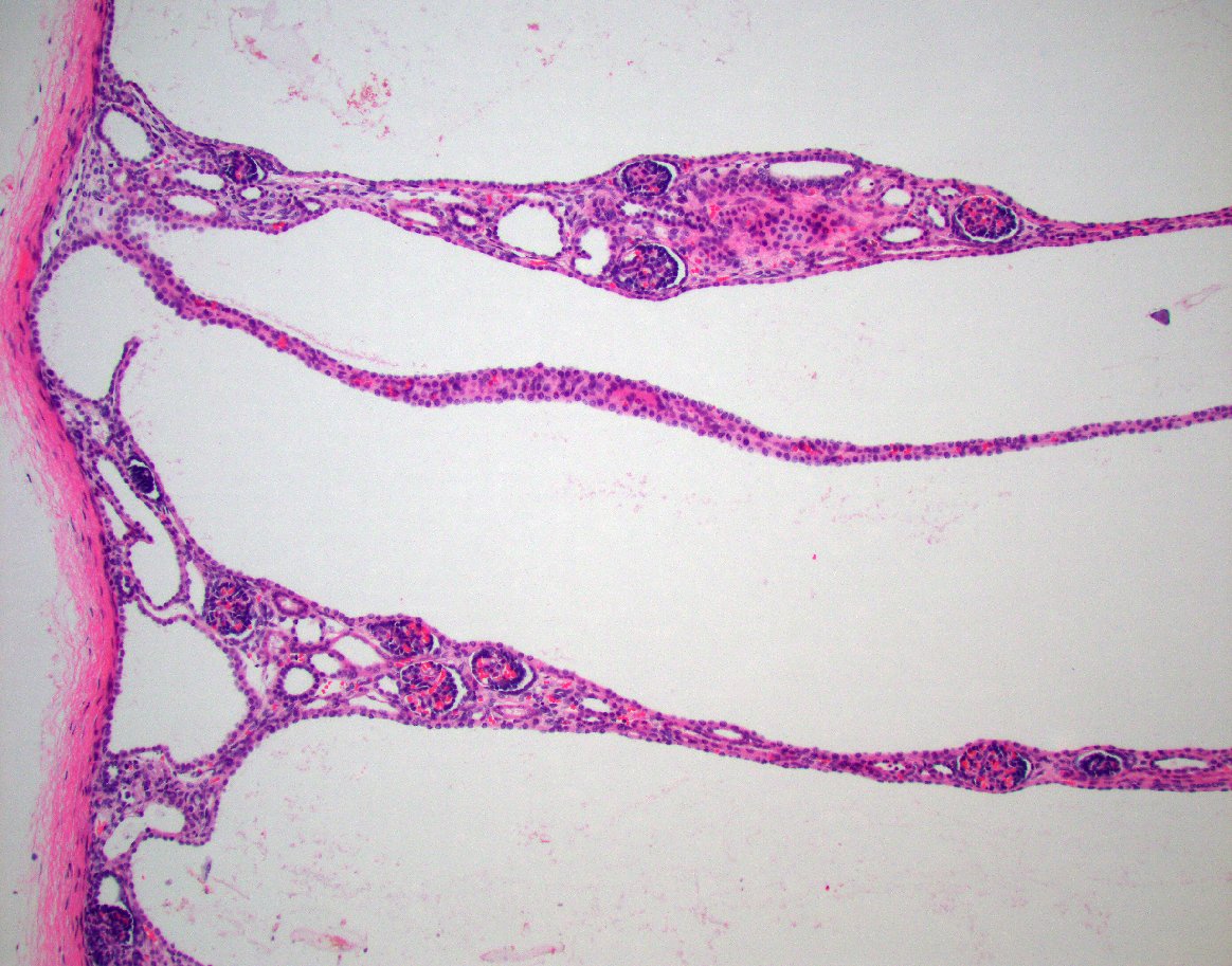
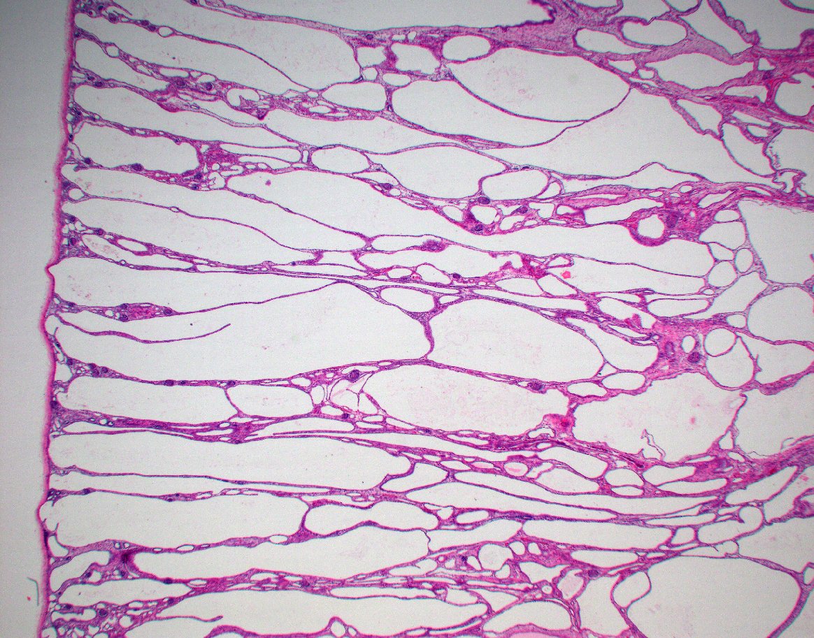
Classic example of autosomal recessive polycystic kidney disease. Large kidneys with poor corticomedullary differentiation radially arrayed cysts. Fusiform dilatation of radially oriented cortical and medullary collecting duct. #renalpath#gupath
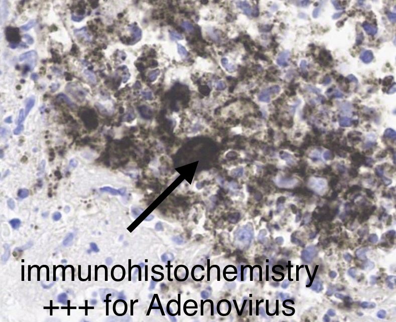
Kidney biopsy: Great teaching case
Severe inflammation and necrosis with abscess formation.
Look carefully=you see cells with large nuclei in the mix of inflammatory cells.
IHC= Positive staining for ADENOVIRUS.
DX: Adenovirus-associated interstitial nephritis.
55-yr old with kidney &
heart transplant, fevers, rise in serum creatinine.
The EM photomicrograph shows capillary walls that are uniform and of normal thickness with no electron dense immune complex-type deposits noted within the capillary walls or mesangium. Additionally, numerous podocyte cell bodies are present and unremarkable and epithelial foot processes show no significant effacement. At the top left of the image you can even see Bowman’s capsule and unremarkable parietal epithelium. These findings are those of a normal glomerulus by electron microscopy. No pathologic changes are present in this image.
#renalpath #kidneypath #pathology #renal #pathtwitter
Rare renal manifestation of malaria-associated AKI.
Kidney biopsy showed collapsing glomerulopathy with severe acute tubular injury in a patient with P. vivax infection. IF negative.
An uncommon but important association.
#NephPath #RenalPath#KidneyBiopsy #Malaria#CollapsingGN
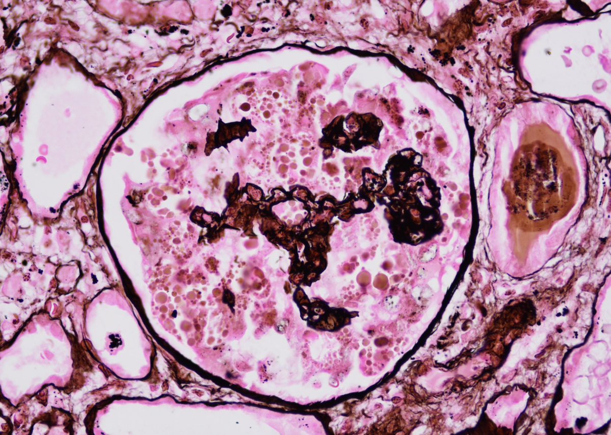
The kidney biopsy showing severe tubular injury, with several foci displaying nuclear cytopathic change typical of BKV infection (arrows). Inset: an SV40 stain for BKV antigen is strongly positive in the abnormal tubular nuclei, confirming the presence of BKV.
🔗Read more👉🏻 https://t.co/S9nU6kDZcP