I'm tapped out for time today, but I'll have some more time later. Here's the general idea that I think would be fun to create a thread for and just let it be a "working" thread continually added to and hopefully even generates public interest for community contributions as it will probably generate "by products" of interest and understandings on baby formula, autism, and disease trajectories indirectly.....
I suspect that "Alzheimer’s p tau" found in newborns vs the older population is a tale of two worlds, one powering up in controlled, progressive fashion and the other powering down as certain functions become in a state of "disrepair" on various levels. Perhapd in the newborn, a controlled "powering" up of the affected p tau cells via stimuli from the gut microbiome via its natural and HEALTHY proliferation to progressively bring the p tau affected cells fully online with an optimally functional cell. This might make sense when one thinks about the limited time windows for decisions on allocations/designations for further compartmentalized activities/specialization for various time alignments...... Honestly, typed this on the fly to respond in a timely manner, but I'm going to double back to more clearly type out the idea by tomorrow. I would absolutely love to generate some community interest/ideas/thoughts for this topic to see what can be pieced together. There's alot to unpack here and iron out, but I'd be excited to see what comes of this.
Peace and love, my friend ♥
@MitoPsychoBio Nice! Can't wait to read. Did you touch on RIG-1 in this? It's fascinating that lithium exposure thresholds have an association in higher autism rates across a population, but when looking at alzheimer's, it's helpful..... RIG-1! RIG-1! I should probably read this now 😆
This crap blew me away yesterday that a facet of stem cell quiescence is active here via the extrachromosomal DNA. It's a reverberation of the problem healthy stem cell lines have in the context of longevity with maintenance of TFAM>sirt3..... but here in this context spermidine, an activator of sirt 3, seems to be their undoing. Went to bed and woke up thinking about how CrAzY this study was in contrast of stem cell activity vs the context here.
Cancer diagnoses in England hit record high of more than 1,000 people a day
“According to the latest cancer registration statistics, more than 373,000 people were diagnosed with cancer in England in 2024, the highest figure on record. On average, NHS England diagnosed 1,021 people a day with cancer in 2024, up from 972 a day in 2023.”
https://t.co/KjXt4BIGsn
DGRs! DGRs!
Bacteroides has been having the run lately on two studies.
Uniformis predominance in colon cancer, and fragilis for pentadecanoic acid generation...... so, good and bad.
🧬🔥 What if the genetic mutations within gut bacteria reveal more about human health than which bacterial species are present?
A new study in Nature introduces a microbiome-wide PheWAS, connecting 12.8 million microbial single-nucleotide variants (SNVs) to human aging, metabolism, cardiovascular traits, and environmental exposures.
🔬 1. Mapping microbial genetics across 10,781 people
Researchers analyzed 11,831 stool metagenomes from six cohorts in the Netherlands and Tanzania.
The study examined 244 host phenotypes and exposures, identifying:
• 49,346 robust SNV–phenotype associations
• 940 independent association signals
• 101 microbial species
• 69 host traits and exposures
Associations were evaluated using population-structure-adjusted linear mixed models and cross-cohort meta-analysis.
⚡ 2. Microbial mutations reveal signals missed by species abundance
Remarkably, 65.7% of independent SNV association signals lacked a corresponding species-abundance association.
Among 533 species–phenotype pairs associated at the SNV level, 460 were detected only through SNV analysis.
This demonstrates why species-level microbiome profiling can miss important genetic variation.
🧬 3. Aging is linked to bacterial glutathione transport
Age showed the largest number of independent associations: 99 signals across 42 species.
One prominent signal involved five missense variants in the sarA_2/oppA gene of Faecalibacterium prausnitzii_I.
The lead variant, F343Y, was associated with approximately 4.9 years higher host age.
Meta-analysis: P = 1.0 × 10⁻¹⁷.
🔋 4. A potential oxidative-stress adaptation mechanism
Structural modeling placed age-associated variants near the substrate-binding cavity of an ABC-type oligopeptide transporter.
Sequence annotation and molecular docking suggested that this complex may transport glutathione.
A proposed mechanism is:
Host aging → Altered intestinal oxidative environment → Selection of microbial transporter variants → Potential changes in glutathione uptake
However, altered transporter function and selective advantage remain unvalidated hypotheses.
🤖 5. Microbial SNVs may help predict age
Machine-learning models trained on microbial SNVs explained 12.8–42.0% of host age variation across external test datasets.
This suggests potential for strain-resolved microbiome biomarkers, although predictive performance requires broader validation.
🎯 6. Implications for precision microbiome medicine
Integrating microbial SNVs with metabolomics, protein structures, and host phenotypes could reveal previously hidden functional pathways relevant to metabolic and inflammatory diseases.
Important limitations include predominantly Dutch cohorts, observational associations, and the absence of experimental confirmation for most predicted functional effects.
💡 Key takeaway: The microbiome's biological relevance extends beyond bacterial abundance. Strain-level genetic variation provides an additional layer connecting microbial adaptation to human aging and metabolic health.
📄 Wang D, Goumagias A, Andreu-Sánchez S, et al.
Microbiome-wide PheWAS links gut microbial SNVs to health and exposures.
Nature (2026).
DOI: https://t.co/PyR2zCrsFB
SNV-MWAS Atlas: https://t.co/QVxd0Q2rsH
Dataset: https://t.co/LrEpKwJ4gI
#GutMicrobiome #MicrobialGenetics #PheWAS #Metagenomics #SNV #Faecalibacterium #Glutathione #HealthyAging #PrecisionMedicine #MetabolicHealth #MicrobiomeResearch
The standard advice on mast cells assumes it’s a 100% immune issue, but your collagen architecture is what actually sets your reaction threshold.
Mast cells do not float freely in isolation. They live anchored inside your connective tissue, surrounded and supported by a matrix of collagen fibers.
Think of a mast cell like a sensitive motion detector mounted to the drywall of a room. If the walls are solid and stable, the detector only trips when an actual intruder walks through the door.
When collagen architecture is abnormal or overly compliant, that structural frame loses its stability. The tissue matrix shifts under everyday mechanical tension, continuously rattling the sensor at baseline.
Because the local micro-environment is physically unstable, the mast cell's activation threshold drops significantly. It begins releasing inflammatory mediators in response to minor everyday exposures, from common foods and fragrances to temperature shifts.
This is why reactivity often persists even after eliminating every possible trigger. The immune cells are responding directly to an unsettled physical foundation that keeps them on permanent high alert.
Calming chronic reactivity requires accounting for that structural environment, rather than treating the response as a purely chemical glitch.
https://t.co/Edvgh6CEKe
@vipintukur I loved looking at this study a few weeks ago.... Bacteroides has been having quite the "run" on studies between uniformis and fragillis. Thanks for giving the study a share. It was a big one.
Bacteroides ❤️
IYKYK
Epigenetic age is not a one-way street for the ovary. Sharif Iqbal and Vadim Gladyshev use pan-mammalian DNA methylation clocks to show ovarian epigenetic age runs in both directions: an old ovary placed in a young mouse becomes epigenetically younger. Conversely, a young ovary in an old mouse grows older. https://t.co/ddt9wHIPZp
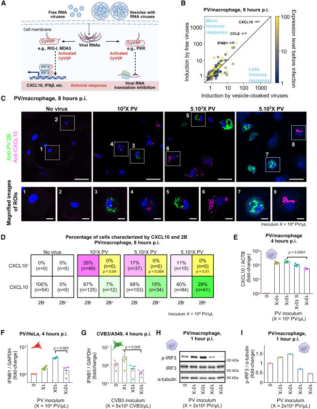
A cell must be infected by significantly more than one viral genome simultaneously for the virus to overcome the cell's innate immune defenses and successfully replicate @CellCellPress@AltanBonnet@NIH@g_altanbonnet
https://t.co/wM2Xp81l7s
🧬🔥 What if the ability to store fat depends on how cells stabilize a single mRNA?
A new study in Nature Communications identifies Star-PAP (TUT1) as a critical regulator of adipose tissue homeostasis, connecting RNA polyadenylation, insulin receptor expression, acetyl-CoA metabolism, and epigenetic control of lipogenesis.
The central discovery: Star-PAP stabilizes insulin receptor (InsR) mRNA, sustaining insulin signaling and adipocyte lipid storage.
🔬 1. Human genetics identifies a rare Star-PAP variant
Whole-exome sequencing of 2,161 lean controls and 1,944 individuals with obesity identified the rare p.D469N variant in six lean individuals and none of the obese cases.
The variant accelerated proteasomal degradation of Star-PAP.
However, its rarity limits conclusions about human metabolic protection.
⚡ 2. Star-PAP deficiency causes severe lipodystrophy
Adipocyte-specific Star-PAP knockout mice developed progressive adipose atrophy.
Fat mass decreased by:
• 29.8% at 5 weeks
• 41.6% at 24 weeks
Under high-fat feeding, fat mass was reduced by 81.6% compared with controls.
Despite reduced adiposity, knockout mice developed severe insulin resistance, hyperinsulinemia, dyslipidemia, and hepatic steatosis.
Less adipose tissue does not necessarily mean better metabolic health.
🧬 3. Star-PAP directly regulates insulin receptor mRNA
Mechanistic experiments demonstrated that Star-PAP binds the InsR mRNA 3′-UTR, supporting polyadenylation and transcript maturation.
Star-PAP deficiency reduced:
• InsR expression
• Insulin-stimulated AKT signaling
• GLUT4 expression
• Lipogenic enzymes ACACA and FASN
Restoring InsR expression partially rescued adipose mass and metabolic abnormalities.
🔥 4. Insulin signaling connects RNA processing to acetyl-CoA metabolism
¹³C-glucose tracing revealed impaired carbon incorporation into citrate, malate, and newly synthesized palmitate.
Star-PAP deficiency reduced intracellular pyruvate, acetyl-CoA, and downstream acyl-CoA metabolites.
The proposed pathway:
Star-PAP ↓ → InsR mRNA stability ↓ → Insulin signaling ↓ → Glucose utilization ↓ → Acetyl-CoA ↓ → Lipogenesis ↓
🧪 5. Acetyl-CoA links metabolism to epigenetic regulation
Reduced acetyl-CoA coincided with decreased histone H3K9/H3K14 acetylation and chromatin accessibility at lipogenic genes.
Remarkably, pyruvate supplementation restored acetyl-CoA availability, partially recovered histone acetylation, and rescued adipocyte lipid accumulation in vitro.
This establishes a mechanistic connection between RNA processing, metabolic substrate availability, and chromatin regulation.
🎯 Therapeutic implications
The Star-PAP–InsR–acetyl-CoA axis represents a potential target for lipodystrophy and metabolic dysfunction.
However, pyruvate rescue was demonstrated in cellular models, not patients, and additional Star-PAP RNA targets may contribute to the phenotype.
Key takeaway: Adipose tissue health depends not simply on fat quantity, but on the molecular machinery that maintains insulin responsiveness and safe lipid storage.
📄 Chen N, Zhao Y, Chen Y, et al.
Stabilization of insulin receptor mRNA by Star-PAP in metabolic homeostasis.
Nature Communications. Published online October 29, 2026.
DOI: https://t.co/UyojYtvPHd
#AdiposeTissue #StarPAP #TUT1 #InsulinResistance #Lipodystrophy #AcetylCoA #Epigenetics #Lipogenesis #MetabolicHealth #RNAProcessing #NatureCommunications
Dysregulation of mTORC2 in intestinal cells can cause neuropsychiatric effects such as depression, anxiety, cognitive impairment due to dysbiosis, chronic visceral pain, and inflammation [126].
https://t.co/cBlQP9cT8r
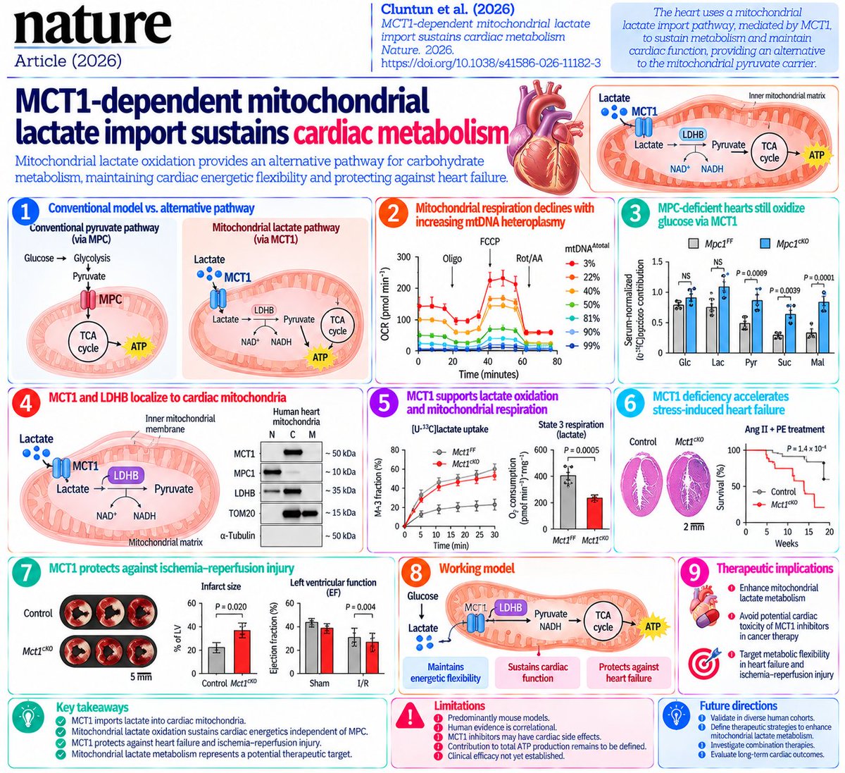
🧬🫀 What if the heart has a second mitochondrial pathway for burning carbohydrates—one that bypasses the mitochondrial pyruvate carrier and protects against heart failure?
A new study in Nature challenges the conventional model of cardiac energy metabolism by demonstrating that MCT1 transports lactate into cardiac mitochondria, where LDHB converts it into pyruvate and NADH to support respiration.
The central discovery: Mitochondrial lactate oxidation provides an alternative pathway for carbohydrate metabolism, sustaining cardiac energetic flexibility during physiological and pathological stress.
🔬 1. Challenging the classical model of cardiac metabolism
The traditional model proposes:
Glucose → Glycolysis → Cytosolic pyruvate → MPC → Mitochondrial TCA cycle → ATP
Lactate is generally thought to require cytosolic conversion into pyruvate before entering mitochondria.
However, using cardiomyocyte-specific Mpc1 knockout mice, researchers observed that glucose-derived carbon continued entering the cardiac TCA cycle despite loss of the mitochondrial pyruvate carrier.
Surprisingly, glucose-derived labeling of several TCA intermediates actually increased in young MPC1-deficient hearts.
This suggested the existence of an alternative mitochondrial carbohydrate-entry pathway.
⚡ 2. MCT1 provides an MPC-independent metabolic route
Researchers identified monocarboxylate transporter 1 (MCT1/SLC16A1) as a critical component of this alternative pathway.
In MPC1-deficient mice, pharmacological inhibition of MCT1 using AZD3965 abolished the residual glucose-derived carbon contribution to cardiac TCA metabolism.
Conversely, deleting MCT1 reduced lactate-derived TCA labeling while largely preserving glucose-derived labeling.
These findings reveal functional redundancy between two mitochondrial substrate-entry systems:
MPC → Pyruvate transport
MCT1 → Lactate transport
This redundancy helps explain how the heart maintains carbohydrate oxidation despite disruption of one pathway.
🧬 3. MCT1 and LDHB form a mitochondrial lactate oxidation system
The investigators detected MCT1 in the inner mitochondrial membrane of mouse and human cardiomyocytes.
They also identified LDHB within the mitochondrial matrix.
The proposed mechanism:
Lactate → MCT1-mediated mitochondrial import → LDHB-mediated oxidation → Pyruvate + NADH
The generated pyruvate can contribute carbon to the TCA cycle, while NADH supplies reducing equivalents for mitochondrial respiration.
This arrangement allows lactate oxidation to occur without competing directly with cytosolic glycolysis for NAD⁺.
Importantly, the study provides biochemical and functional evidence supporting the long-debated mitochondrial lactate shuttle hypothesis.
🔍 4. Direct evidence from mitochondrial isotope tracing
Researchers combined several complementary approaches:
• [U-¹³C]glucose tracing
• [U-¹³C]lactate tracing
• [2-²H]lactate tracing
• Rapid mitochondrial isolation using MITO-Tag
• Proteinase K protection assays
• Isolated mitochondrial respirometry
The results demonstrated that MCT1 deficiency impaired mitochondrial lactate entry and reduced lactate-supported oxygen consumption.
By contrast, MPC1 deficiency selectively impaired pyruvate-supported respiration while preserving lactate-supported respiration.
Experiments using isolated human cardiac mitochondria further supported MCT1-dependent lactate oxidation.
🔥 5. Lactate oxidation can support respiration independently of TCA-cycle carbon oxidation
One particularly intriguing observation involved inhibition of pyruvate dehydrogenase and α-ketoglutarate dehydrogenase using CPI-613.
Although CPI-613 markedly suppressed lactate-derived citrate labeling, lactate-supported mitochondrial respiration remained largely unchanged.
This suggests that LDHB-generated NADH can contribute to respiratory electron transport independently of complete lactate-carbon oxidation through the TCA cycle.
The distinction is important:
Lactate is both a carbon substrate and a source of reducing equivalents.
This expands the conventional understanding of lactate as merely a precursor for mitochondrial acetyl-CoA production.
🫀 6. MCT1 deficiency accelerates stress-induced heart failure
Under baseline conditions, cardiomyocyte-specific MCT1 knockout mice maintained relatively normal cardiac function and exercise capacity.
However, under pathological stress, the phenotype changed dramatically.
Following angiotensin II plus phenylephrine treatment, MCT1-deficient mice developed:
• Greater cardiac hypertrophy
• Reduced left ventricular ejection fraction
• Reduced stroke volume and cardiac output
• Increased mortality
Similar functional deterioration occurred following transverse aortic constriction.
Thus, mitochondrial lactate metabolism appears particularly important when the heart faces increased energetic demand.
⚠️ 7. Loss of MCT1 worsens mitochondrial damage
Electron microscopy revealed that MCT1-deficient cardiomyocytes exhibited abnormal mitochondrial morphology and reduced cristae density.
Under neurohormonal stress, these abnormalities became more pronounced.
MCT1 deficiency was also associated with:
• Impaired lactate-derived TCA metabolism
• Altered myocardial nucleotide metabolism
• Increased mitochondrial oxidative stress
• Abnormal mitochondrial calcium handling
These findings suggest that MCT1-dependent lactate oxidation contributes to mitochondrial structural integrity and metabolic resilience.
🩸 8. MCT1 protects against ischemia–reperfusion injury
In mouse models of myocardial ischemia–reperfusion injury, MCT1 deficiency increased myocardial necrosis.
Following injury, control mice recovered systolic function toward baseline.
In contrast, MCT1-deficient mice exhibited persistent impairment in left ventricular function during follow-up.
These findings identify mitochondrial lactate metabolism as a potential determinant of myocardial recovery after ischemic injury.
🧪 9. Human heart failure samples support biological relevance
Researchers examined myocardial samples from non-failing donors and patients with advanced heart failure.
MCT1 was detectable in purified human cardiac mitochondria.
Interestingly, mitochondrial MCT1 abundance was increased in failing hearts compared with donor controls.
This may represent a compensatory response to impaired mitochondrial metabolism.
However, whether increased mitochondrial MCT1 is protective, maladaptive, or simply associated with disease progression remains unresolved.
🎯 10. Therapeutic implications
The study identifies several potential research directions:
Enhancing mitochondrial lactate utilization
Could increasing mitochondrial MCT1 activity improve energetic flexibility in stressed cardiomyocytes?
Preserving mitochondrial LDHB activity
Could optimizing matrix-localized lactate oxidation support NADH production and respiratory function?
Targeting metabolic redundancy
Could coordinated modulation of MPC and MCT1 improve adaptation to pathological cardiac stress?
Reconsidering systemic MCT1 inhibition
MCT1 inhibitors are being investigated in oncology. These findings raise questions about potential cardiac consequences, particularly in patients with pre-existing cardiovascular disease.
Importantly, the study does not establish a clinically validated MCT1-targeted therapy.
⚠️ 11. Important limitations
The causal cardiac injury experiments were predominantly performed in mice.
Human evidence supports mitochondrial MCT1 localization and lactate oxidation but does not establish therapeutic efficacy.
MCT1 also functions at the plasma membrane, and its mitochondrial and cytosolic transport roles cannot be completely separated in the in vivo knockout experiments.
The relative contribution of mitochondrial lactate oxidation to total ATP production under different human physiological conditions remains uncertain.
The broader implication
Lactate is not merely a glycolytic waste product.
It is an important oxidative fuel and redox substrate.
This study establishes an MCT1–LDHB-dependent mitochondrial pathway that allows the heart to oxidize lactate independently of the conventional mitochondrial pyruvate carrier.
The findings provide a new mechanistic framework for understanding cardiac metabolic flexibility, mitochondrial redox homeostasis, and susceptibility to heart failure.
The next challenge is determining whether selective enhancement of mitochondrial lactate metabolism can improve cardiac recovery without disrupting systemic lactate transport.
📄 Cluntun AA, Visker JR, Velasco-Silva JN, Carrington J, et al.
MCT1-dependent mitochondrial lactate import sustains cardiac metabolism.
Nature. 2026.
DOI: https://t.co/Uswn3v480c
#CardiacMetabolism #Mitochondria #Lactate #MCT1 #SLC16A1 #LDHB #MPC1 #HeartFailure #CardiovascularResearch #MetabolicFlexibility #IschemiaReperfusion #MitochondrialMetabolism #PrecisionMedicine
Source: Uploaded Nature original research article, particularly Figures 1–4 and Extended Data Figures 1–12.
s41586-026-11182-3.pdf
s41586-026-11182-3.pdf
🧬🫀 What if a mitochondrial metabolic defect—not cholesterol alone—drives atherosclerosis?
A new study in Nature Communications identifies DDIT4 (REDD1) as a metabolic–epigenetic regulator of vascular smooth muscle cell (VSMC) plasticity, revealing a potential therapeutic strategy for atherosclerosis and restenosis.
The central discovery: DDIT4 directly disrupts the pyruvate dehydrogenase complex (PDC), reducing acetyl-CoA production and histone acetylation, thereby driving VSMCs from a contractile to a proliferative, synthetic phenotype.
🔬 1. DDIT4 increases during vascular remodeling
Researchers identified DDIT4 through integrated transcriptomic analysis of human coronary artery smooth muscle cells.
• PDGF-BB and IL-1β stimulation increased DDIT4 expression.
• Repsox-induced differentiation suppressed DDIT4.
• DDIT4 increased in mouse neointimal lesions and atherosclerotic plaques.
• Higher DDIT4 correlated with greater plaque burden.
In Western diet-fed Apoe⁻/⁻ mice:
DDIT4 vs. plaque area: r = 0.8619
DDIT4 vs. ACTA2: r = −0.7514
These observations link DDIT4 expression to loss of the contractile VSMC phenotype.
⚡ 2. DDIT4 promotes pathological phenotypic switching
DDIT4 overexpression in human coronary artery smooth muscle cells caused:
• Contractile markers ACTA2, CNN1, TAGLN, and MYH11 ↓
• Synthetic phenotype markers KLF4 and SPP1 ↑
• Cell proliferation ↑
• Extracellular matrix remodeling programs ↑
Conversely, DDIT4 knockdown restored contractile markers and suppressed proliferation.
Interestingly, DDIT4 did not significantly alter mTOR phosphorylation in these VSMCs, suggesting a mechanism distinct from its classical mTOR-inhibitory function.
🧬 3. A newly identified DDIT4–PDC interaction
Using BioID2 proximity labeling, mass spectrometry, and co-immunoprecipitation, researchers identified interactions between DDIT4 and three PDC components:
PDHA1 | PDHB | DLAT
DDIT4 overexpression disrupted interactions among PDC subunits and reduced enzymatic activity.
Two DDIT4 mutants, C140A/G141A and K219A/Y222A, showed reduced PDC binding and diminished inhibition of PDC activity.
DDIT4 also increased inhibitory PDHA1 phosphorylation and weakened the PDP1–PDHA1 interaction.
The metabolic consequences were:
PDC activity ↓ → NADH ↓ → Acetyl-CoA ↓
🔥 4. Metabolic dysfunction rewires the epigenome
Acetyl-CoA is not simply a mitochondrial metabolic intermediate.
It also supplies acetyl groups for histone acetylation, linking cellular metabolism to gene expression.
DDIT4 overexpression reduced:
• Global histone H3 acetylation
• H3K9ac
• H3K27ac
CUT&Tag analysis demonstrated reduced acetylation at regulatory regions of contractile phenotype-associated genes, including SRF, CNN1, and CCN5.
The proposed mechanism:
DDIT4 ↑ → PDC inhibition → Acetyl-CoA ↓ → H3K9ac/H3K27ac ↓ → Contractile gene repression → Synthetic VSMC transition
This establishes a mechanistic connection between mitochondrial metabolism and vascular cell identity.
🧪 5. Restoring metabolic–epigenetic signaling rescues VSMCs
Researchers tested whether restoring PDC activity or histone acetylation could reverse the effects of DDIT4.
PDP1 overexpression:
• Reduced inhibitory PDHA1 phosphorylation.
• Restored contractile proteins.
Acetate supplementation:
• Restored histone acetylation.
• Increased SRF, CNN1, ACTA2, and related markers.
• Suppressed DDIT4-associated VSMC proliferation.
The HDAC inhibitor trichostatin A also restored histone acetylation and contractile protein expression.
These rescue experiments support the functional importance of the PDC–acetyl-CoA–histone acetylation axis.
🩸 6. Genetic targeting of DDIT4 reduces atherosclerosis in mice
The investigators used vascular injury and atherosclerosis models to test DDIT4 function in vivo.
DDIT4 overexpression:
• Increased neointimal hyperplasia.
• Increased atherosclerotic lesion burden.
• Reduced VSMC contractile markers.
VSMC-specific Ddit4 deletion:
• Reduced neointimal formation.
• Reduced aortic plaque burden.
• Restored ACTA2, CNN1, and TAGLN expression.
Importantly, these effects occurred without significant changes in circulating cholesterol, triglycerides, or body weight.
This suggests that vascular DDIT4 influences disease progression independently of major systemic lipid changes.
💊 7. Acetate partially reverses DDIT4-driven vascular disease
In Apoe⁻/⁻ mice with VSMC-targeted DDIT4 overexpression, sodium acetate supplementation:
• Reduced aortic atherosclerotic lesions.
• Increased H3K9ac and H3K27ac.
• Restored contractile phenotype-associated proteins.
These results support metabolic–epigenetic rescue as a potential intervention.
However, acetate has additional biological actions, and these experiments do not establish that its protective effects are exclusively mediated through histone acetylation.
🔍 8. Human vascular specimens support clinical relevance
Analysis of human atherosclerotic arteries revealed:
• DDIT4 ↑
• Inhibitory phospho-PDHA1 ↑
• ACTA2, SRF, and CNN1 ↓
• H3K9ac and H3K27ac ↓
DDIT4 expression also correlated positively with coronary artery stenosis severity in the examined patient cohort.
Public transcriptomic datasets independently supported increased DDIT4 expression in atherosclerotic plaques, particularly advanced lesions.
These human findings are consistent with the experimental mechanism, although they do not establish causality in patients.
🎯 9. Therapeutic implications
The findings suggest several potential strategies:
• Selective inhibition of DDIT4 in vascular smooth muscle cells.
• Disruption of the DDIT4–PDC interaction.
• Restoration of PDC activity.
• Modulation of acetyl-CoA availability.
• Preservation of contractile gene-associated histone acetylation.
An especially interesting possibility is targeting DDIT4–PDC protein interactions rather than globally inhibiting DDIT4, potentially reducing interference with its other stress-response functions.
⚠️ Important limitations
The therapeutic evidence remains preclinical.
The study did not identify a clinically validated DDIT4 inhibitor.
The DDIT4 mutants may affect additional binding partners.
Only selected histone acetylation marks were investigated.
The long-term safety of manipulating PDC activity or histone acetylation in vascular tissues remains unknown.
The broader implication
Atherosclerosis is not exclusively a disorder of lipid accumulation and inflammation.
It is also a disease of metabolic reprogramming and cellular identity.
DDIT4 connects mitochondrial pyruvate metabolism to chromatin regulation, determining whether vascular smooth muscle cells maintain their contractile identity or adopt a pathological synthetic phenotype.
Targeting this metabolic–epigenetic interface could provide a complementary therapeutic approach to conventional lipid-lowering treatments.
📄 Li F, Yue B, Xia L, et al.
Smooth muscle DDIT4 exacerbates vascular remodeling in atherosclerosis and vascular injury by impairing PDC activity.
Nature Communications. 2026. Article in Press.
DOI: https://t.co/oqiCo8cF20
Research datasets:
RNA-seq: GSE311133, GSE310361
CUT&Tag: GSE311116
Proteomics: PXD083369
#Atherosclerosis #DDIT4 #REDD1 #VascularSmoothMuscle #PDC #PDHA1 #AcetylCoA #Epigenetics #HistoneAcetylation #VascularRemodeling #CardiovascularResearch #MetabolicReprogramming #PrecisionMedicine
Source: Uploaded original research article, particularly Figures 1–10, including the DDIT4–PDC interaction experiments, metabolic–epigenetic rescue studies, genetic mouse models, and human vascular specimens.
s41467-026-79145-w_reference.pdf
Have you seen section 3 in @stephanieseneff 's new preprint? This is so much deeper than a simple perspective of the body choosing protection mech like glutathione over ops like methylation. You look at that section and really process what's there mechanistically and it's overwhelming.
This is a very important point. It gets to the interaction between physical embodiment and the ingressing patterns. I say "free lunches" but technically, it isn't free - you need the embodiment (at least, for us to be able to see it) and that always costs. My point is that as much as entropy tries to steal our effort, there's a phenomenon aiming in the other direction which gives us more than we paid for. Upkeep is important, as is metamorphosis and change, but I think it's part of the same overall scheme: making something, maintaining it against entropy, using it as a stepping stone to improve and change, etc. - all cost, and all (I think) get the benefit of something we don't pay for the way we pay costs on our embodiment features. The quality/depth/size/magnificence of the ingressions you're able to host is definitely something - maybe it's the parameter that grows when systems enlarge their cognitive light cone and go up the cybernetic hierarchy. Maybe it's just related to it. Works in progress...
@vipintukur The lactate driven response within immune cells is amusing. I thought it strange that it took until over a year ago to narrow down a lactate mechanism responsible for immune cell response..... but the paradox is forgivable given the deeper level of immune remodeling in TB.
@grok@whohead1@TravisAbbott429@kacdnp91@grok can you explain how nitrous oxide depletes b12 and how this potential depletion can influence immune function relevant to flu vaccination?