#JADEL@CASEfromASE
Baseline & follow up TEE after warfarin demonstrates native AoV thrombosis should be considered in pts with recurrent events despite DOAC Rx.
Repeat antiphospholipid Ab testing required for formal APS classification.
https://t.co/yoUPOcSfo4
@ASE360
🌎 ¡Sumate al CCAS LATAM-SIG!
Si trabajás en anestesia cardiovascular pediátrica en Latinoamérica, ¡unete!
🌎 Junte-se ao CCAS LATAM-SIG!
Se você trabalha com anestesia cardiovascular pediátrica na América Latina, junte-se!
📩 Para participar, DM
#CCAS#LATAMSIG
Dear cardiology fellows 🫀
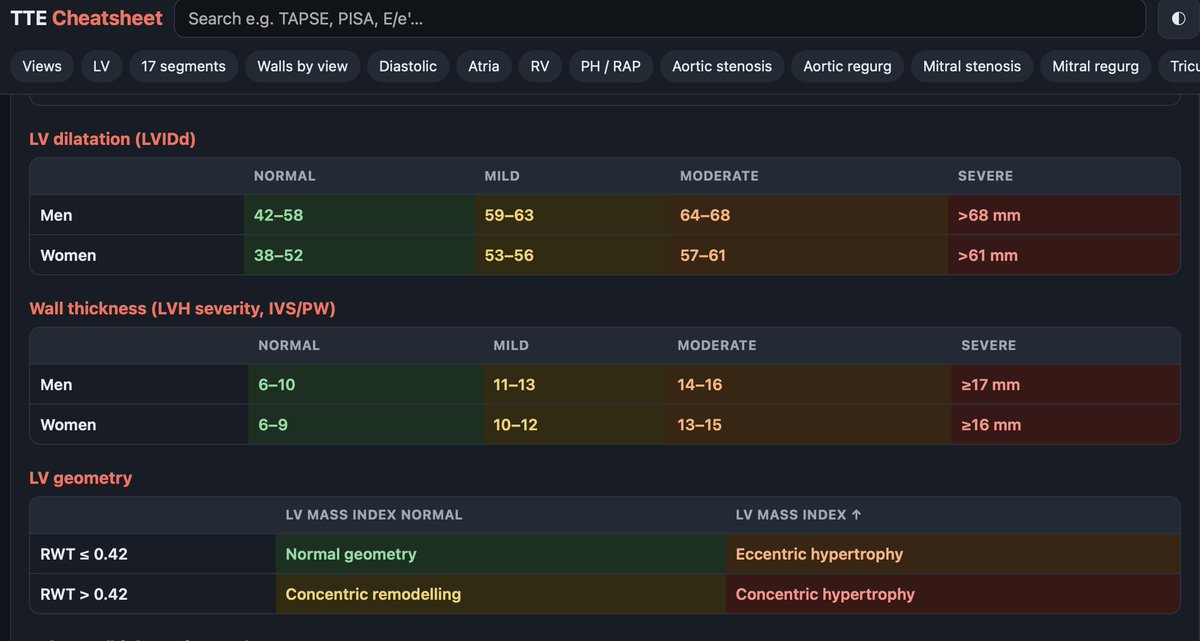
I built a free TTE cheatsheet with @claudeai I keep open while reading echos: guideline cutoffs, 17-segment walls by view, valve grading and 15 built-in calculators (AVA, SV/CO).
Check it out 👇
https://t.co/MZzWwYgeMe
#EchoFirst#CardioTwitter
Zverev gave an emotional speech after winning the U.S. Open about his mom and how she helped him achieve his dreams despite struggling with diabetes
“I wanna thank one more person who’s never actually watching my matches but is the most important person in my tennis life and in my life in general.
When your 4 year old son gets diagnosed with diabetes, and doctors in the office tell you that life and sports will be very very limited for that child, it takes a very very strong mother to walk out of those offices and say ‘My son will be wherever he wants to be.
If he wants to be a tennis player he’ll be a tennis player. If he wants to do something else, he’ll do something else. But this illness will not define us.’ My mother is the most important and strongest person I know. It takes an unbelievably strong woman to do what she did.” 🥹
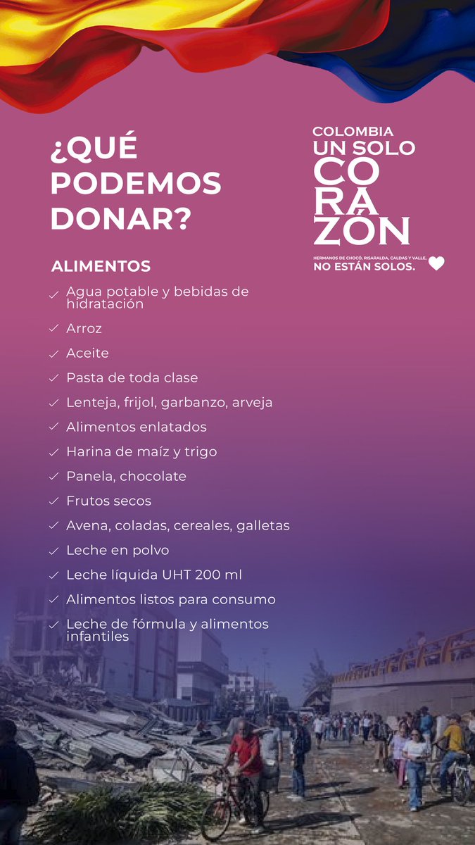
Nuestras primeras damas Ana Lucía Pineda y Tatiana Céspedes lideran COLOMBIA UN SOLO CORAZÓN ♥️
Acá encontrarán los elementos que se están necesitando para los damnificados y las cuentas bancarias donde pueden consignar.
En hilo encontrarán los puntos de acopio.
🟥Acoplamiento Ventrículo-Arterial izquierdo [VAC]. 🔎🫀💥
🔴El VAC describe que tan bien están coordinados el ventrículo izquierdo y el sistema arterial durante la eyección. Esta relación se expresa como: VAC = Ea/Ees.
🔺️Ea (elastancia arterial): representa la carga arterial global o poscarga que enfrente el VI. Integra la resistencia vascular, distensibilidad arterial y FC. Una Ea ⤴️ sugiere mayor carga para el VI.
🔺️Ees (elastancia telesistólica): representa la capacidad contractil del VI para generar presión. Una Ees ⤵️ sugiere menor capacidad contractil.
📐Actualmente puede estimarse de forma no invasiva mediante ecocardiografía, utilizando el método se "un sólo latido", descrito por Chen et al.
📊Valores normales:
🔺️Ea: 2.2 ± 0.8 mmHg/mL.
🔺️Ees: 2.3 ± 1.0 mmHg/mL.
🔺️Ea/Ees: 1.0 ± 0.36
*En sujetos sanos, el sistema tiende fisiológicamente hacia un VAC ~0.5, asociado con una eficiencia metabólica óptima y una FEVI ~66%.
✅️VAC ~0.5 = máxima eficiencia energética. 👌🏻💓
⚠️VAC ~1.0 = máximo trabajo sistólico.
🚫VAC >1.3-1.4 = desacoplamiento.
https://t.co/rvQd0x1XtY
How is a MitraClip implanted?
Transcatheter edge-to-edge repair (MitraClip) is performed under echocardiographic guidance to reduce mitral regurgitation without open-heart surgery.
The procedure follows these key steps:
Cross the interatrial septum to access the left atrium with the delivery system
⬇️
Advance the MitraClip into the left atrium and position it above the mitral valve
⬇️
Open the clip arms and align them perpendicular to the mitral valve coaptation line
⬇️
Advance the clip into the left ventricle through the area of the largest regurgitant jet
⬇️
Retract the clip to grasp both the anterior and posterior mitral leaflets
⬇️
Close the clip, creating a double-orifice mitral valve and reducing mitral regurgitation
Throughout the procedure, transesophageal echocardiography (TEE) is essential for guiding clip positioning, confirming adequate leaflet grasp, and assessing the final reduction in mitral regurgitation.
Reference: Catherine M. Otto, Textbook of Clinical Echocardiography.
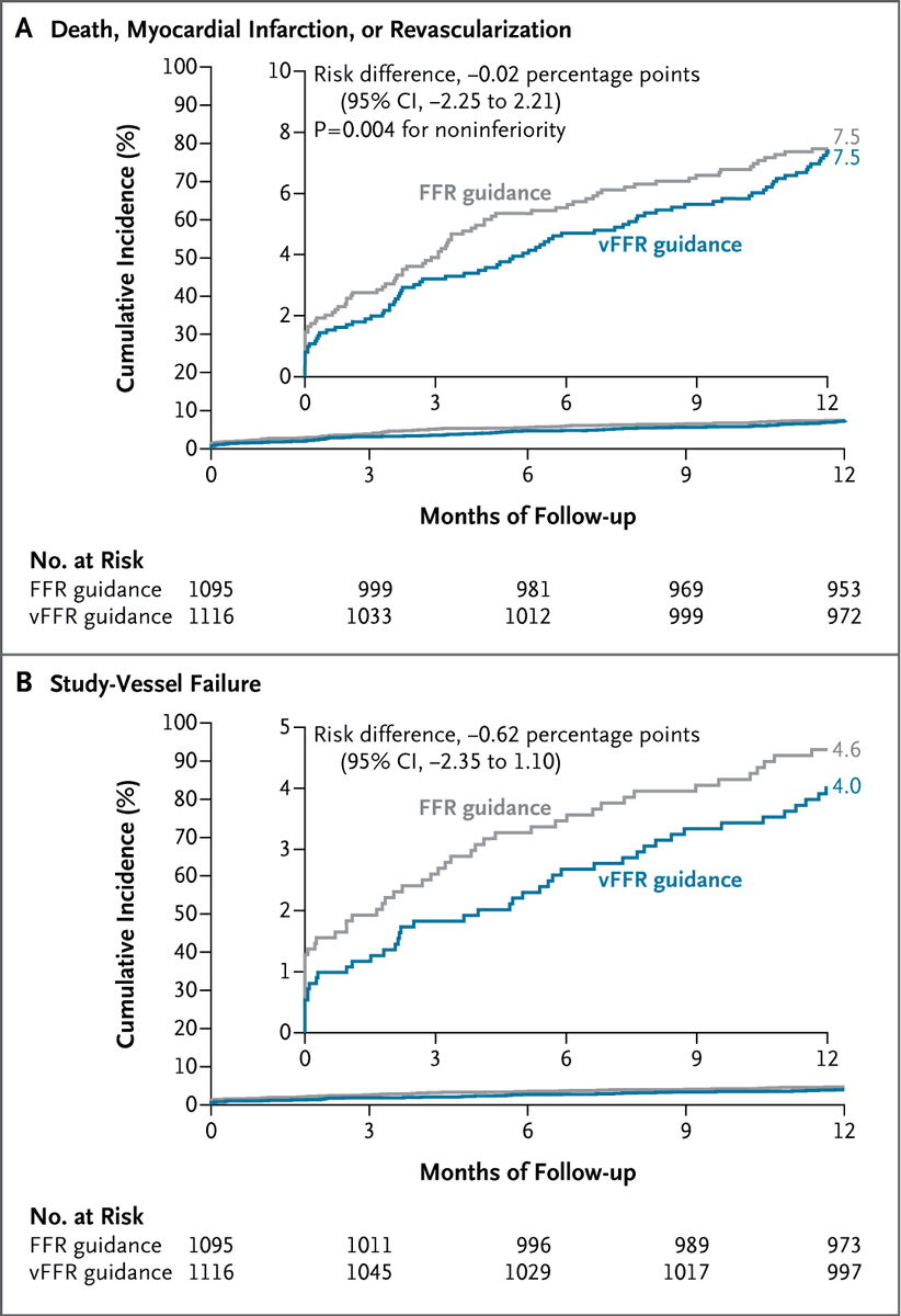
Original Article: Angiography-Based Physiology to Guide Coronary Revascularization (FAST III trial) https://t.co/xunm2zaent
Editorial: Redefining Physiology in the Cardiac Catheterization Laboratory https://t.co/HQNHdF0MYm
Science behind the Study: Physiological Assessment of Coronary Artery Disease https://t.co/t0qS5itiXq
#Cardiology
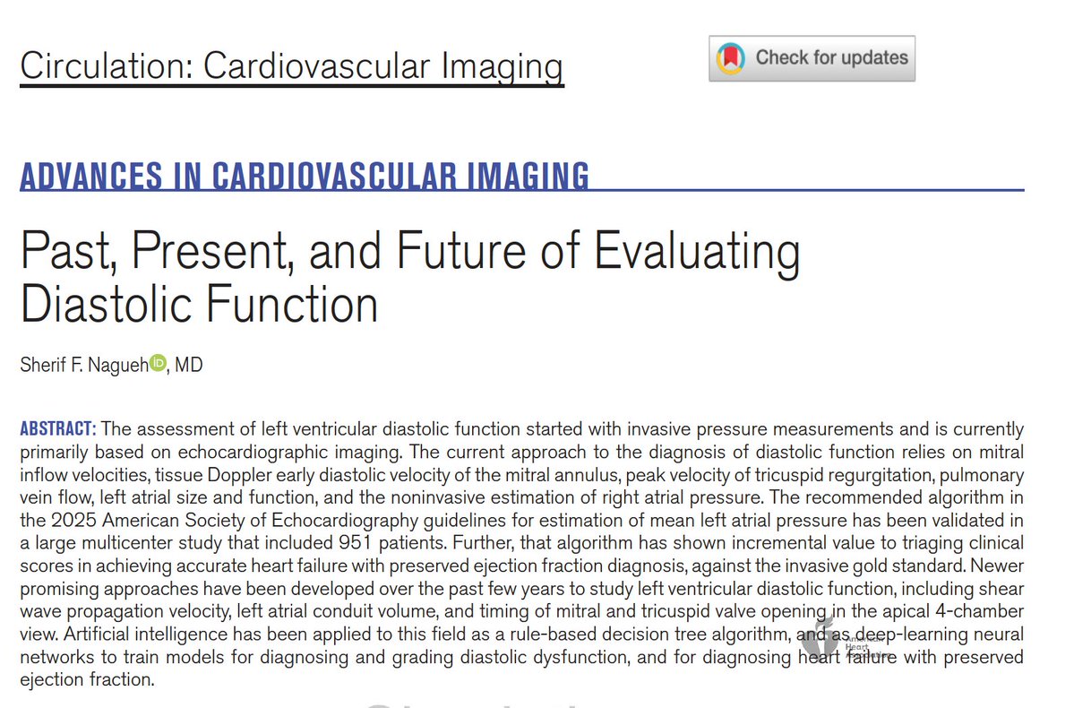
Past, Present and Future of Evaluating Diastolic Function
A new review by Sherif F. Nagueh provides a comprehensive overview of how the assessment of left ventricular (LV) diastolic function has evolved—from invasive haemodynamic measurements to advanced echocardiographic and AI-driven approaches.
Historically, evaluation relied on cardiac catheterisation and direct measurements of filling pressures. The introduction of Doppler echocardiography transformed clinical practice, allowing non-invasive assessment through mitral inflow, pulmonary vein flow, tissue Doppler imaging, and later, left atrial (LA) function analysis.
The review highlights the major advances incorporated into the 2025 ASE diastolic function guidelines, which simplify assessment and improve diagnostic accuracy. The new algorithm uses age-adjusted e′ velocity, E/e′ ratio, pulmonary artery systolic pressure (PASP), pulmonary vein flow, LA reservoir strain (LARS), LA volume, and IVRT to estimate left atrial pressure and grade diastolic dysfunction. Importantly, validation studies demonstrated excellent performance with very low rates of indeterminate classification.
The article also discusses practical aspects of implementation, emphasizing the importance of high-quality image acquisition, dedicated LA views, and careful interpretation of variables that may be affected by specific clinical conditions.
Looking ahead, emerging techniques such as shear-wave propagation imaging, novel markers of atrial function, and artificial intelligence-based algorithms may further improve the diagnosis of diastolic dysfunction and heart failure with preserved ejection fraction (HFpEF), moving the field towards more accurate, reproducible, and automated assessments.
#CardioTwitter #Echocardiography #Diastology #HFpEF #CardiacImaging #ASEGuidelines #ArtificialIntelligence #CardiologyResearch