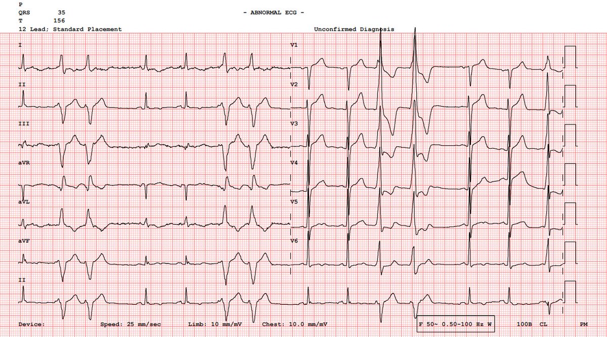
54M, history of CABG×4 performed 3 months ago. He presented to the outpatient cardiology clinic for a routine follow up visit. He is asymptomatic and in good general condition.
1 Pre CABG ECG and 2 outpatient follow up ECG:
Would these post ECGs concern you?
@Frances98392343 We share the same view. We believe this is congenital complete AV block. Given the absence of symptoms, excellent exercise capacity, an appropriate chronotropic response, preserved ventricular function and narrow QRS we favored conservative management over pacemaker implantation
We performed a treadmill exercise stress test. The patient remained completely asymptomatic throughout the test, achieving an excellent exercise capacity of 19 METs with an appropriate heart rate and blood pressure response to exercise.
@4eyedoracle Regarding IVUS, yes, it demonstrated thrombus overlying a ruptured atherosclerotic plaque. Another colleague raised the same possibility of a PFO related embolism, but the IVUS made a primary coronary embolism much less likely, so we focused our work up on thrombophilia instead
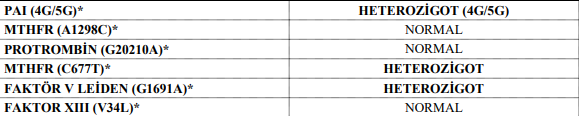
Serial hs-troponin levels were 287, 786, 958, 839, 690, 412, 200, and 99 ng/L. The patient has remained clinically stable with a normal LVEF and continues to do well on dual antiplatelet therapy. He is still undergoing evaluation for an underlying hypercoagulable state (5/5)
@4eyedoracle Thanks for the question. We did not consider thrombectomy because there was no flow limiting lesion and by the time we performed angiography the patient was completely symptom free. We first administered an IC GP IIb/IIIa inhibitor and continued with an intravenous infusion.
@koopenthusiast Very thoughtful point. I can't believe I forgot to mention this but IVUS actually showed a thrombus overlying a ruptured atherosclerotic plaque. So this was not a pure coronary embolism.
Repeat angiography after 48 hours demonstrated persistent thrombus without significant resolution. We therefore proceeded with IVUS-guided PCI and stenting of the proximal LAD, successfully completing the procedure (3/5)
Large, non flow limiting thrombus in the ostial LAD (it was a little hard to spot!). Given the patient's age, absence of ongoing chest pain and preserved coronary flow, we decided to treat him with 48 hours of IV GP IIb/IIIa inhibitor therapy before considering stenting. (2/5)
POCUS showed no findings suggestive of ACS. Subtle yet bulky precordial T waves, terminal QRS distortion in V5–V6, STD in aVL, together with the clinical presentation were sufficient for us to activate the cath lab. We proceeded directly to CAG based on the very first ECG. (1/5)
Pre-ablation ECGs showed identical positive P waves in V1, but opposite P-wave polarity in the inferior leads. My best guess is a scar related change in the preferential exit site rather than a different focus @Frances98392343
EPS showed a focal AT arising from the LAA in a scarred LA. Initial RF applications at the earliest site failed to terminate the tachycardia, so a staged cryoballoon LAA isolation was performed. AT terminated during cryo and LAA isolation was confirmed
Some arrhythmia fun: Location, Location, Location.
A 72 year old woman presented to the cardiology clinic with a long history of palpitations. Her episodes last up to 30 minutes.
What's going on?