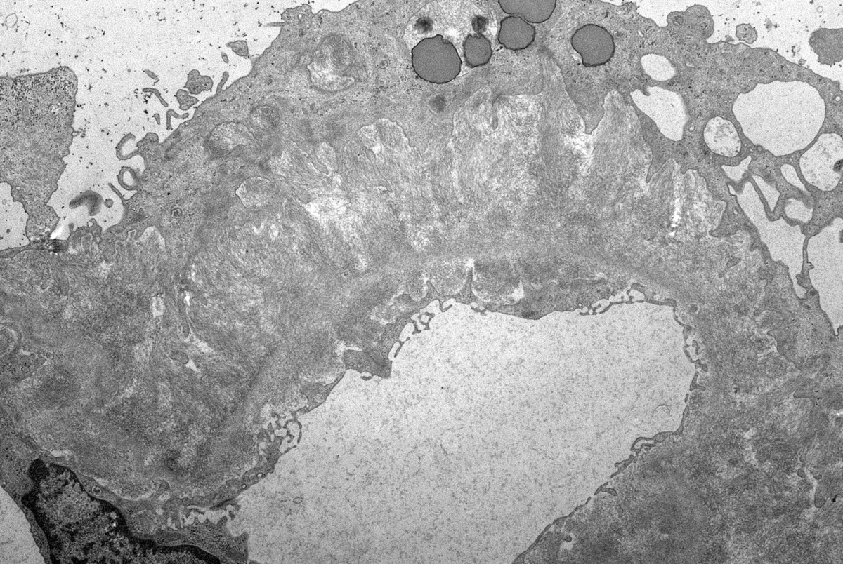
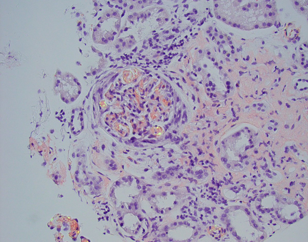
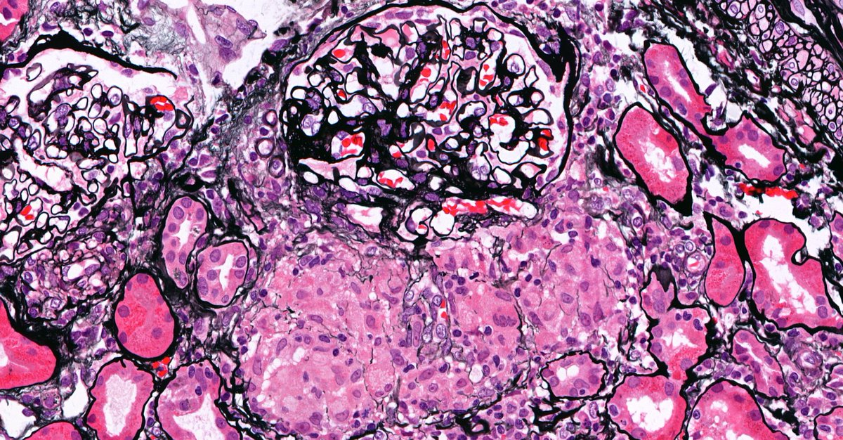
Another interesting case with crescents but this time due to amyloid. A few small cellular crescents in this case of AL-amyloidosis. Massive capillary wall amyloid infiltration. #renalpath#pathtwitter#nephrology
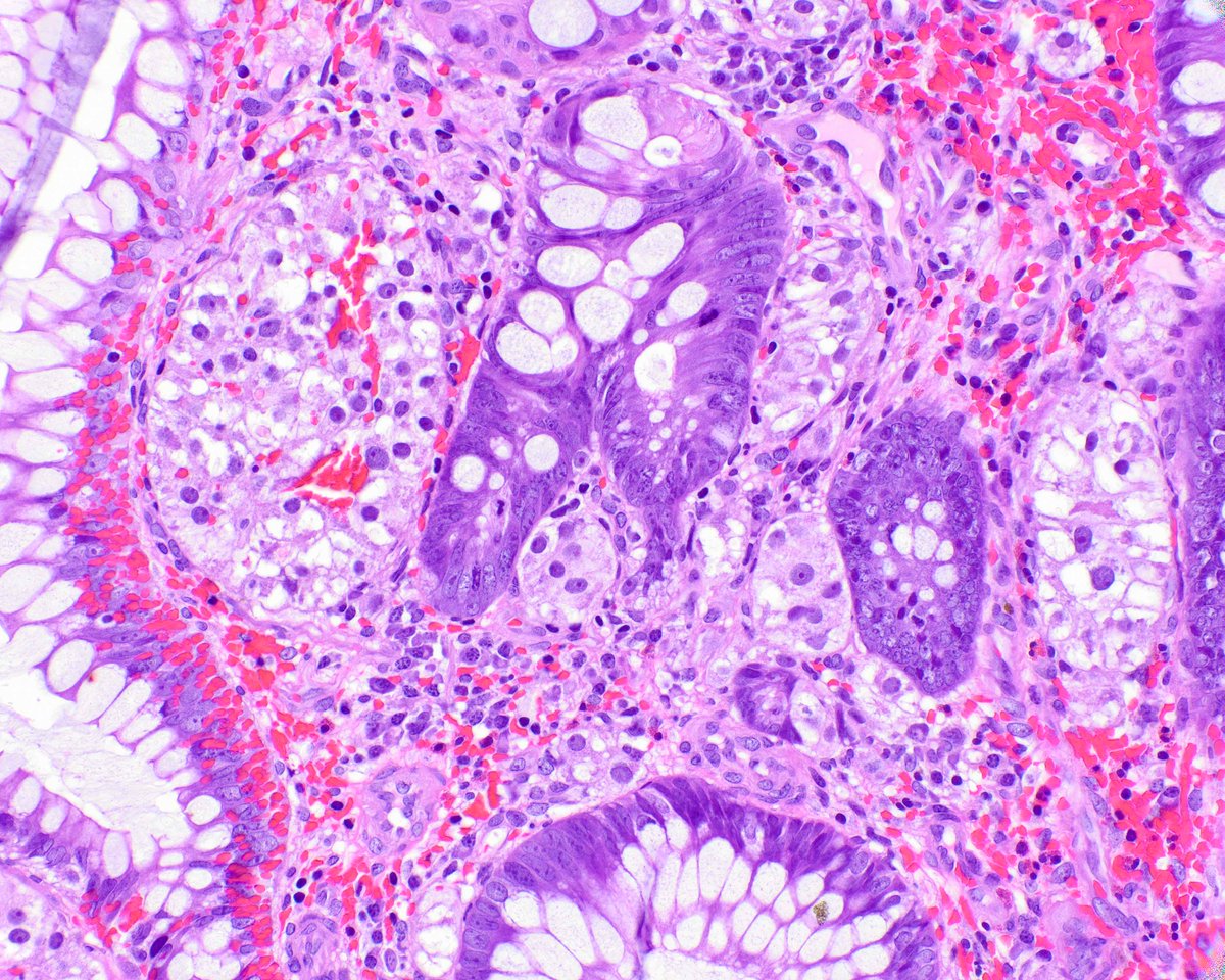
Never seen this: ? Whipple’s disease in the kidney.
Patchy accumulation of aggregates of PAS-positive rods in Bowman’s space, few capillary loops, interstitium & tubules.
Initially thought of crystal storing histiocytosis but IF negative for Ig. EM showed bacteria in tubules.
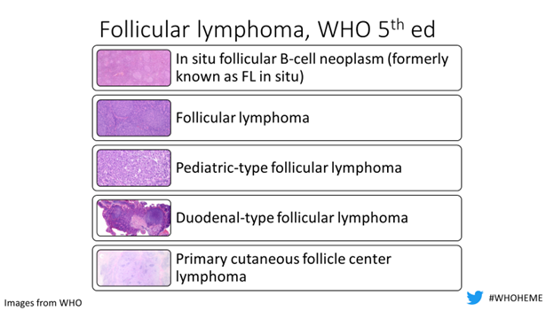
Back with another episode of #WHOHEME this time covering what’s new (or old but gold) in Follicular Lymphoma #hemepath#LymSM … diagnostic pearls 🔬
@WHO 5th ed recognizes 5 flavors of FL… tune in & remember ISFBN, pediatric, duodenal and primary cutaneous are very indolent 1/
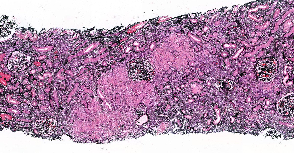
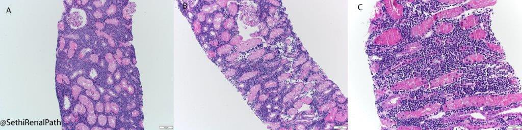
ALL: Never quite seen anything like this in a COVID patient and AKI. I was expecting just ATN. Instead saw a sea of blue cells separating tubules. Work-up revealed acute lymphoblastic lymphoma/leukemia (ALL). 35-yr old previously healthy man with recent history of COVID pneumonia
Location is key to understanding pathogenesis of immune-complex (IC) GN. Subendothelial deposits result from circulating IC (lupus, PIGN). On the other hand, Subepithelial deposits result from in-situ formation of IC (membranous). The location then dictates inflammation: + vs -
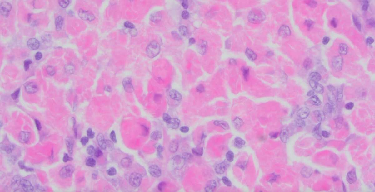
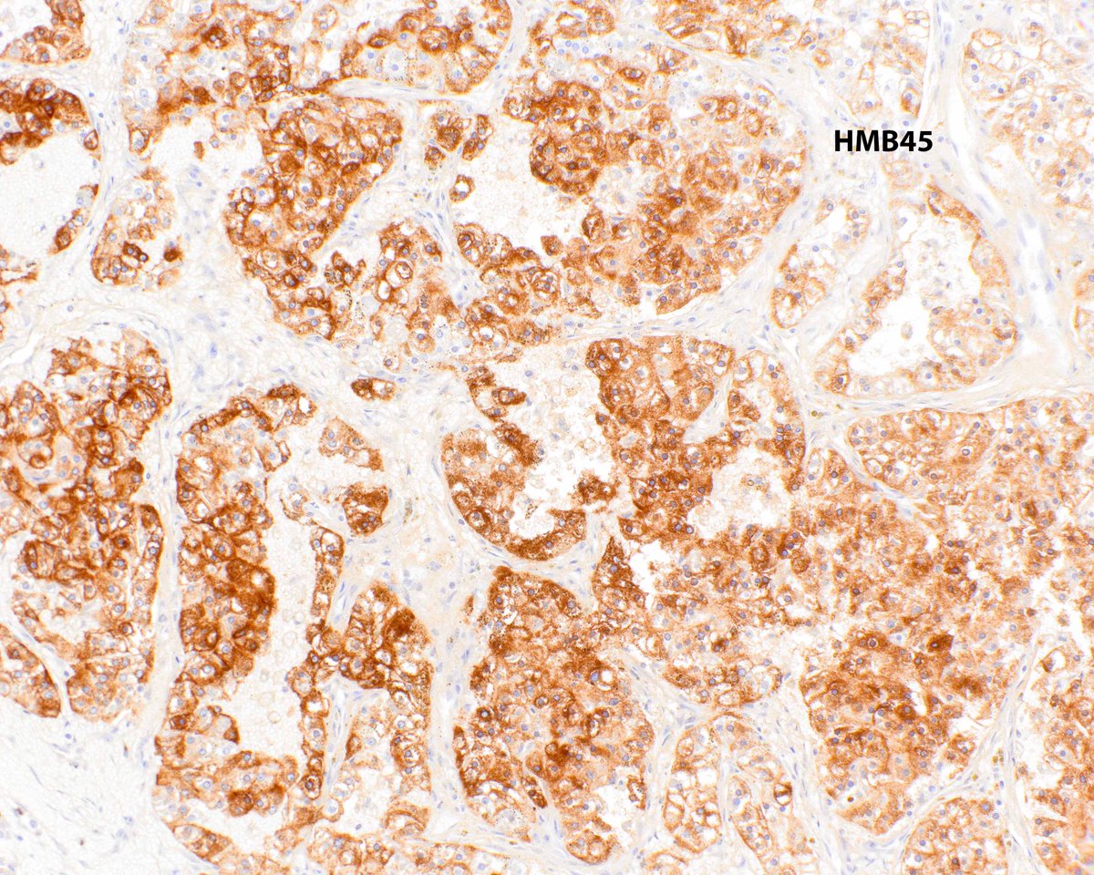
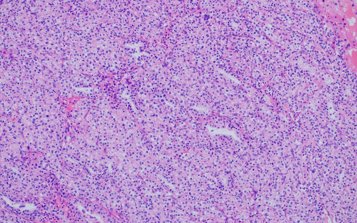
(1) Our paper on BAP1-mutated clear cell RCCs came out, so of course I had a case today! Think of BAP1 mutation in CCRCC with high grade, eosinophilic area with cytoplasmic granules/globlules adjacent to traditional component. https://t.co/0WXKFnyo5N