Non-invasive cardiologist with special interest in cardiosleep and advanced cardiac imaging, cardiac CT and MR. Views are my own. Bleed orange and blue.Go UVA!
Dear cardiology fellows 🫀
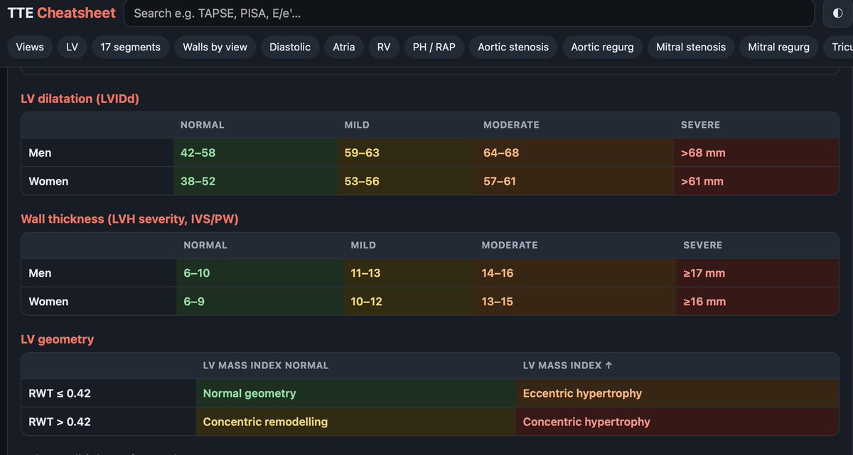
I built a free TTE cheatsheet with @claudeai I keep open while reading echos: guideline cutoffs, 17-segment walls by view, valve grading and 15 built-in calculators (AVA, SV/CO).
Check it out 👇
https://t.co/MZzWwYgeMe
#EchoFirst#CardioTwitter
@FreddyLA7 Dear Freddy,
I am so sorry about the loss. But think about how you brought joy and smile to so many people from different backgrounds, proving we are all very similar. Best of luck in your future adventure and people like you make WC an amazing experience.
@presjimryan Thank you President Ryan. You are the best thing that has happened to UVA in years. You will be greatly missed and today is truly a sad day in the history of this institution. Wishing you the best in your next chapter in life.
This is an amazing story on the power of the American dream.
I still remember when Kevin gave this interview — he worked hard and I knew it would be good. Proud to have been a small part of your journey, Kevin. Keep going!
Let’s talk about the Top 10 cardiology papers of 2024 that shook up the field!
Hard science, no BS, 🚀 and yes — some practices WILL change.
🫀 Let’s dive in: a tweetorial thread!
#CardioTwitter#MedEd#Cardiology
@DGlaucomflecken He loves to live on controversy and number of likes. Thank you for pointing at the problems of insurance companies after your own struggle with sudden cardiac death. We need more people like you in this field.
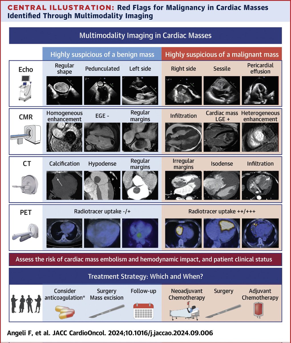
How to differentiate between malignant and benign cardiac masses?
Look for red🚩at #echofirst
Go for #whyCMR or #yesCCT for further characterisation
Go for PET by inconclusive findings
https://t.co/B0nKedmozr
Sunday #cardiooncology read 👇
Excellent updated review on MAD and how to use #cvimaging to identify pts at risk in #JACCImaging.
https://t.co/lRbxRFM9IK
Few key messages for the imagers:
🔻 MAD is infrequently observed in the LV posterior wall and often missed when the diagnosis relies on 2D PLAX -> #3Decho is superior than 2D to detect MAD
🔻 Echo resolution may not allow distinguishing pseudo MAD (posterior MV leaflet abutting the LA wall during systole) from true MAD (superior displacement of posterior leaflet in the LA) -> #WhyCMR may help
🔻 #YesCCT with systolic scans can detect millimetric MAD (3 mm) in 96% of normals that is "invisible" to echo and CMR -> longer MAD length (8-10 mm) may be relevant in MVP as risk marker
🔻 high FR imaging is essential for measuring MAD length, using dynamic frame-by-frame analysis of the mitral annulus throughout the cardiac cycle -> use narrow sector/volume to 🔼 FR
🔻 "double-peak" strain of inf-lat basal segment due to PSS -> risk marker for VAs
🔻 🔼 mechanical dispersion and posterobasal LV wall thickening associated with MAD -> additional risk markers to report
Congratulations @KristinaHaugaa@EssayaghBen@Cristinabasso64@ThorEdvardsen@NAjmoneMarsan
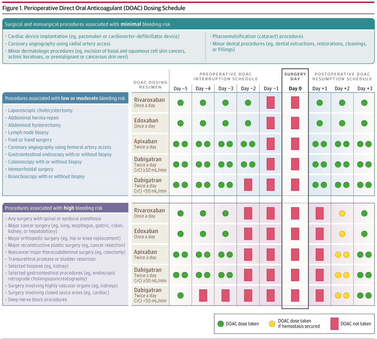
Narrative review explores how to manage use of DOACs—apixaban, rivaroxaban, edoxaban, and dabigatran—in patients undergoing surgical and nonsurgical procedures to decrease risks of bleeding and thromboembolism. https://t.co/HUra5cUmAB
Pectus Excavatum and RV Compression in a Young Athlete with Syncope led by @PennMedicine CV fellow Alex de Feria, published in @amjmed
Has anyone pursued chest wall reconstruction surgery in an adult with severe pectus deformities? @RonBlankstein
https://t.co/QvExftd975