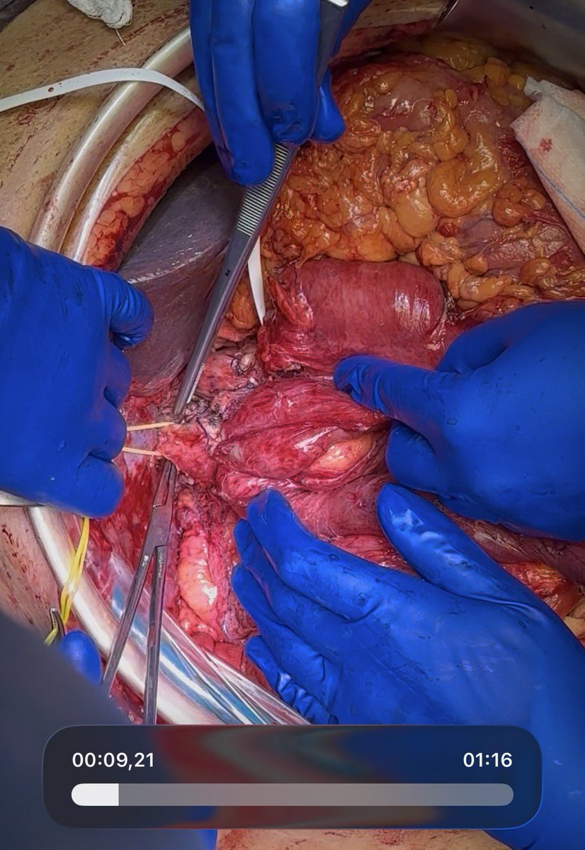
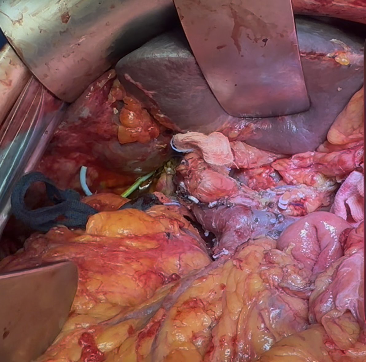
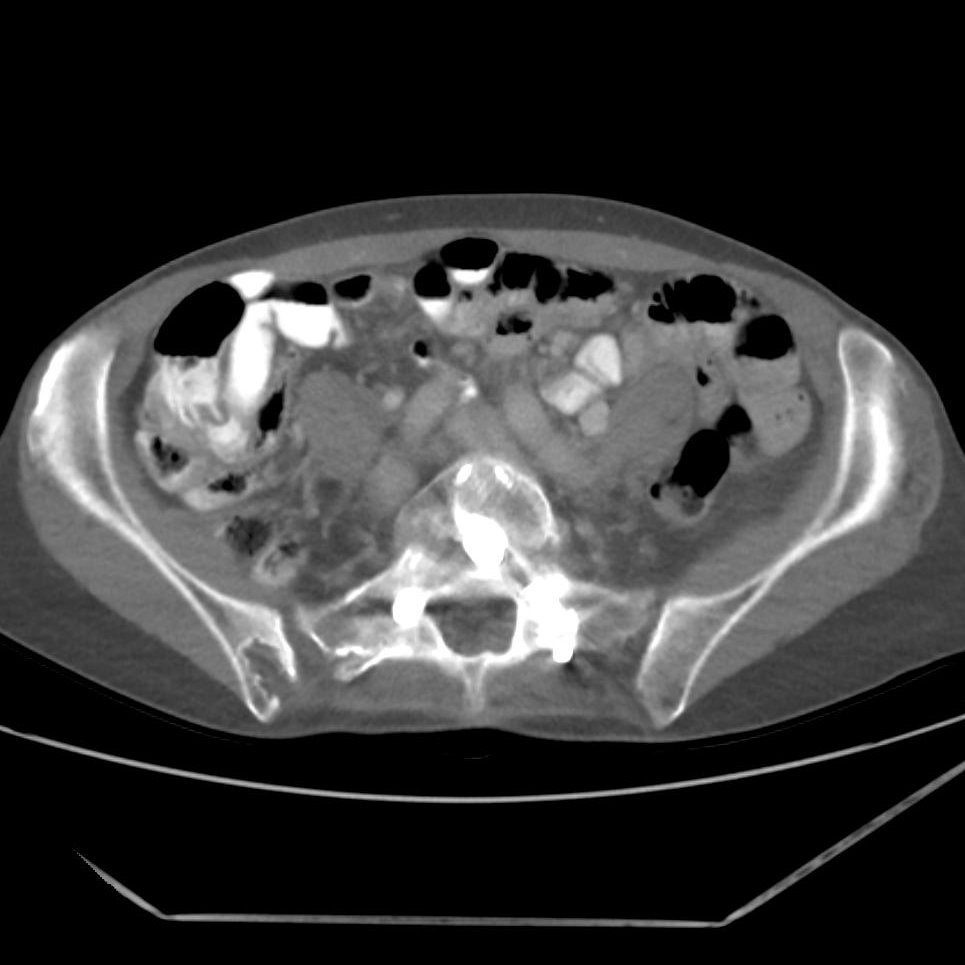
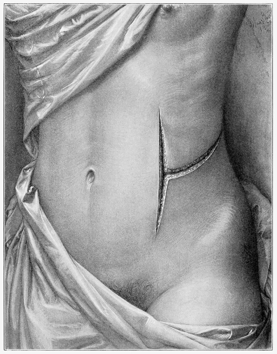
𝙀𝙢𝙚𝙧𝙜𝙚𝙣𝙩 𝙇𝙖𝙥𝙖𝙧𝙤𝙨𝙘𝙤𝙥𝙞𝙘 𝙄𝙣𝙛𝙧𝙖𝙘𝙤𝙡𝙞𝙘 𝙉𝙚𝙘𝙧𝙤𝙨𝙚𝙘𝙩𝙤𝙢𝙮
Patient Selection:
🔵Central, infracolic collections that are dominantly solid (WON) often not amenable to perc. drainage and/or narrow window of access for transgastric approach
🔵Too unwell for concomitant cystjejunostomy
🔵Septic
🔵3-4 weeks into acute attack of SAP
Approach:
🔴Initial identification of bulge in transverse mesocolon
🔴Location confirmed with IOUS
🔴Window created into infracolic compartment
🔴Necrosectomy and washout performed
🔴2 x 18F Robinsons drains placed adjacent to cyst
Key Points:
🟢Timing of intervention critical
🟢Surgical approach allows 𝘀𝗶𝗻𝗴𝗹𝗲 𝘀𝘁𝗮𝗴𝗲 𝗰𝗹𝗲𝗮𝗿𝗮𝗻𝗰𝗲 of WON which avoids multiple hits of sepsis
🟢Concomitant cholecystectomy can be undertaken in select cases
🟢Patient selection and discussion in benign specialist MDT key
Link to our paper in @JournalofGISurg below:
https://t.co/kcdwWFwSjz
#FOAMed #GITwitter #surgery #SoMe4Surgery #MedEd #hpb
Technically challenging Whipple for Distal Biliary carcinoma arising on a previous choledoco-duodenal anastomosis done 30 years ago for a biliary iatrogenic lesion
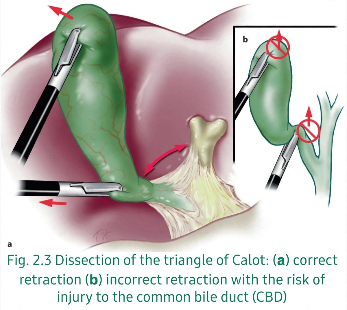
🔑 Retraction is dissection
✅ Correct (a): Fundus up ⬆️ + infundibulum lateral ➡️ Opens Calot's triangle and gives you a real Critical View of Safety
❌ Wrong (b): Pulling the infundibulum up ⬆️ Tents the CBD right into line with the cystic duct → classic bile duct injury
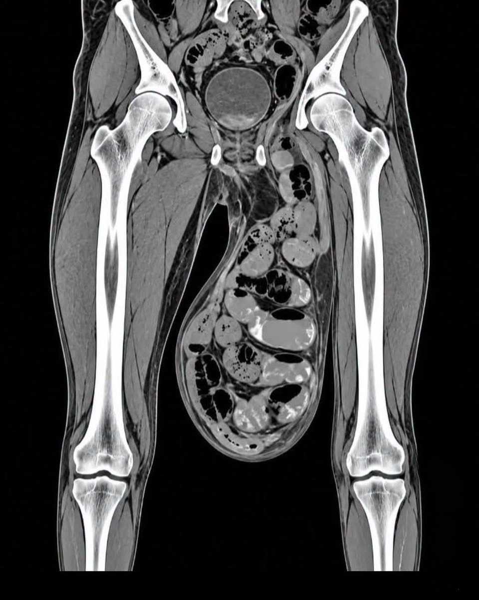
أمس رأيت أشعة لشاب تظهر كيس الصفن (Scrotum) متمدد بشكل مخيف مثل ما ترون.
هذا الشاب رياضي٫ ياكل كل شي صحي٫ ليس عنده أي مرض أدى لها أو ورم.
لكنه كان يفعل عادة واحدة خبيثة أدت إلى نزول الخصيتين وتمددهم بهذا الحجم٫ العادة كانت:
Comma-tail tumor.
Would a root diving into the parenchyma stop you from trying to spare the kidney?
Laparoscopic enucleation.
Followed the tail. Coagulated the bed.
A 28-year-old man is taken to surgery after four days of worsening right lower abdominal pain and fever. During laparoscopy, the surgeon finds the appendix buried in a dense mass of inflamed omentum and adherent bowel loops.
The surgeon pauses before attempting to separate them.
What is this inflammatory mass called?
🔺 HEPATOCYSTIC TRIANGLE (CALOT’S TRIANGLE)
Boundaries:-
• Medial - Common hepatic duct.
• Inferior/Lateral - Cystic duct.
• Superior - Inferior surface of liver.
Contents:-
• Cystic artery
• Cystic lymph node (Lund’s node)
• Lymphatics & connective tissue
EXAM TRAP:-
Calot’s original triangle (1891) was bounded by the cystic duct, common hepatic duct and cystic artery.
Modern hepatocystic triangle uses the inferior surface of the liver as its superior boundary.
SURGICAL Importance is 👇
Critical View of Safety (CVS) during laparoscopic cholecystectomy requires:-
1. Clearance of the hepatocystic triangle
2. Separation of the lower ⅓ of gallbladder from liver bed
3. Confirmation that ONLY TWO structures enter the gallbladder i.e cystic duct & cystic artery.
🎯 Remember:- CVS helps prevent bile duct injury.
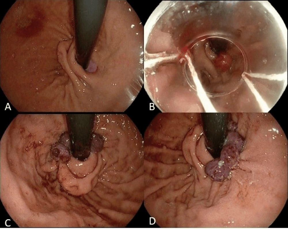
Endoscopic GERD therapy, by mechanism:
Radiofrequency: Stretta
Plication: TIF (EsophyX, MUSE), GERDx
Resection: ARMS, ESD-G
Constriction: CLEAR band ligation
Ablation: ARMA
None is as established as laparoscopic fundoplication, and most have not been evaluated extensively.
A quick look at the two-incision technique used in lower leg fasciotomy to release pressure in the four compartments of the leg.
To access full video, start your free trial at https://t.co/yZdFnplAid
#orthopaedics#trauma#Fasciotomy#leg#orthotwitter#orthoX