Artificial light at night doesn't just brighten coral reefs—it disrupts sleep and damages DNA.
In reef damselfish, nighttime light increased aggression and nocturnal feeding, reduced sleep, and was linked to greater neuronal DNA damage. Another reason to protect natural darkness.
https://t.co/WeT3hL50q4
#Sleep #CircadianRhythms #LightPollution #MarineBiology #BrainHealth
Obesity doesn't just affect the waistline—it reshapes the brain. Emerging evidence suggests chronic metabolic overload disrupts neurovascular coupling, blood–brain barrier integrity, cerebrospinal fluid dynamics, and myelination, creating conditions that may accelerate neurodegeneration. As obesity and neurological disorders rise in parallel, understanding these interconnected pathways could reveal new strategies to protect brain health across the lifespan. #Neuroscience #Obesity #BrainHealth
https://t.co/jb4TtNV1xu
Delighted to see our metabolomics lab work, how nanoplastics affects the brain metabolism, featured in different National news. I sincerely thank to @Ratnasekhar_Ch sir for his invaluable guidance. Sincere gratitude to our director @PrabodhTrivedi sir, for support and guidance.
Congratulations! Dr. Ratnasekhar CH and team for the publication titled “Brain lipidomics identifies mitochondrial redox dysfunction and metabolic trade-offs associated with Parkinson’s disease-like pathology induced by Nanoplastics exposure” has been published in Free Radical Biology and Medicine (I.F. 8.2), a leading journal in redox biology. (https://t.co/PJprWDERQt).
@PrabodhTrivedi@CSIR_IND@DrNKalaiselvi@DrJitendraSingh
Congratulations ! @PriyaRatho98796@ashushamal from Metabolomics lab for publication inFree Radical Biology and Medicine (I.F. 8.2), (https://t.co/saivCSKL30).
Thank you so much @Ratnasekhar_Ch sir. It has been an incredible experience working on this research question. Grateful for your constant support and guidance throughout this journey.
Congratulations @Ashushamal@PriyaRatho98796 for the publication from our #metabolomics lab in Neurotoxicology, demonstrating disrupted lipid mediated crosstalk between Organelles underlies Parkinson’s pathology studied using Drosophila (https://t.co/VdqEJuXL5d)
We are pleased to share that the research team led by Ratnasekhar has published a significant study in the journal NeuroToxicology, uncovering how disrupted lipid-mediated crosstalk between key cellular organelles—particularly mitochondria and peroxisomes—contributes to Parkinson’s disease-like pathology.
Using a rotenone-induced Drosophila melanogaster model, the study demonstrates systemic lipidome perturbations that mirror critical aspects of human Parkinson’s disease.
🔗 [https://t.co/AhSOjJ3Su7](https://t.co/AhSOjJ3Su7)
#CSIRCIMAP #ResearchHighlight #NeuroToxicology #ParkinsonsDisease #Neuroscience #Lipidomics #Mitochondria #Peroxisomes #Drosophila #ScientificResearch
@CSIR_IND@DrJitendraSingh@DrNKalaiselvi@zabeerahmedb
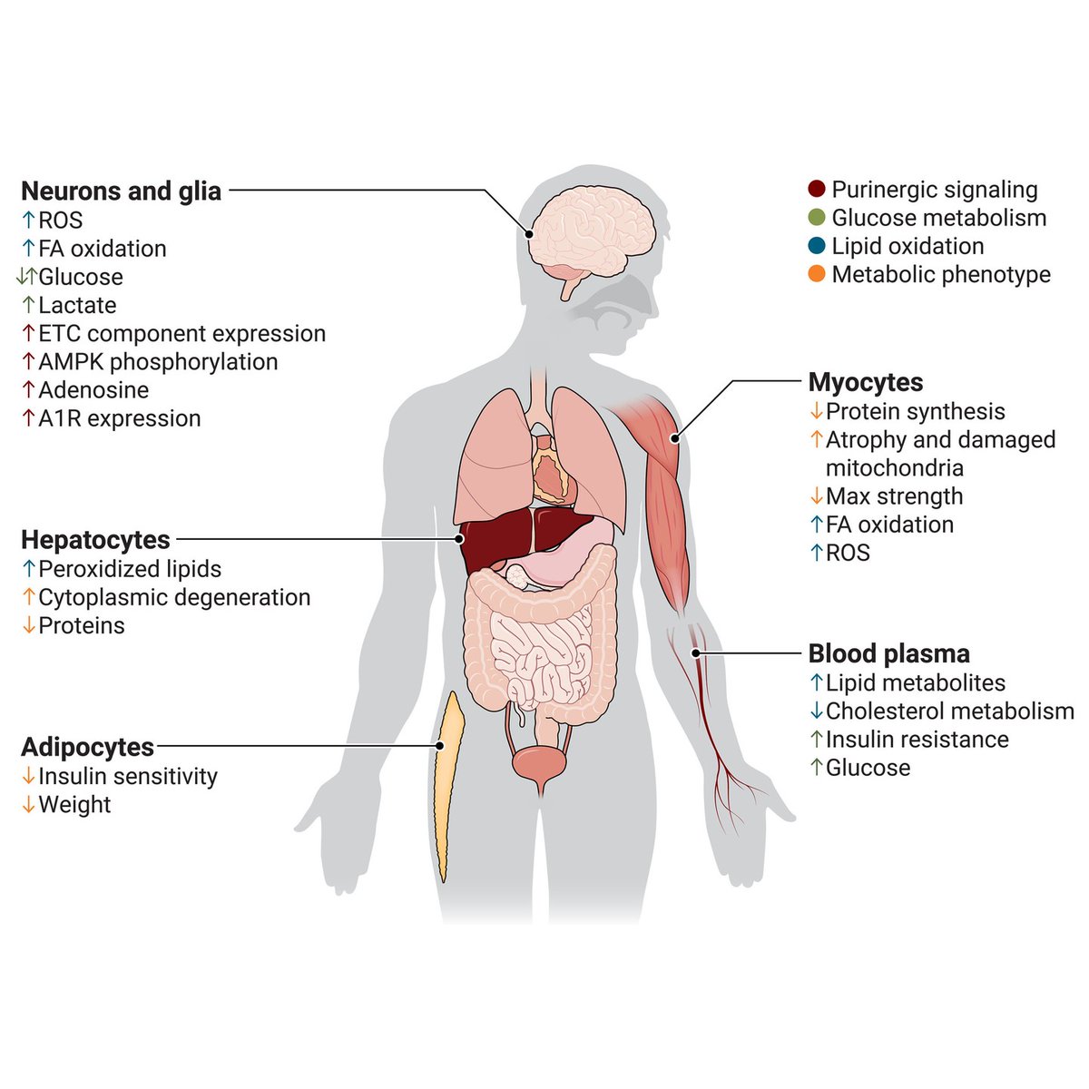
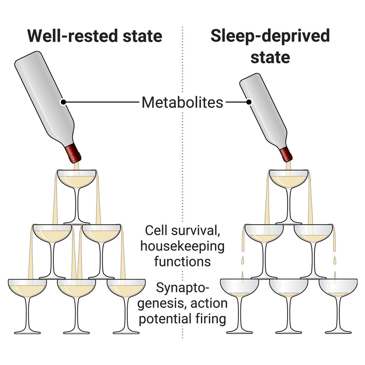
In a 2025 @SciSignal Review, researchers discussed an emerging body of research on the metabolic effects of sleep loss, which disrupts the natural balance of energy within neurons.
The authors highlighted how the loss of sleep forces neurons to shunt resources to cell survival pathways, at the expense of more long-term, energy-demanding processes such as cognition and memory formation.
Learn more on #WorldSleepDay: https://t.co/L5u4AQ5ytL
Celebrating #NationalScienceDay with our latest publication in Molecular and Cellular Endocrinology. 🔬🥼Our findings demonstrate how Withaferin A attenuates saturated fatty acid–induced ER stress, mitigates insulin resistance, and restores bone homeostasis.
Proud of our team. 👏
Clocking Parkinson’s disease: Our lab has received a ₹1.5 Cr ICMR Research Grant to study phytochemical derivatives targeting biological clock pathways. We thank ICMR for supporting this work.
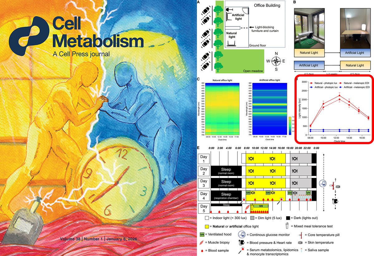
Exposure to natural daylight that is ~5 times brighter and have plenty of light in 460-520nm spectrum than the typical fluorescent or LED light in offices improves metabolism. @Cell_Metabolism
https://t.co/0IU2lrO7n2
@CSIRCIMAP successfully organized National Metabolomics Workshop (9–11 Dec) with participants from across India. Grateful to the Director @PrabodhTrivedi sir, for his unwavering support in making it a success.
Congratulations to the Whole metabolomics team for the successful completion of National Metabolomic workshop 2025. Finally we have got wonderful experience with several challenges while the workshop. Many thanks to @Ratnasekhar_Ch sir for this wonderful informative initiative.
How your body moves electrons from food to energy
This figure explains how the human body acts like an electrical circuit—moving electrons extracted from food all the way to oxygen to generate energy. Every meal you eat feeds an invisible current that powers your cells through a continuous flow of electrons inside the mitochondria.
1���⃣ Food as an electron source
Carbohydrates, fats, and proteins are broken down into molecules like glucose and fatty acids that release electrons during oxidation. These electrons are captured by carrier molecules such as NAD⁺ and FAD.
🟢 Example: One molecule of glucose donates enough electrons through NADH and FADH₂ to drive the production of about 30 ATP molecules.
2️⃣ Electron delivery to mitochondria
Nutrients are converted into acetyl-CoA, which enters the TCA cycle in mitochondria. Each turn of the cycle generates high-energy electron carriers that feed into the electron transport chain.
🟢 Example: When oxygen is limited, cells divert pyruvate to lactate to keep glycolysis running and prevent a bottleneck in electron flow.
3️⃣ The electron transport chain
Electrons move through a series of protein complexes embedded in the inner mitochondrial membrane. As they flow, energy is released to pump protons across the membrane, creating an electrochemical gradient.
🟢 Example: This “proton motive force” is the voltage that powers ATP synthase, the enzyme that produces ATP from ADP and phosphate.
4️⃣ Oxygen as the final electron acceptor
At the end of the chain, oxygen captures electrons and forms water, completing the circuit. Continuous oxygen flow keeps the system balanced and prevents electron buildup.
🟢 Example: When oxygen supply drops, excess electrons can leak, generating reactive oxygen species that damage cells.
5️⃣ ATP as usable energy
The proton gradient drives ATP synthase to generate ATP, the chemical energy currency used for everything from muscle contraction to DNA repair.
🟢 Example: Tissues with high energy demand, such as the brain and heart, contain dense mitochondrial networks to maximize electron throughput.
In essence, metabolism is electricity at the molecular level. Food provides the electrons, mitochondria manage their flow, and oxygen completes the circuit—turning chemical energy into the electrical current that sustains life.
🚀 Announcing!
CSIR-CIMAP, Lucknow is hosting a 3-Day National Workshop on Hands-on LC-MS based Metabolomics 🧪
📅 9–11 Dec 2025
🔬 Practical training + expert insights
⚡ Limited seats available!
https://t.co/L4wQbpyKEE
https://t.co/UuZzoNF7vz
#Metabolomics#LCMS#CSIRCIMAP