Finally out! 🥳We added a lot of rescue experiments. We now show that Arl13b needs to locate to commissural neurons primary cilium for proper rostral turning of axons. We also further characterized Shh retrograde transport to the soma of comm. neurons.
https://t.co/LL7srqxc2k
Delighted to share our latest preprint entitled « A cell-autonomous role for #primarycilia in long-range commissural #axonguidance ». We propose a mechanism involving the neuronal primary cilium during axon guidance at an intermediate target. https://t.co/0CkvBNk0QS 🧵(1/7)
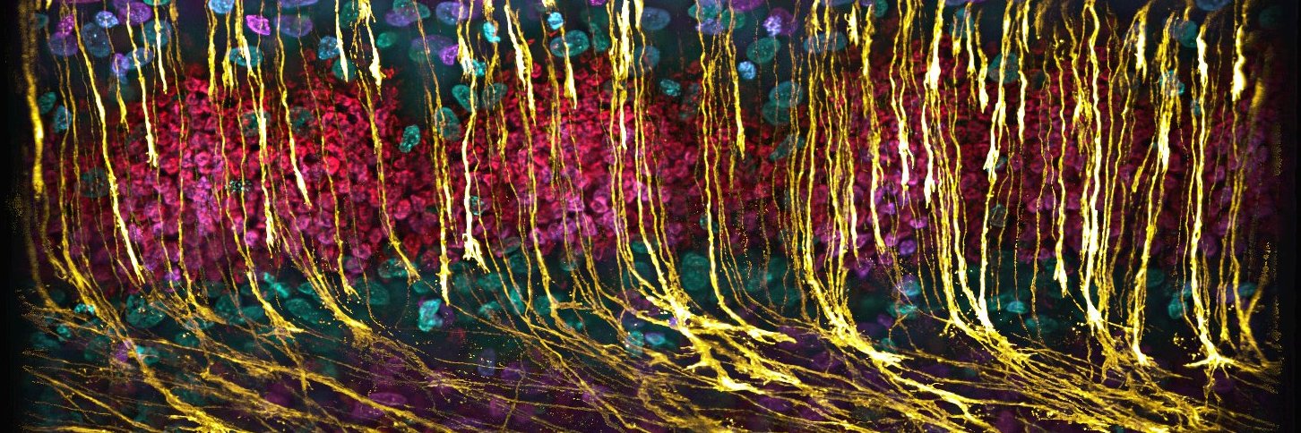
The tissue shown here is an ex vivo culture of intact spinal cord from a🐣embryo, allowing us to observe these processes in a controlled yet physiologically relevant setting.What looks like a simple crossing event emerges from complex and tightly regulated cellular processes.
During development, these neurons extend long projections that grow toward the midline, cross it, and then turn to grow along the length of the spinal cord. This coordinated behavior is essential for establishing connectivity between the two sides of the nervous system.
These are dI1 neurons, a well-studied class of spinal interneurons that have been used for more than 30 years to uncover fundamental mechanisms of axon guidance and neural circuit formation.
This Is What Drives Axon Growth. Axon growth is powered by the actin cytoskeleton at the growth cone.
Here, filamentous actin dynamics drive fast, directed axon extension in real time. #Fluorescencefriday#science#neuroscience
Thrilled to collaborate with @ZhixingChen2 Lab @PKU1898!
We tested new PK Mem dyes—gentle, photostable tools to label the plasma membrane in live neurons, making it easier to track growth cone motility & axonal transport. #liveimaging#neuroscience
https://t.co/KLsx2cA5Fn
This time-lapse captures 17h of axonal growth from a chick dorsal root ganglion explant, seen through the actin cytoskeleton using live imaging. I just submitted this video to the Nikon Small World in Motion competition. Today is the last day to upload yours!😉 #neuroscience
Come and join the Williams lab @OfficialUoM@FBMH_UoM@The_MRC We are using single-cell Multiomics and in vivo CRISPR approaches to understand the molecular mechanisms underlying lineage segregation from the neural plate border 🐣https://t.co/41X4A6oRIY
I’m very excited to finally present something we’ve been working on for the past couple of years – a spatial proteome map of primary cilia, released in @ProteinAtlas today. Amazing teamwork led by @jn_hansen
Check out our preprint https://t.co/uhauaTmCz2
Baby you’re a fiiiiirework!!!
Explanted Xenopus neural crest cell, microtubules (green) and actin (magenta). Imaging in frogs rocks- this is done room temp, no incubation, no problem. 🐸🔬✨
By Micaela Lasser, Helen Willsey @goodfrognosis Lab
@zeiss_micro LSM980 fast airyscan
@lreymond2 Thanks Luc!
It is again the Pkmem 555 probe (magenta) and a mStayGold fusion protein (green). I don't have to correct my recordings for breaching anymore! there is no apparent bleaching to be seen...🤯🤯🤯
Time-lapse recording of floorplate cytonemes (green) interacting with axons (magenta). 1 image taken every second for 10 min (played at 30 fps). Cytonemes love interacting with axons! 💚#FluorescenceFriday
Retinoic acid, an essential component of the roof plate organizer, promotes the spatiotemporal segregation of dorsal neural fates
Read this #OpenAccess Research Article by @DinaRekler, Shai Ofek, Sarah Kagan, Gilgi Friedlander & @ChayaKalcheim@HebrewU:
https://t.co/aMQVD5pBMh