A stroke in the internal capsule produces a distinctive clinical picture, specific enough that a physician can often localize the lesion on exam alone. #StanfordMed25's guide walks through the related anatomy & exam findings: https://t.co/jsY9CmKsNb #StrokeAwarenessMonth
#POCUS#VExUS#Nephpearls
This figure depicts the renal venous stasis index (RVSI). Basically, more interruption of venous flow during a given cardiac cycle = more congestion.
One of the key conceptual advances of RVSI is that waveform categories alone may miss subtle progression within the same pattern. RVSI quantifies the proportion of the cardiac cycle without venous outflow, so you can still comment on improvement or worsening even if the waveform remains monophasic.
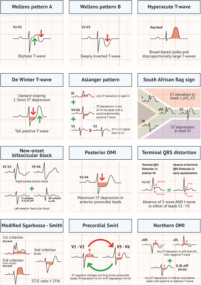
Acute MI is not always STEMI.
A major 2025 review highlights why relying only on classic STEMI criteria can miss dangerous coronary occlusions.
A thread on OMI (Occlusion Myocardial Infarction): 🧵
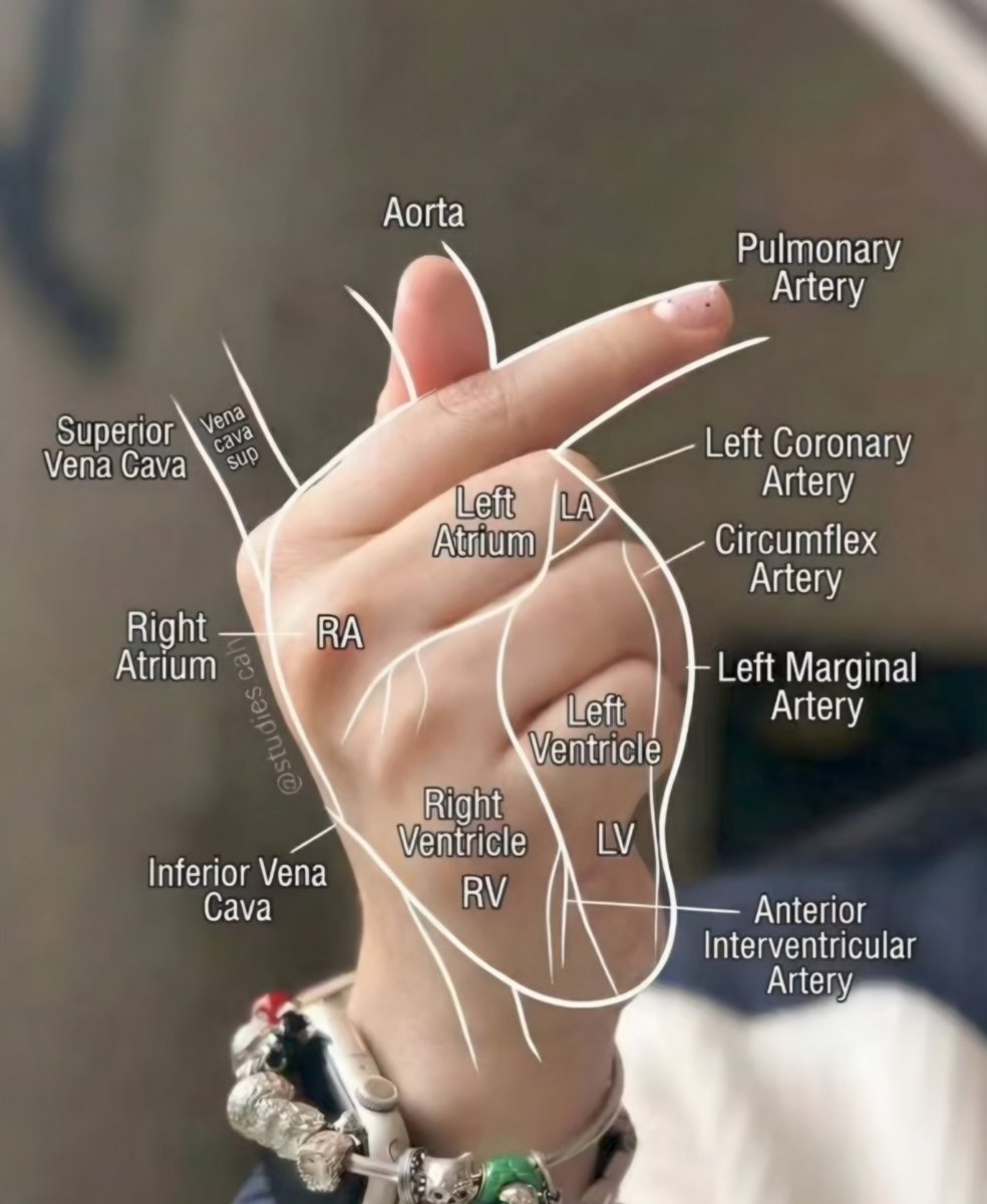
Blood Supply of the Cardiac Conduction System — Summary
■ SA node:
▪︎ 60% from Right Coronary Artery.
▪︎ 40% from Left Circumflex Artery.
■ AV node:
▪︎ ~85–90% from Right Coronary Artery.
▪︎ ~10–15% from Left Circumflex Artery.
■ Bundle of His:
▪︎ Proximal: Right Coronary Artery.
▪︎ Distal: Septal branches of Left Anterior Descending Artery.
■ Right bundle branch:
▪︎ Septal branches of Left Anterior Descending Artery.
■ Left bundle branch:
▪︎ Septal branches of Left Anterior Descending Artery.
✅ Key pearl:
● RCA infarction → sinus node dysfunction & AV block.
● LAD infarction → bundle branch blocks.
#cardiology
LV Diastolic Filling – What Echo Reveals
This diagram explains how LV volume changes relate to Doppler diastolic filling patterns:
- E wave: Early rapid filling (passive, LV relaxation dependent)
- Diastasis: Minimal flow phase
- A wave: Atrial contraction (active filling)
Clinical interpretation:
- Normal: E > A
- Impaired relaxation (Grade I): E < A
- Pseudonormal (Grade II): E/A appears normal but abnormal LA pressures
- Restrictive filling (Grade III): E >> A, short deceleration time.
Source: Catherine M. Otto, Textbook of Clinical Echocardiography
#Cardiology #Echo #LVFilling #DiastolicDysfunction #CardioTwitter #MedX
Let me know if you'd like this expanded into a thread with one tweet per stage or dysfunction grade.
Mitral Valve: Structure & Function
The mitral valve is a bicuspid (two-leaflet) valve that regulates blood flow between the left atrium & left ventricle, ensuring one-way circulation and preventing backflow.
It consists of:
✔️ Leaflets (Anterior & Posterior)
✔️ Annulus (fibrous support ring)
✔️ Chordae tendineae (tether leaflets to muscles)
✔️ Papillary muscles (Medial & Lateral, preventing prolapse)
🔹 Anterior leaflet = longer, but attaches to a smaller annular portion
🔹 Posterior leaflet = shorter, but divided into three scallops (Medial-M, Central-C, Lateral-L)
🔹 Both leaflets attach to both papillary muscles, ensuring efficient closure & preventing regurgitation
A vital structure in maintaining proper circulation.
#Cardiology #MitralValve #HeartHealth #MedicalEducation