@Flipkart Had a really bad experience with purchase of MacBook. They are not sending invoice of the product since last one month. Have been wasting lot of time running behind the customer service for bill/invoice. Have decided not buy anything from Flipkart. What else to do ?
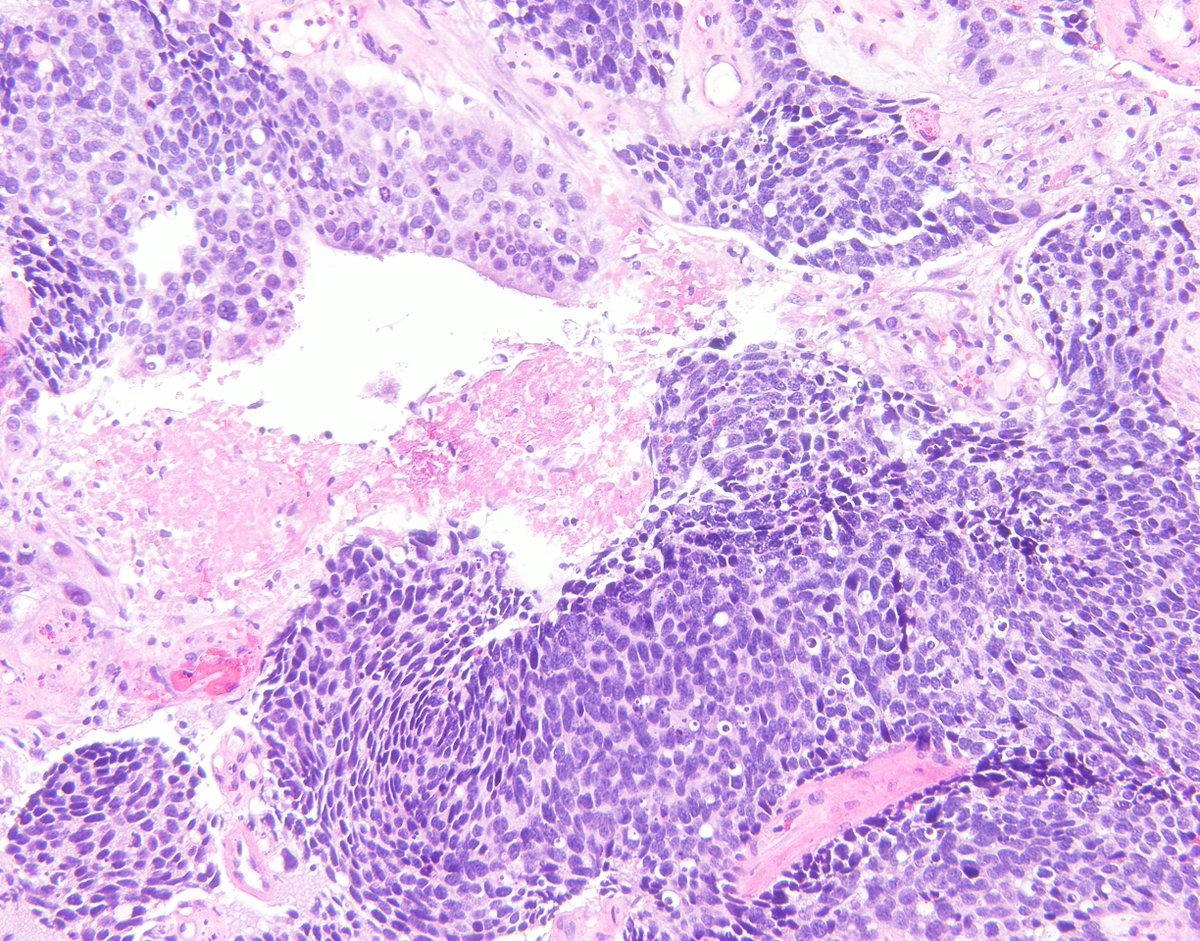
🔥A TIP for diagnosing MALT Lymphoma starts with low magnification.
🎯I search for the muscularis mucosa and see it at the highest magnification.
👉Physiologic lymphoid collection typically does not infiltrate the muscularis mucosa.
Here is a gastric MALT Lymphoma.
#gipath
Sunday musings. Five things I have learned over the years, cemented by the consult service, that every pathologist should consider when releasing diagnoses into the wilderness.
1. Pathologists are morphologists, yes, but much more importantly, they are consultants. Findings that are beside the point and clinically insignificant need not clutter our microscopic description or diagnosis unless they are relevant to how the diagnosis was made.
2. Because pathologists are consultants, we need to know the clinical implications of our diagnosis. And if both entities on the differential are treated the same way, a differential is perfectly fine as a diagnosis.
3. Pathologists should remember what clinical finding prompted a biopsy. As obvious as this may seem, we sometimes report calcifications and forget that the clinical finding was a mass. If a mass forming lesion is not seen, then the biopsy may be non representative.
4. Immunohistochemistry should be spare and targeted. It should answer a morphologic question instead of serving as a small flash light in a vast and uncharted darkness.
5. Contrary to popular belief, a pathologist’s desire to be definitive is ever present, and always fueled by pressure from clinicians. It is ok not to know, it is ok to seek help, it is ok be inconclusive when facing too little material, as long as we try to provide a plan of action. A tiny bit of ugliness does not a monster make, and one small assumption can wreak irreversible havoc on a patient’s life.
Pathology Report for Fungal Infections #tweetorial
1/9 Histopathological diagnosis of an invasive fungal infection should be PRIMARILY DESCRIPTIVE and must include:
Follow the thread to discover.
📌 The order can vary but try to include them all.
#DCF_path#PathTwitter
Great learning resource: new website with interactive unknowns https://t.co/xdWpD7vgY9. You can work through interesting cases and test your IHC and molecular knowledge. The platform is presented in @ArchivesPath article. #pathtwitter#path2path#pathology#pathresidency#MedEd
@Path_Matt@smlungpathguy A must read article on this topic: "Mitotic Figures-Normal, Atypical, and Imposters: A Guide to Identification" This article provides morphologic criteria for mitosis identification and for distinguishing from atypical mitosis and mitotic-like figures. https://t.co/NXNhlAH53M
#breastpath with another interesting case. This Triple negative tumor in young female is CK7, CK20 negative. E-cadherin with membranous positivity while Keratin and GATA-3 are positive too. Diagnosis?
This is a great point that we often forget. Never let your ego cloud your judgement. Be ready to accept your mistakes, we are humans.
Beautifully depicted in this scene from movie Bahubali. #MustWatch for Indian movie buffs🤩. @01sth02@yMDPhD#PathTwitter#pathresidents
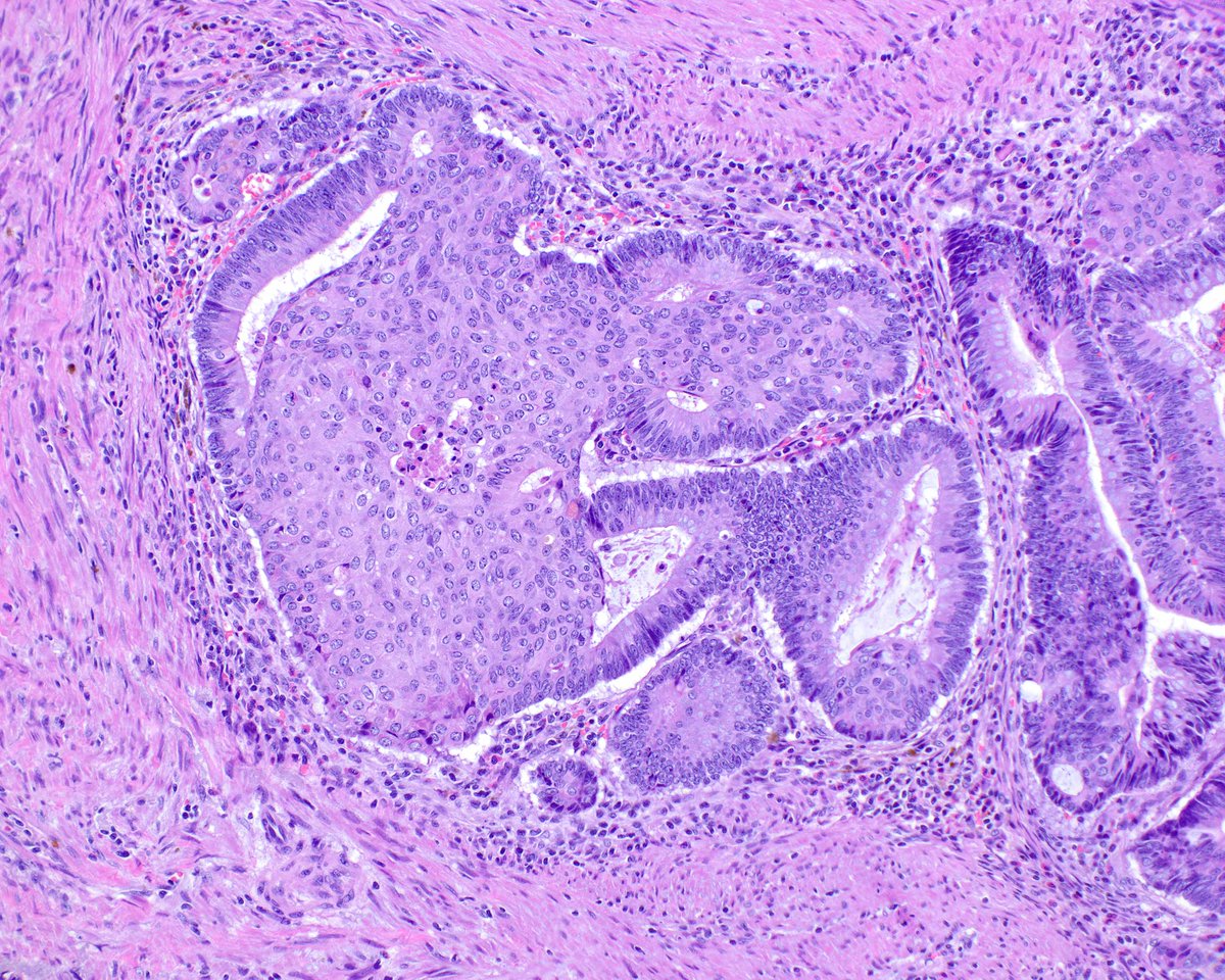
Squamous morules (some call them "microcarcinoids" because they can express neuroendocrine markers as well as squamous ones) in colorectal adenomas are fun to see. This adenoma has lots of pseudo-invasion, so the nests are invested in their lamina propria.
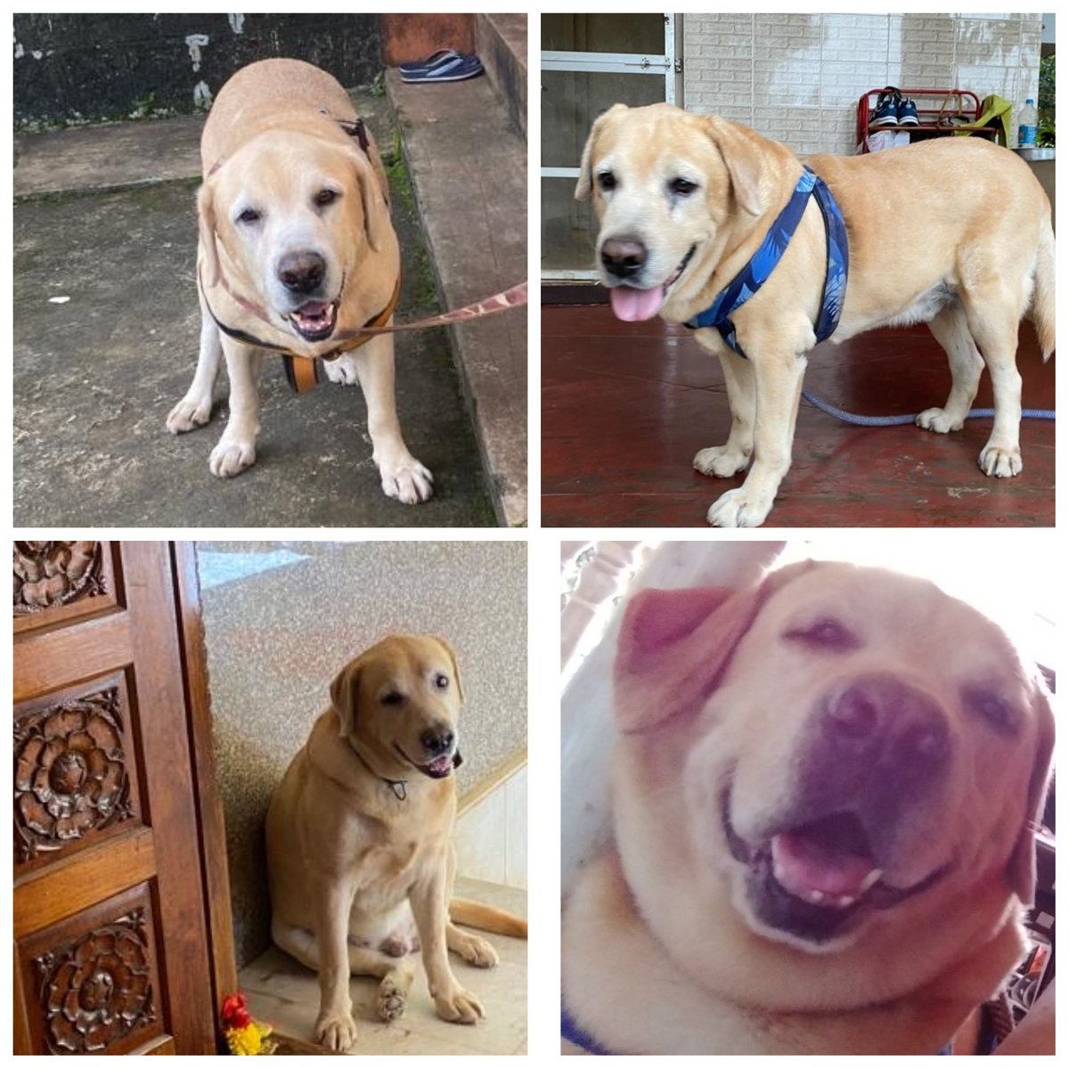
#777charlie must for non-dog lovers(how can u not love them?), loved every bit of it. @rakshitshetty the bond onscreen shared by u with charlie its amazing. This is our 9yr old Maxie, all he also does is eat, walk and sleep.
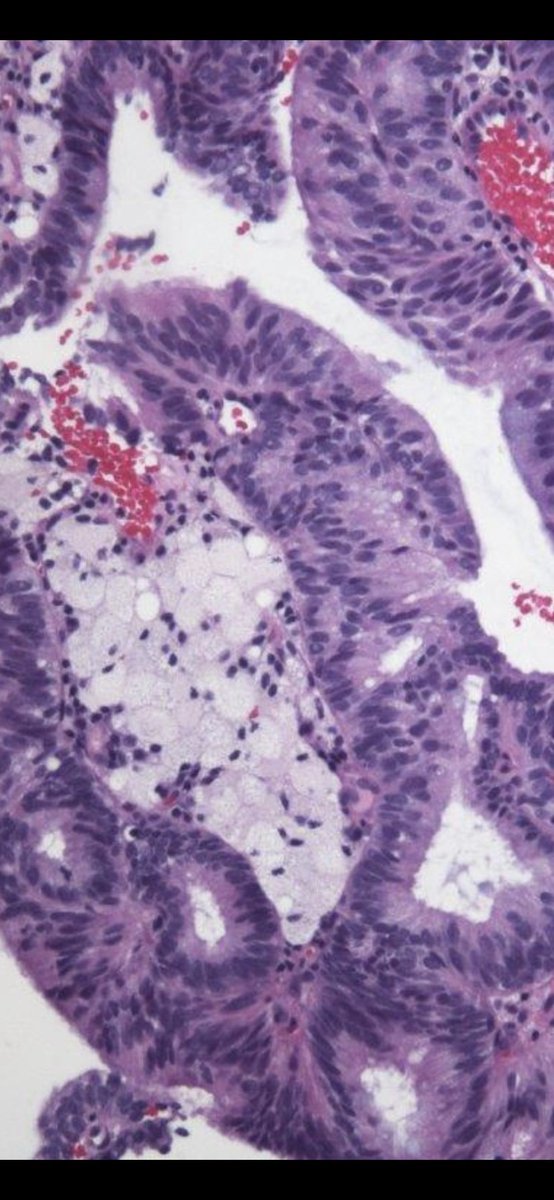
Presence of histiocytes in Pap- test of post-menopausal women should not be ignored. Example of histiocytes
associated with endometrial adenocarcinoma,
https://t.co/ztul0HLQKq