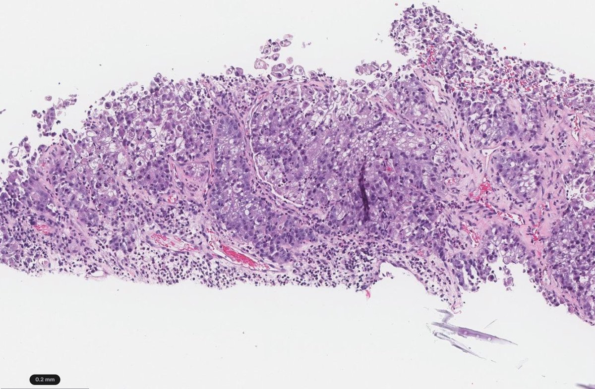
Crowded Gleason pattern 3
Glands are back-to-back and can look cribriform
Pitfall: calling it pattern 4
A keratin stain will highlight the architecture and show they are individual glands
Dr. McKenney. Prostatic Adenocarcinoma Grading: 2026 Update #USCAP#pathology#PathX
Prostate - Gleason pattern 3
Well-formed individual glands; some may have a small lumen (pic 1, yellow arrow)
Occasional glands without a lumen and focal branching are acceptable
Evaluate at low power
Dr. McKenney. Prostatic Adenocarcinoma Grading: 2026 Update #USCAP #pathology #pathX
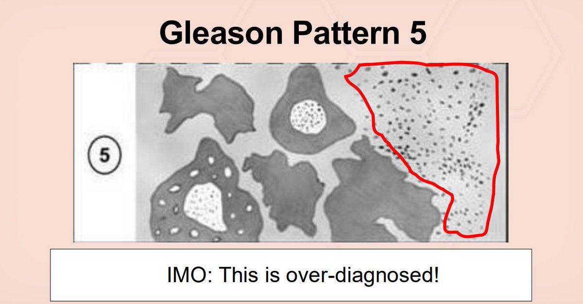
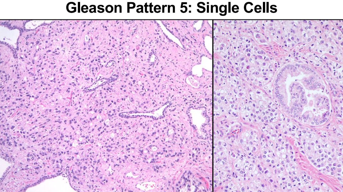
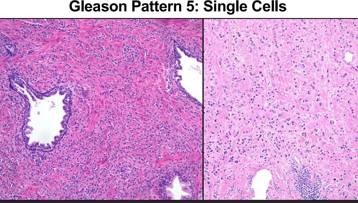
Gleason pattern 5-Single cells
This pattern is overdiagnosed
If you call it, the field should look like lobular breast cancer: a sheet of discohesive single cells
Do not pick out isolated cells sitting between individual glands
Dr. McKenney. Prostatic Adenocarcinoma Grading: 2026 Update #USCAP #PathX #pathology
Advance your cytology skills with ASC’s 3-day virtual Comprehensive Review Course for Cytologists.
Join experts Sept. 26, Oct. 3 & 10, 2026, and earn up to 23.75 CMLE credits.
Register today: https://t.co/w94eFdux8G
#Cytology#Cytopathology#ASC
Prostatic stromal tumor of uncertain malignant potential (STUMP).
Variable morphology: This one show atypical stromal cells, some with degenerative change, proliferating around crowded, architecturally complex benign glands.
Common pitfall: BPH stromal nodules are bland and multinodular, with prominent thick-walled vessels.
Hypercellularity, marked atypia, increased mitotic activity, atypical mitoses, necrosis, or invasion favor prostatic stromal sarcoma; exclude other sarcomas and sarcomatoid carcinoma. Ref: WHO 5th ed. #DxMindMap #ProstatePath #GUPath #PathTwitter @JHUpath
What’s New in the Classification of Bladder Neuroendocrine Tumors?
I am delighted to share our comprehensive review article published this week in Endocrine Pathology @EndoPath, written by Dr. Anandi Lobo@loboAnandi
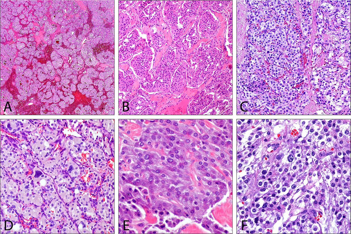
Our review provides an updated overview of neuroendocrine neoplasms of the urinary bladder, including small cell and large cell neuroendocrine carcinoma, mixed neuroendocrine neoplasms, well-differentiated neuroendocrine tumors, and paraganglioma. It highlights their distinctive histologic and immunophenotypic features, diagnostic pitfalls, clinical behavior, and emerging molecular biology.
Emerging molecular data suggest that morphology alone may not fully capture their biological complexity. Most bladder neuroendocrine carcinomas are highly aggressive and frequently show TP53/RB1 inactivation, TERT promoter mutations, APOBEC-related changes, and lineage plasticity. Emerging transcriptomic subtypes defined by ASCL1, NEUROD1, and POU2F3 may have prognostic and therapeutic relevance. In this review, we also propose a more refined, bladder-specific classification framework that integrates histopathology, biomarkers, and molecular features.
Some key highlights:
•TP53 and RB1 co-inactivation is a molecular hallmark of bladder neuroendocrine carcinoma, accompanied by frequent TERT promoter alterations and evidence of lineage plasticity.
•Transcriptomic studies are identifying lineage-associated subgroups characterized by ASCL1, NEUROD1, and POU2F3, with potential prognostic and therapeutic relevance.
•Recognition and quantification of mixed neuroendocrine–non-neuroendocrine components remain important for diagnosis and management.
•We discuss a more refined, bladder-specific classification framework integrating morphology, proliferative activity, and molecular alterations, with the goal of improving diagnostic reproducibility and facilitating biomarker-driven therapy.
Full article is available:
https://t.co/pLBmNKJYSk
https://t.co/i9WDNnV2CC
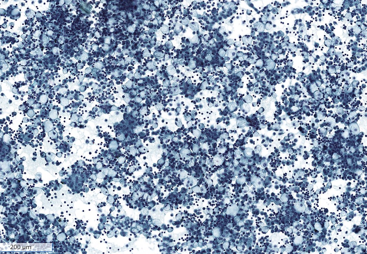
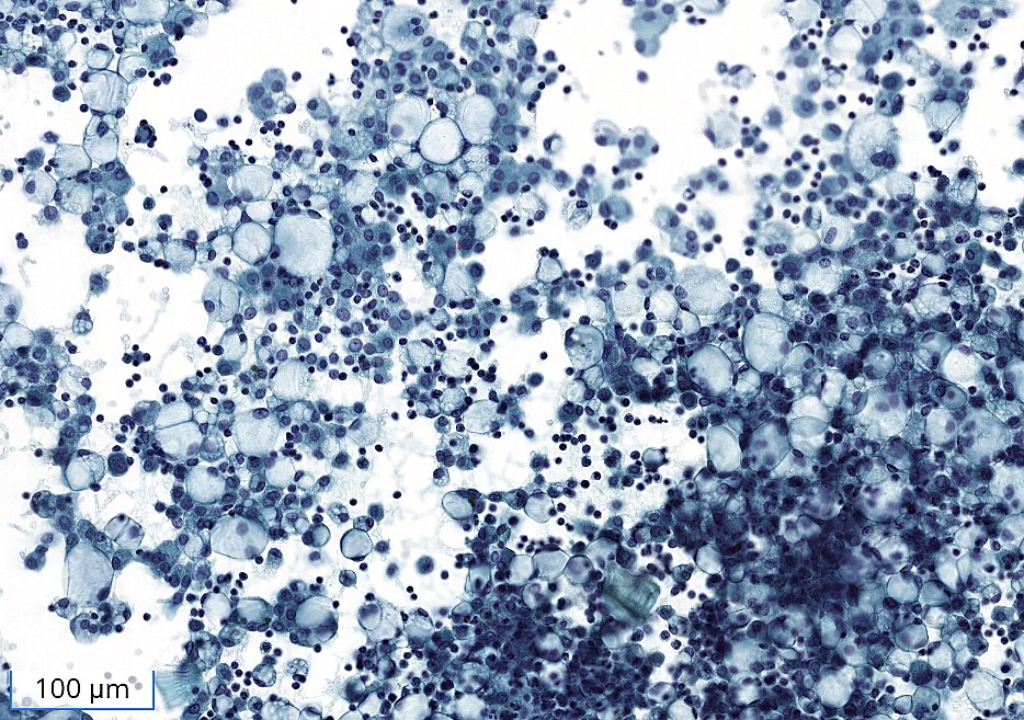
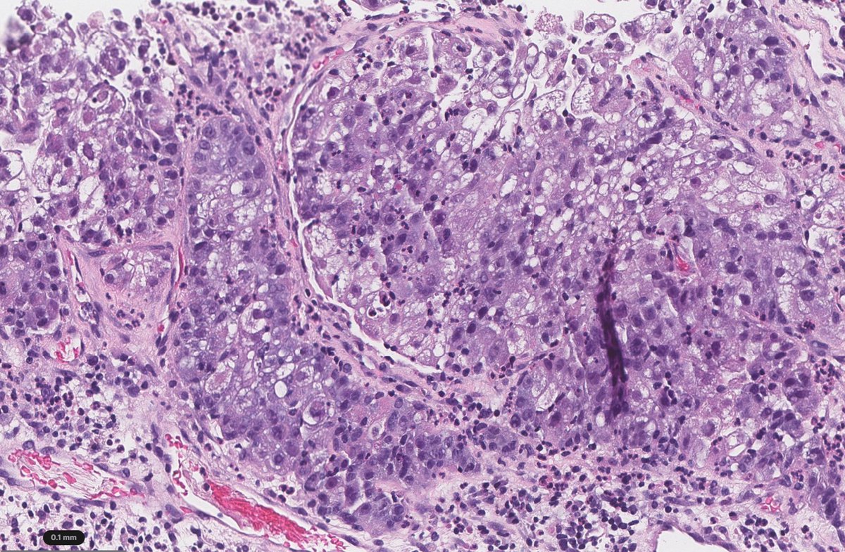
“Pseudo–signet ring cells in ascitic fluid: a deceptive look that can lead you straight to the wrong diagnosis.”
Hydropic degeneration is known to occur in histiocytes and mesothelial cells.
(Cytospin preps)
Brave New World of Breast Pathology 🧑🏫
We’ll discuss breast tumors defined by recurrent alterations—how to recognize them on H&E and triage ancillary testing, including Pan-TRK 👇.
#PathX#PathTwitter#breastpath#USCAP2026
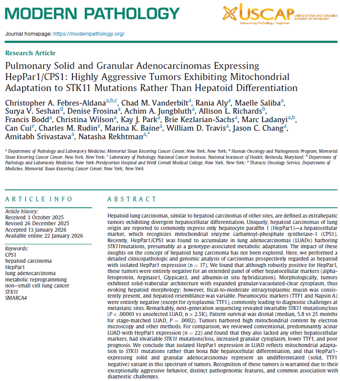
Final version of this article is now posted!
Granular/oncocytic lung carcinomas: what these are, and how they can cause diagnostic problems.
https://t.co/3saxnQzEEl
https://t.co/23ieW6eoET
The newly-described solid and granular/oncocytic lung adenocarcinomas (SAGAs). Recent case (adrenal met) illustrating their distinct morphology: granular-vacuolated-clear cytoplasm + macronucleoli. Focal mucin is common (best by mucin stains).
https://t.co/3saxnQzEEl
Dear all, we are proud to announce that the latest issue of the IAC-@IARCWHO -@WHO Reporting Systems for Cytopathology has just been published. The topic: Soft Tissue Cytopathology https://t.co/6KPYoR7FEc #SoftTissue#Cytopath
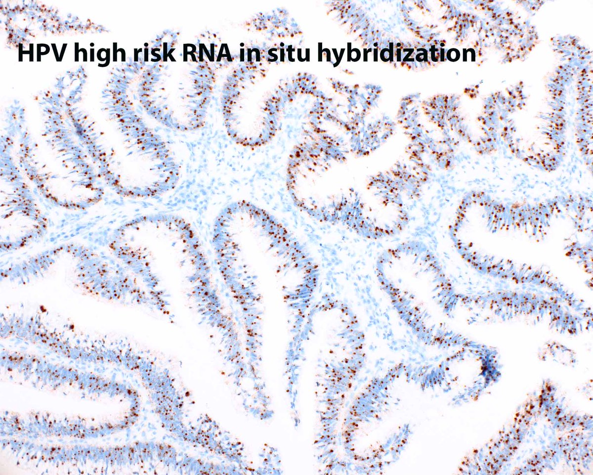
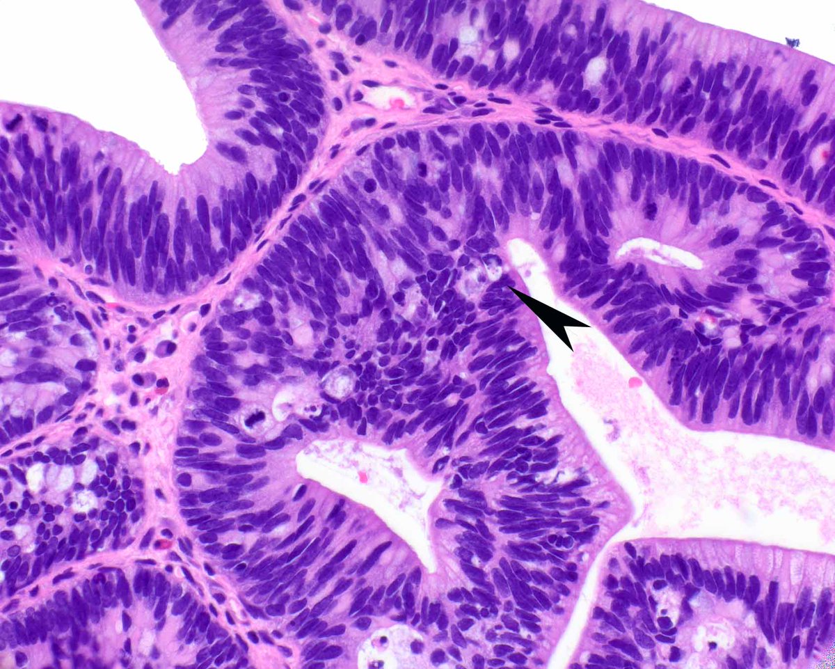
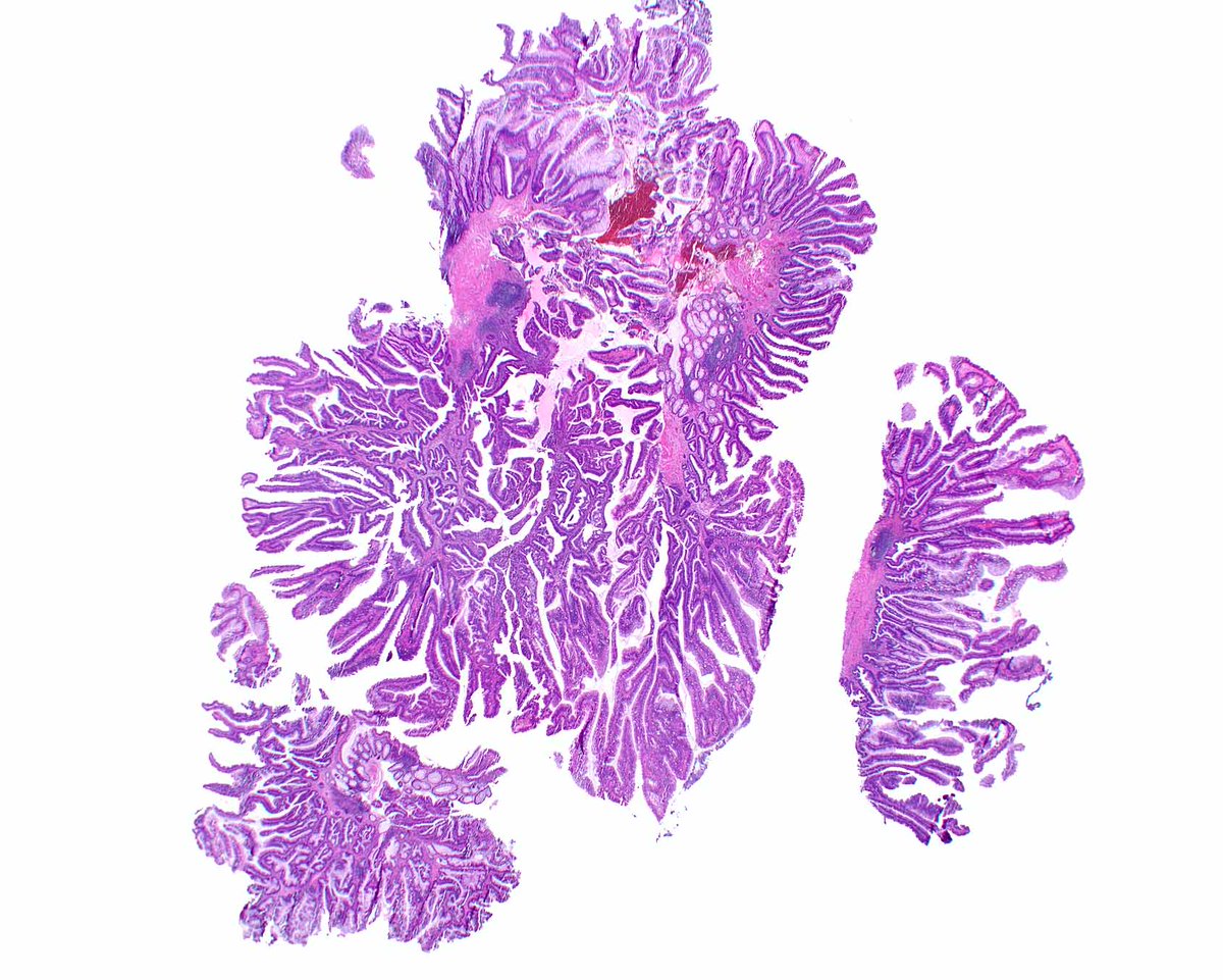
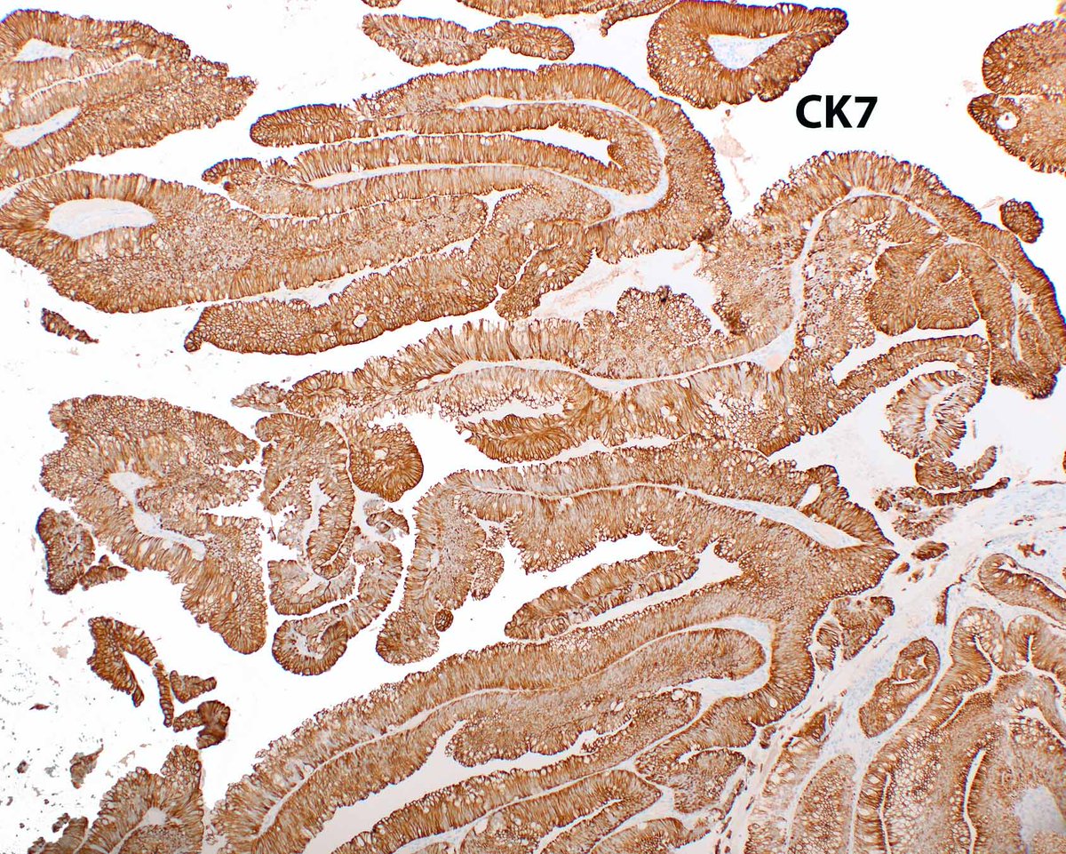
Distal rectal polyp - HPV-associated glandular neoplasms are rare and mimic tubulovillous adenomas. The mucin is peculiar and apical apoptotic bodies can be plentiful (arrow).
Voltaggio L, et al. Mod Pathol. 2020 May;33(5):944-952. PMID: 31857682.
Isolated, bizarre, giant and pleomorphic naked nuclei are a common cytopathologic feature of "Malignant PECOMA". (FNA, Neck mass)
See our previously published article - Wangsiricharoen S, et al. Cytopathology of extra-renal perivascular epithelioid cell tumor (PEComa): a series of 7 cases and review of the literature. J Am Soc Cytopathol. 2021 PMID: 33162379.
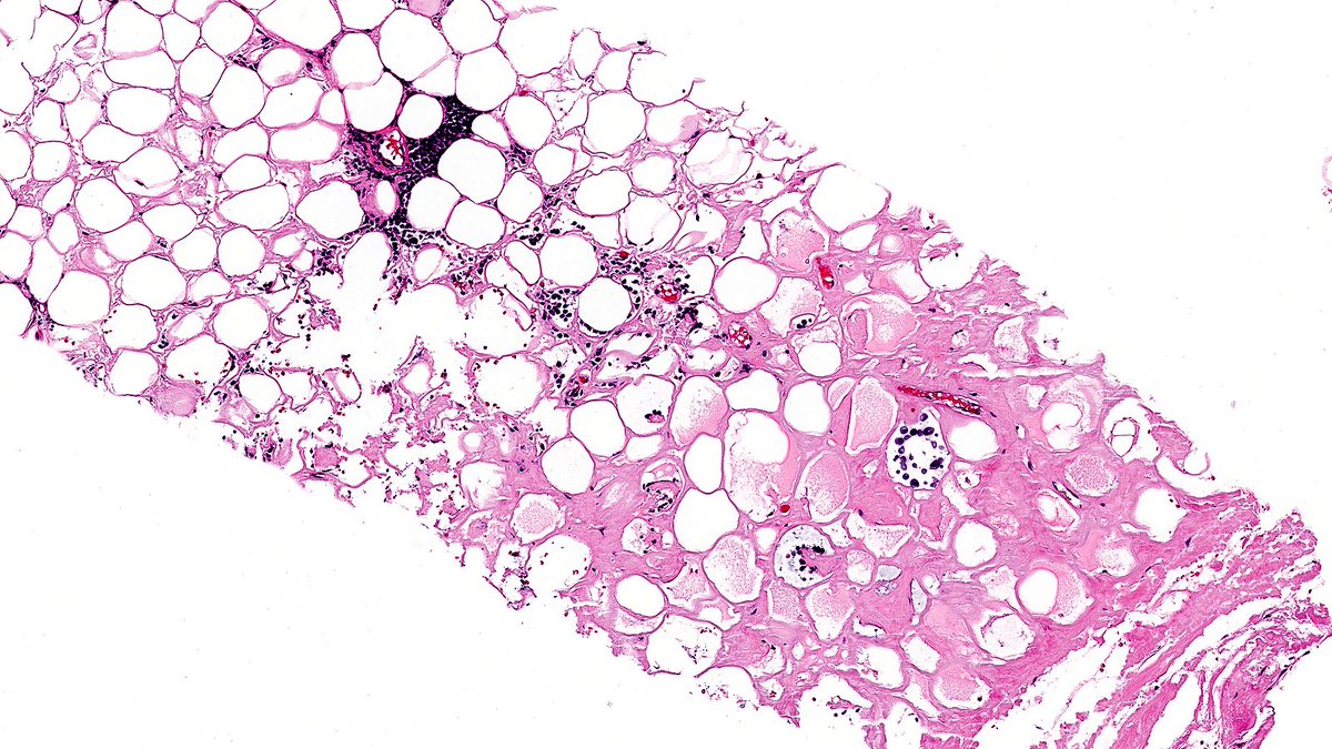
Case of Amyloid Deposition in Breast 🤒🔬
Breast amyloidosis is rare and can represent localized disease, a manifestation of systemic inflammatory disorders, or even be associated with underlying lymphoid neoplasia.
#PathX#PathTwitter#breastpath
Gastric glomus tumour with positive DOG1 and synaptophysin expression: a diagnostic challenge https://t.co/LBursRctPz #Pathtwitter@Pathoutlines@diagnexia