New preprint: We have developed an integrated pipeline to i) image gastruloids in toto with dual-view 2-photon🔬, ii) quantitatify cell and tissue-scale patterns of gene expression and morphometric features, iii) interact with the data in @napari_imaging. https://t.co/NvexmiWnHY
Introducing #NovaFLIM: our new #fluorescence lifetime imaging analysis software. It is designed for an efficient and multi-faceted analysis of #FLIM, FLIM-FRET, and #anisotropy. ➡ Learn more: https://t.co/xGpOrnyDYX
📢Our preprint list is now up on FocalPlane. This list is focussed on new research in bioimage analysis. Drop us a message if there is a preprint that we've missed!
https://t.co/NzqzHmmjQX
#Gastruloids have many uses as #models for: early mammalian development, #SelfOrganization, mapping signalling/gene expression, morphogenesis. Here @Equipe_lenne use’m to develop gral tools to image gene/protein expression at diff scales https://t.co/9NbKx1YWpM #InNumbersWeTrust
📡🧵 Excited to release our preprint: Ultrack: Pushing the Limits of Cell Tracking Across Biological Scales
https://t.co/KN5Hw3BpI0
A tour-de-force by @jobragantini, #Ultrack is a versatile, highly accurate, and fast ILP-based cell tracking software for 2D, 3D, and multicolor datasets. It has convenient @napari_imaging and @FijiSc plugin interfaces and built-in HPC cluster support.
https://t.co/DvKyowFbbi
#cell_tracking #microscopy #devbio @czbiohub
A first step towards a basis for research on #StemCell based #EmbryoModels
https://t.co/hhQTCVBiVM @NatureCellBio
We hope that this will be useful in this new field where quality control and appropriate criteria are needed
Happy that I could contribute 🙂 Flipper-TR also works well with 2-photon excitation 👇. It will be interesting too see in different systems what the probe can tell, and on which time scales. In this context, fast FLIM will be useful. Our attempt here: https://t.co/zdJ0yc6VKQ
Super happy to share with you a detailed tutorial covering everything you need to know about membrane mechanosensitive Flipper probes (🐬) using fluorescence lifetime microscopy. Accessible here: https://t.co/ya89JmzXpg See 🧵 below
All what you wanted to know about using flipper probes @spirochrome@matile_group for cell biology and didn’t dare asking is in: https://t.co/MOC98W09ld
Thanks to all co-authors and colleagues, thanks to @CRoffay & @vincent81196538 for their great work!
New preprint: We have developed an integrated pipeline to i) image gastruloids in toto with dual-view 2-photon🔬, ii) quantitatify cell and tissue-scale patterns of gene expression and morphometric features, iii) interact with the data in @napari_imaging. https://t.co/NvexmiWnHY
Interested in diffusion and oligomerization? Our brightness-transit statistics pipeline based on sFCS acquisitions just came out in @NatureComms 🥳 Start correlating! Great collab with @pfcespedes@KaredlaNarain@FritzscheLab@MichaelLDustin
https://t.co/biJiDFwEUn
Great to be back in Basel for #BaCell3D. I will present a poster (#216) tomorrow on in toto imaging of organoids, which we use to investigate patterning mechanisms in gastruloids. Looking forward to discuss.
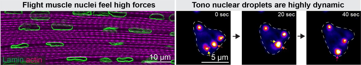
PhD position in Mechanobiology in Marseille! Are you excited to study how mechanical forces impact the transcription of muscle nuclei as well as muscle metabolism? Join us at our fantastic campus in Marseille. https://t.co/zGdtbTdQTM @Fly_EDS@IBDMmarseille@CNRSbiologie#muscle
I’m so happy to share another story from my PhD with @ErcanLab and @preibischs, where we studied the gene regulation in the dosage compensation system of C. elegans using smFISH! Here are a few highlights…🧵 https://t.co/uUu2g8UkU3
🔬🌟Join us for the 3rd @centuri_ls Hackathon for Quantitative Biology in Marseille!💥Both wet and dry lab people are welcome! Dive into amazing projects and experience this year stunning location!🌍 Registration now open!🚀💻#HackBio#QuantBio
⬇️Don't miss out, sign up now!⬇️
Very happy that I could contribute to this work led by @PRecouvreux. It has always fascinated me how cells coordinate using morphogens. When I started to get interested in biology, C.elegans was the first organism that we imaged at a🔬. Here, both came together with in vivo FCS.
1/n
We are very pleased to see our study on Wnt spreading in C. elegans embryos published in @CurrentBiology
https://t.co/MLJocpw6ZF .
The outcome of an exciting collaboration between Vincent Bertrand's team and our team. A short thread on our main results!