🔸 Desde la #SERAM, tras consultar con nuestros socios, nos hemos posicionado sobre el borrador del nuevo Estatuto Marco del personal estatutario de los servicios de salud
Manifestamos nuestro apoyo a las acciones reivindicativas convocadas en las próximas fechas con el objetivo de reformar el actual anteproyecto para adecuarlo a las necesidades de nuestra profesión y nuestra especialidad.
Consulta nuestro posicionamiento completo aquí 🔗 https://t.co/mODN35wUDa
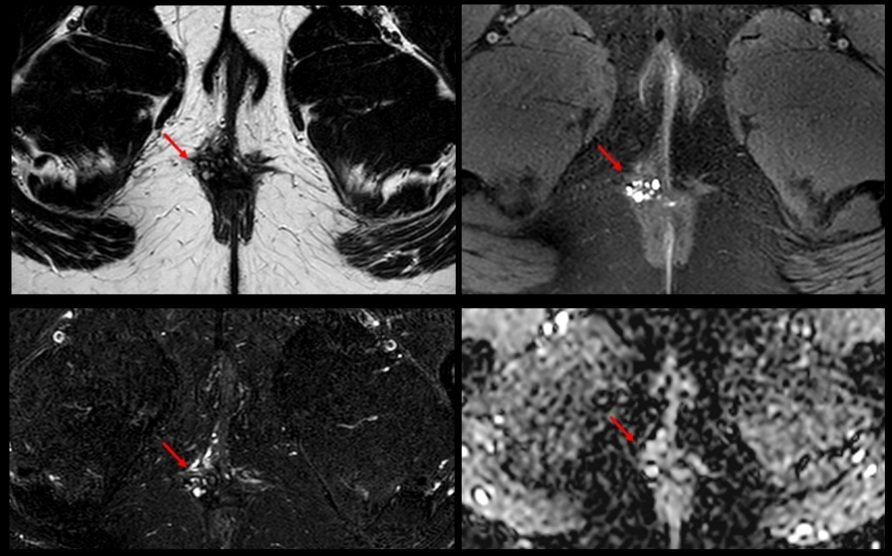
💥 New YE case. Courtesy of Dr. Canales, submitted by @AnaVillanuevaCa
♀️ 27 yo. Perianal pain of 2 years, recently admitted to ED with diagnosis of perianal abscess
🔴 Multilocular hyperintense T2 lesion, hyperintense on T1 fat sat and not restricted difussion
🧵..
Have MULTIPLE questions about MULTIPLE sclerosis?
Having trouble seeing neuromyelitis optica?
In a fog about MOG?
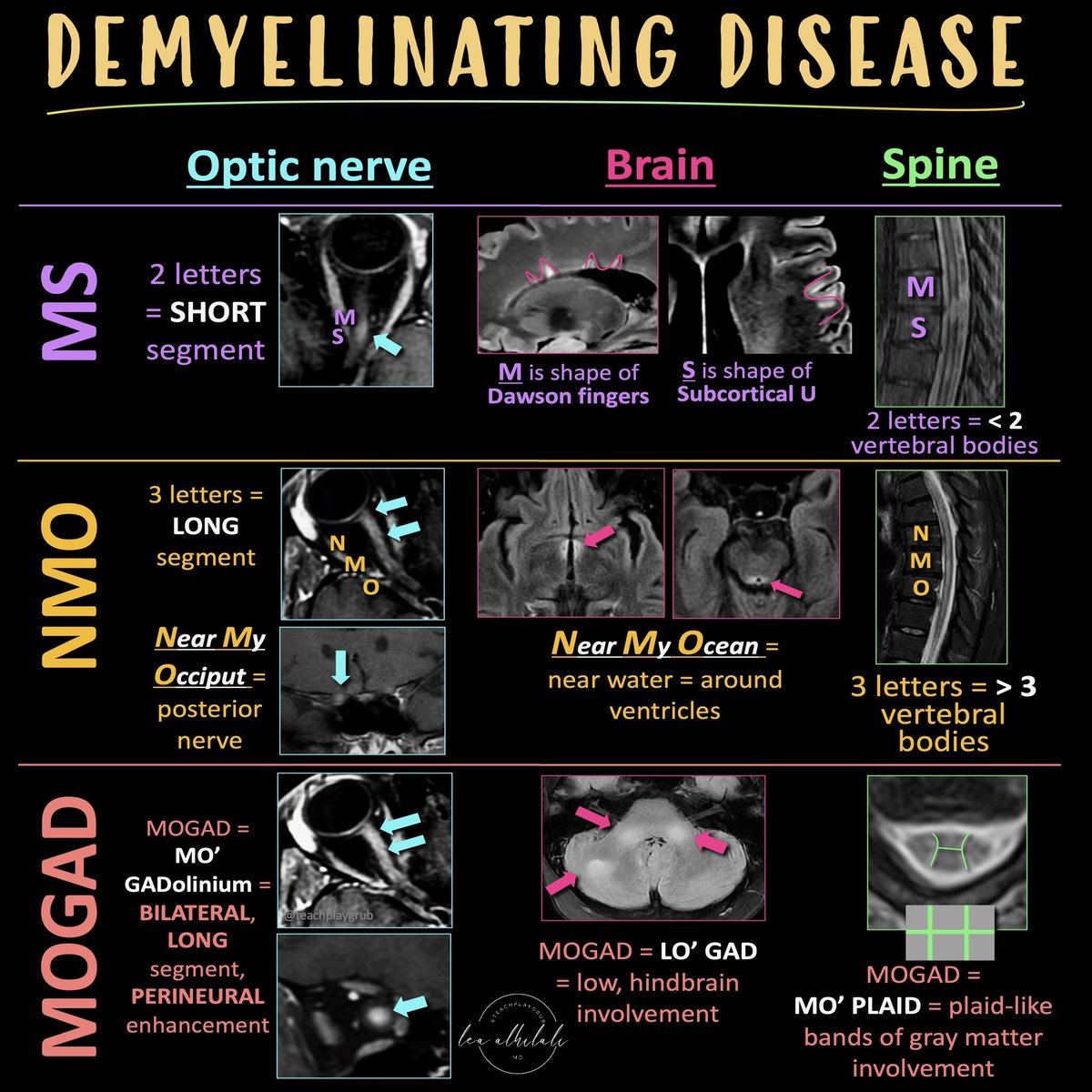
Here’s the cheat sheet you NEED to distinguish the demyelinating diseases!
Demyelinating diseases predominantly involve the optic nerves, brain, & spine.
The three main chronic demyelinating diseases are Multiple sclerosis (most common), neuromyelitis optica (NMO), & myelin oligodendrocyte glycoprotein (MOG) antibody associated disease or MOGAD
Each has its own features in the optic nerve, brain, & spine. Here’s how to remember them!
MS
Optic nerve:
MS only has 2 letters, so MS involvement of the optic nerve tends to be short segment
Brain:
Letter M makes the shape of the perivascular distribution of lesions along the ventricles (Dawson’s fingers)
Letter S makes the shape of the subcortical U fiber involvement
Spine:
MS is only 2 letters, so lesions are usually less than 2 vertebral bodies in length
NMO
Optic nerve:
NMO is a longer abbreviation, three letters, so longer involvement
NMO can stand for Near My Occiput. Occiput is posterior, so more posterior nerve involvement
Brain:
NMO can stand for Near My Ocean. What is your brain’s ocean? The ventricles. NMO lesions are all periventricular
Spine:
NMO is 3 letters, so lesions usually more than 3 vertebral bodies in length
MOGAD
Optic Nerve:
Remember MO’ GAD-olinium. So things that cause more regions of enhancement. MOGAD lesions are commonly bilateral & long segment & enhancement can extend perineural
Brain:
Remember LO’ GAD. MOGAD typically involves the lower areas of the brain
Spine:
Remember MO’ PLAID. MOGAD can give a plaid-like H shape in the cord from predominantly gray matter involvement
Hopefully, this cheat sheet will help you remember how to distinguish the demyelinating diseases! It ain’t lyin’ about diseases of myelin!
New #lirads treatment response algorithm includes simpler definitions, optional use of ancillary features and a separate algorithm for radiation therapy. Read 👇for more information! @LIRADS5@RadiologyACR
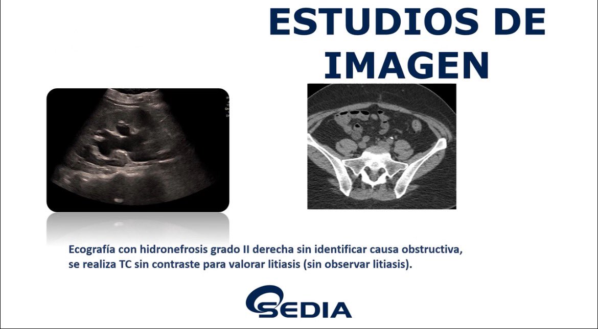
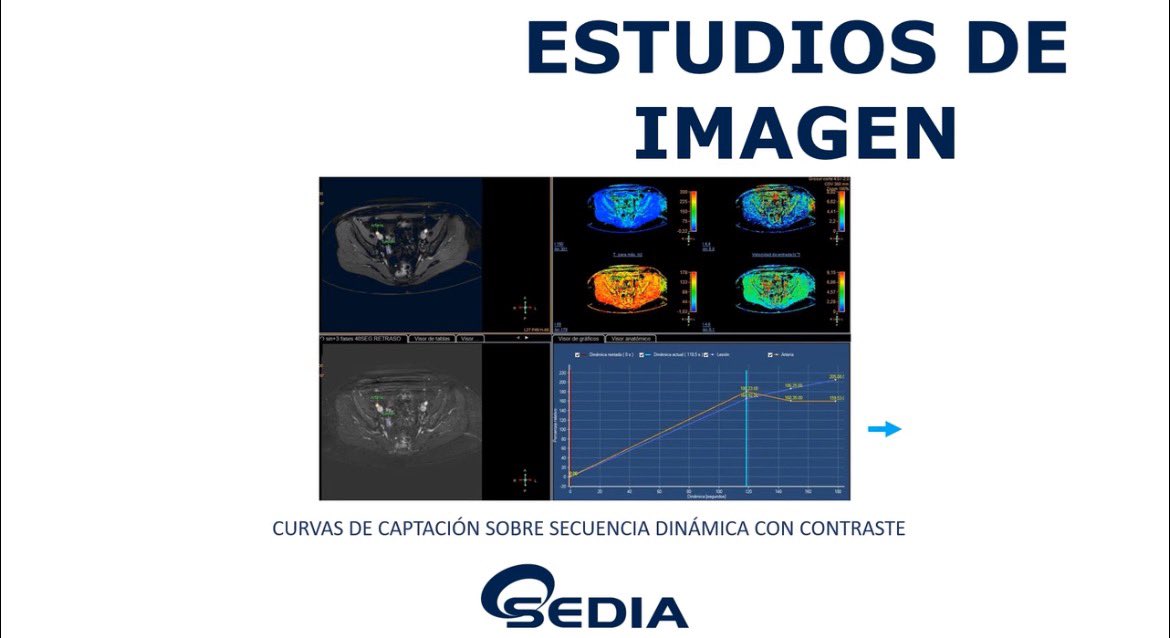
Nuevo #casosedia de noviembre:
“Mujer 52años con dolor en hipocondrio derecho sin fiebre”
Puedes verlo en https://t.co/ZpZZw3t1LU.
✍️Envía tu respuesta a [email protected] y empieza a participar para conseguir gratis tu inscripción para el #congresoSEDIA2026
Finally, the Shoulder Ultrasound course is ready!!
1st calling on 06/24/24, open access for 3 months.
Discount coupon SHOULDER15 during the first month!
You can sign up now, all the information here:
https://t.co/NJRefm2mVT
Another free livestream in support of #Radiopaedia2024 coming up this Friday!
Watch @DocLasker’s entertaining lecture on the imaging of gym injuries. Hosted by @francisdeng.
🔔 https://t.co/6gNQuUPOha
#foamrad#mskrad
New @AJR_Radiology:
"New LI-RADS Ultrasound Surveillance v2024: A Conversation With Working Group Cochair Dr Aya Kamaya"
By Dr @ayakamaya@StanfordRad
https://t.co/6OZIattSYG
dry needling: MRI findings
Patient with pain in the long head of the biceps femoris muscle and a suspected fibrillar tear, for whom an MRI of the thigh was requested.
The MRI shows a patchy increase in signal in the distal third of the long head of the biceps associated with hyperintense linear paths, which correlate with the history of dry needling performed three days before the radiological study.
Dry needling is an invasive technique used in physiotherapy to eliminate muscle pain points (or trigger points), which can be generated due to overload.
It is imperative not to do this treatment during the five days before performing an MRI because it can make the diagnosis of muscle injury difficult or impossible, mainly grade I.
How many routes do you see in the video?
New @AJR_Radiology Accepted Manuscript:
"Beyond the AJR: Repeat Screening With PSA and MRI Identifies Few Prostate Cancers"
By Drs Glazer & @FennessyFiona@BrighamRad
https://t.co/Ib6oykl1Uj
“Double beak sign” of closed loop obstruction.
Commonest cause of closed-loop obstruction is adhesions (this case also).
Volvulus and Internal hernia can also result in closed-loop obstruction and similar morphology.
When performing contrast-enhanced ultrasound using perfluorobutane to diagnose HCC, Kupffer phase images may be optional when recognizing features of non-HCC malignancy and incorporating transitional phase washout.https://t.co/bPxmbzbl36
The broken-fat pad sign helps clinicians distinguish between knee rheumatoid #arthritis and osteoarthritis, allowing for early accurate diagnosis and proper treatment. (Qizheng Wang et al.)
#InsightsIntoImaging
🔗 https://t.co/R3sFiSvqi0
New @AJR_Radiology Accepted Manuscript:
"Beyond the AJR: The Burden of Multidisciplinary Tumor Boards"
By Drs @Vplanz & @olgabrook @VUMCradiology @BIDMCRad
https://t.co/gqQxDkrTLC
Endometriosis: lo que el radiólogo debe de saber
🔎 Este #PiperSERAM explica los distintos tipos de endometriosis y los principales hallazgos visualizados en #Ecografía y resonancia magnética #RM
Accede aquí 🔗 https://t.co/NW2GoesogP
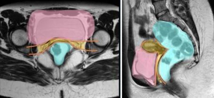
Educational Review: MRI anatomy of the rectum - key concepts important for #RectalCancer staging and treatment planning. (Nino Bogveradze et al.)
#InsightsIntoImaging#RadiologyHeadToToe
🔗 https://t.co/ABuCy3vKTn