Cardiac sonographer. Accredited in echo CHD by EACVI-ESC and BSE. Intensive care, mechanical circulatory support and LVAD. Royal Brompton Hospital Hospital.
How to do SVC #POCUS 📹 + an illustration of abnormal SVC Doppler patterns.
#VExUS#eVExUS#Nephpearls
From 🔗J Am Soc Echocardiogr. 2023;36(5):447-463. doi: 10.1016/j.echo.2023.01.017
Complete AVSD Repair — Two-Patch Technique✌️🎥
👉Ventricular component closure: anchor the VSD patch while preserving adequate LAVV orifice size and respecting the displaced conduction axis.
👉LAVV cleft closure: restore leaflet coaptation and control AV valve regurgitation without creating left AV valve stenosis.
👉Primum ASD patch closure: complete atrial septation while avoiding injury to the displaced AV node and conduction tissue.
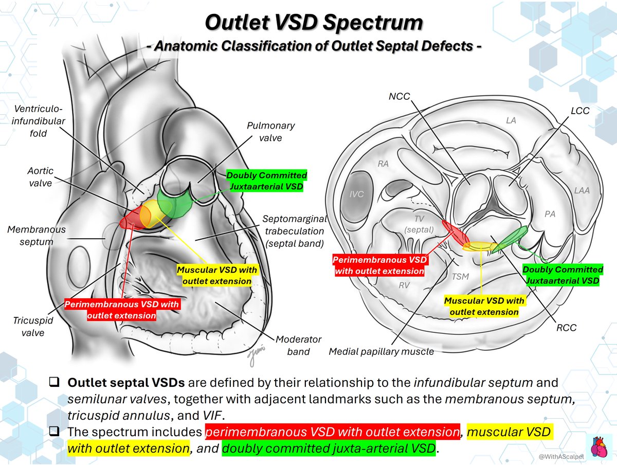
Outlet VSD Spectrum📝
👉Outlet septal VSDs include three main anatomic types: perimembranous with outlet extension, muscular with outlet extension, and doubly committed juxta-arterial VSD.
👉The short-axis echo view is especially helpful.
ECO SKILLS | Venas pulmonares
La valoración ecocardiográfica de las venas pulmonares aporta información clave sobre la función diastólica, presiones de llenado y severidad de valvulopatías.
Analizar su patrón de flujo permite complementar la evaluación del paciente
LV Distension on VA-ECMO: The Hidden Load
👉Peripheral VA-ECMO raises aortic afterload → suppresses LV ejection and risks AoV closure, promoting LV distension and pulmonary congestion.
👉Early assessment is critical—confirm AoV opening and initiate LV unloading (LA vent/Impella) before thrombus or myocardial injury develops.
Fetal Circulation #2 — Preferential Streaming of Oxygenated Blood
👉Oxygenated placental blood returns via the umbilical vein and is shunted through the ductus venosus into the IVC, where the Eustachian valve directs it across the foramen ovale.
👉This preferential stream supplies the left atrium, left ventricle, and ascending aorta, optimizing cerebral and coronary perfusion.
👉In contrast, SVC-derived blood enters the right ventricle and, with high fetal PVR, is diverted through the ductus arteriosus to the descending aorta.
At first glance, this appears to be a large mid-muscular VSD. Look closer: an angled VSD patch incorporates part of the RV cavity into the systemic ventricle, surgical anatomy redefining physiology. #ACHD ❤️🩹
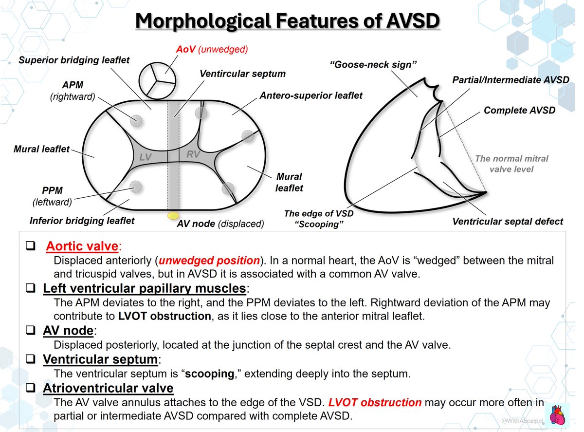
AVSD: Unwedged Aortic Valve and LVOTO
👉The AoV is displaced anteriorly in an unwedged position, no longer seated between the two AV valves.
👉This characteristic geometry elongates the LVOT and can contribute to LVOTO.
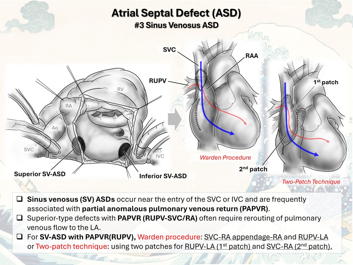
Atrial Septal Defect (ASD): Four Types
👉Primum ASD: Low ASD near the AV valves, within the AVSD spectrum.
👉Secondum ASD: Fossa ovalis defect and the most common ASD type.
👉Sinus venosus ASD: Venous inflow defect near the SVC/IVC, often with PAPVR.
👉Coronary sinus ASD: An unroofed coronary sinus, often associated with LSVC.
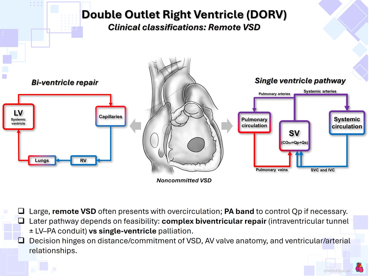
DORV #2 — Clinical Classification
DORV is clinically grouped by physiology driven by VSD position and RVOT obstruction:
👉VSD-type (overcirculation, treat like large VSD ± PA band, plan LV–Ao baffle),
👉TOF-type (PS → cyanosis, TOF repair, RVOT relief),
👉TGA-type (subpulmonary streaming, ASO+VSD closure),
👉Remote VSD (complex BiV vs SV).