The self driving multi-scale microscope from @Daetwyler_St is now on BioRxiV:
https://t.co/RClR6BEmbz
Here some immune - cancer cell interactions. The whole Zebrafish is imaged as well during this time lapse acquisition.
1/ Excited to share our new paper with @SihanSean@utswcancer@CRI_UTSW and Michael Lawrence @MGH_RI, now online @CD_AACR. Led by Shreoshi Pal Choudhuri, we found that ecDNAs with MYC paralogs drive acquired cross-resistance in small cell lung cancer #SCLC
https://t.co/MraxEM6PKb
Out now! @HFarsibaf, @MegDrisc, @DanuserLab and colleagues propose a framework to assess the spatial scales of molecular organization at the cell surface in a cell-morphology invariant manner. https://t.co/rb6kojKSi5
👉https://t.co/A69JUwnF9U
Excited to finally see this work out in @JCellBiol !!! Check it out if you're interested in:
✅ #zebrafish cancer models
✅ quantitative in vivo imaging assays
✅plasticity in Ewing Sarcoma
1/n
How sensitive is the analysis to the cell morphology? ZERO!!
We simulated polka dot patterns to validate that the framework introduces the spatial scale signature in a shape-invariant mode.
6/6
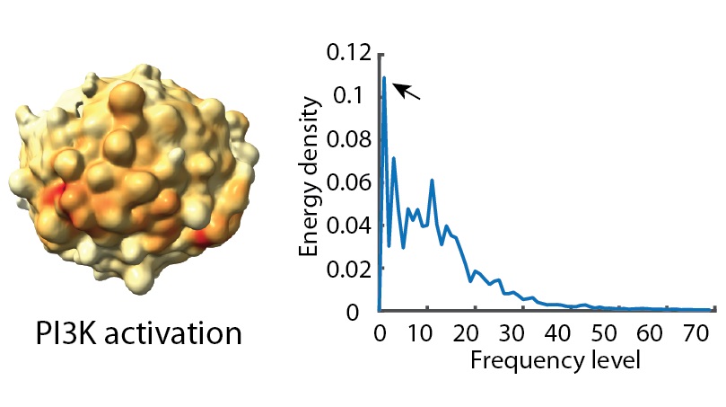
The organization of molecules on the cell membrane is crucial for regulating cell functions. Here we introduce a new pipeline for analyzing the molecular patterns in a shape-agnostic mode: https://t.co/Ruw17ZfiWB @MegDrisc@gdanuser1@jennyqzou#CellularHarmonics,#uSignal3D
1/6
Using this basis set, we parameterized the molecular signal pattern and computed the energy density spectra to define the spatial scale signature of a given pattern.
5/6
Woo hoo! Excited to finally share a big chunk of my postdoc work with this (updated) preprint:
In vivo profiling of site-specific human cancer cell states in zebrafish
https://t.co/LB0vrG3apJ
Wanna hear more? Let's roll👇
And the bleb saga continues. @MuthrNaturzSon has worked on this for quite a while, and I am glad it is out now. Also happy to see 3D microscopy and 3D shape analysis leveraged in this work.
Congrats to all involved!