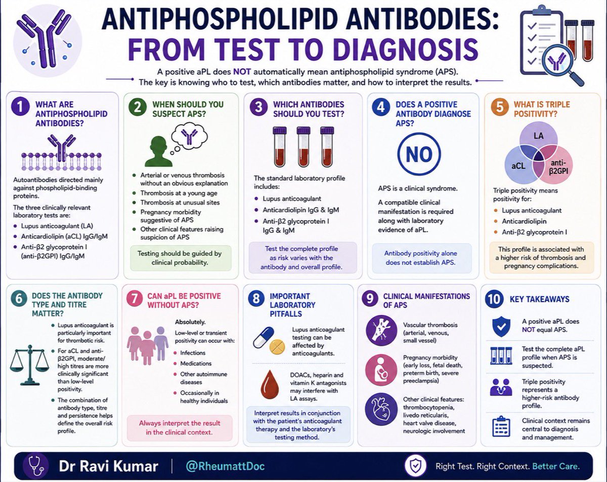
🧵 Antiphospholipid Antibodies: From Test to Diagnosis
A positive antiphospholipid antibody does not automatically mean antiphospholipid syndrome (APS).
The key is knowing who to test, which antibodies matter, and how to interpret the results. 👇
#RheumattDoc#MedTwitter #RheumTwitter #Medicine #rheumatology @docakx@IhabFathiSulima@CelestinoGutirr@DurgaPrasannaM1
ఆషాఢ బోనాలను పురస్కరించుకుని కోరిన కోరికలు తీర్చే కల్పవల్లిగా, నగర ప్రజలను సదా చల్లగా కాపాడే జగజ్జనని లాల్దర్వాజా శ్రీ సింహవాహిని మహంకాళి అమ్మవారిని దర్శించుకున్నాను.
అనంతరం ఉత్సవాల నేపథ్యంలో ఆలయ పరిసర ప్రాంతాల్లో చేపట్టిన పటిష్ఠ భద్రతా ఏర్పాట్లను పరిశీలించాను.
@hydcitypolice@CPHydCity
#LalDarwazaBonalu #Bonalu2026
Identification & Holistic Management of CKD
✅ Test at-risk adults (HTN, diabetes, CVD, obesity + others) for eGFR + uACR
✅ Diagnose CKD when abnormal for >3 months
✅ Use KDIGO heatmap to classify risk & set monitoring frequency
✅ Know your refer-to-renal red flags
Epicardial Fat in HFpEF: From “Bystander Fat” to a Potential Therapeutic Target
Based on:
🔥 Why this review matters
HFpEF is increasingly viewed as an inflammatory–metabolic syndrome, rather than simply a disorder of impaired LV relaxation. Epicardial adipose tissue (EAT)—the visceral fat depot directly surrounding the myocardium—may be an important link connecting obesity, insulin resistance, inflammation and myocardial dysfunction. Pathological EAT expansion has been associated with HFpEF and adverse outcomes, although causality is not yet established.
🔹 CME INDIA Clinical Pearls
1. Epicardial fat is not simply “bad fat.” In the healthy heart, EAT serves as an energy reservoir and fatty-acid buffer and produces cardioprotective, anti-inflammatory and antiatherogenic adipokines. Brown/beige EAT may additionally provide thermogenic and antioxidant benefits.
2. The problem is pathological transformation—not merely the amount of fat. With aging and HFpEF, EAT can undergo beige → white adipose transformation, accompanied by loss of UCP-1 and mitochondrial density. Thus, the biology or “quality” of EAT may ultimately matter more than its volume.
3. A useful mechanistic sequence is:
EAT expansion → ↑ lipolysis/free fatty acids → myocardial lipotoxicity + ↓ glucose uptake/insulin resistance → mitochondrial dysfunction/oxidative stress → inflammation → fibrosis → diastolic dysfunction/HFpEF. The schematic on page 5 beautifully summarizes this metabolic-inflammatory pathway.
4. EAT is associated with subclinical myocardial dysfunction. Increased EAT thickness/volume has been linked with worse global longitudinal strain, adverse ventricular mechanics, atrial dysfunction and arterial stiffness; EAT thickness has also emerged as an independent predictor of HFpEF in several studies.
5. But association ≠ causation. Some studies have found no relationship—or even lower EAT volume—in HFpEF, and UK Biobank analyses suggest that associations weaken substantially after accounting for overall visceral adiposity. Therefore, EAT should not yet be considered a proven causal therapeutic target.
6. “How much epicardial fat?” may be the wrong question. There is currently no universally accepted normal EAT volume or density. The review emphasizes standardization of measurement and suggests that EAT quality/density reflecting inflammation, metabolism and browning/whitening may provide more clinically meaningful information than volume alone.
7. SGLT2 inhibitors and GLP-1 receptor agonists are particularly interesting. Both drug classes have demonstrated reductions in EAT thickness/volume. Whether this simply reflects overall fat/weight reduction or includes a direct EAT effect remains unresolved; receptors relevant to these pathways are expressed in human heart/EAT. The review therefore places these agents at the forefront of currently available systemic approaches potentially modifying EAT biology.
8. Dapagliflozin provides an intriguing metabolic clue. Experimental human EAT observations cited in the review suggest dapagliflozin may increase GLUT4-mediated glucose uptake, reduce EAT volume and reduce secretion of pro-inflammatory chemokines. This raises the possibility that restoring glucose utilization could interrupt the vicious cycle of insulin resistance, excess lipolysis and lipotoxicity.
9. “Re-browning” epicardial fat is an exciting concept—but not clinical therapy yet. Converting pathological white EAT toward metabolically healthier beige/brown adipose tissue could theoretically restore thermogenesis and improve metabolic function. However, EAT re-browning has not yet been tested as a therapeutic strategy in HFpEF.
https://t.co/a7AKKS1qom
The forests have protected our Tribal communities for generations, and in return, they have protected the forests as guardians of nature, culture and ancient wisdom. On the International Day of the World’s Indigenous Peoples, I extend my heartfelt greetings to all my Tribal brothers and sisters.
This year’s theme, “Honouring Indigenous Midwives: Safeguarding Life and Well-being,” reminds us of the invaluable traditional knowledge passed down through generations and the vital role Indigenous women and communities play in protecting life, health, culture and heritage.
My bond with our Tribal communities is deeply personal. Their affection and trust have always strengthened my resolve to work for their dignity and welfare. Through initiatives such as Adavi Thalli Baata, improved connectivity to remote habitations, livelihood opportunities, and stronger protection of forests and wildlife, we are working to ensure that development reaches every Tribal family while their identity and traditions remain protected.
Their trust is a responsibility. Their heritage is our pride. Their progress is our commitment.
Let us walk together towards a future where every Tribal family lives with dignity, opportunity, security and hope.
Jai Hind!
- @PawanKalyan@TribalAffairsIn@PIB_India@IPR_AP@pibvijayawada
🧠 Algoritmo diagnóstico de la meningitis
🚨 Sospecha clínica: fiebre + cefalea + rigidez de nuca ± alteración del estado mental.
🔹 Estabiliza al paciente (ABCDE).
🔹 Toma hemocultivos antes de iniciar antibióticos, si es posible.
🔹 No retrases el tratamiento antibiótico ante la sospecha de meningitis bacteriana.
🔹 Realiza punción lumbar inmediata, excepto si existen criterios para tomografía previa (déficit neurológico focal, convulsión reciente, papiledema, alteración importante del estado de conciencia, lesión ocupante de espacio o inmunosupresión grave).
🔹 Analiza el LCR: presión de apertura, celularidad, glucosa, proteínas, Gram, cultivo y PCR.
📌 Claves del LCR:
🦠 Bacteriana → PMN↑, glucosa↓, proteínas↑.
🦠 Viral → Linfocitos↑, glucosa normal, proteínas normales o ligeramente elevadas.
🦠 Tuberculosa/Fúngica → Linfocitos↑, glucosa↓, proteínas↑↑.
⏱️ Cada hora cuenta: el diagnóstico y tratamiento tempranos reducen significativamente la mortalidad y las secuelas neurológicas.
#Meningitis #Urgencias #MedicinaInterna #Emergencias #Infectología #EducaciónMédica #DrSoquendo
Today's Paper of the Day is:
Point-of-Care Echocardiography in the Difficult-to-Image Patient in the ICU: A Narrative Review
https://t.co/JKgcYjlUQ5
Join us to read 1 paper per day and stay up-to-date as we cover the spectrum of critical care across 2026
🦠 Helicobacter pylori es una de las principales causas de gastritis crónica, úlcera péptica y un importante factor de riesgo para cáncer gástrico. Las guías ACG 2024/JAMA 2026 recomiendan como tratamiento de primera línea la terapia cuádruple con bismuto durante 14 días, reservando otros esquemas según el contexto clínico y tratamientos previos. Además, todo paciente debe confirmar la erradicación con prueba de aliento o antígeno fecal al menos 4 semanas después de finalizar el tratamiento. ¡Diagnosticar y tratar oportunamente reduce complicaciones y mejora el pronóstico! 📚🩺
#HelicobacterPylori #Gastroenterología #MedicinaInterna #ÚlceraPéptica #Gastritis #EducaciónMédica #DrSoquendo
🫀🫁 CME INDIA CLINICAL PEARLS
Liver for the Cardiologist: Is It Time to Move From CKM → CKML?
Based on: Liver for the Cardiologist: Why the Liver Belongs in the Cardiometabolic Workflow and the 2026 AHA/ACC/ADA/ASN CKM Guideline. The 2026 guideline now explicitly places MASLD assessment inside cardiovascular-kidney-metabolic care, rather than treating fatty liver as a separate hepatology issue.
🔥 The paradigm shift
The old cardiometabolic conversation was Heart–Kidney–Metabolism. The emerging concept is Heart–Kidney–Liver–Metabolism.
The reason is simple: MASLD is not merely a liver complication of obesity or diabetes. It is a systemic cardiometabolic phenotype and an independent marker of cardiovascular risk. Advanced hepatic fibrosis identifies an especially high-risk population.
🫀 Pearl 1 — In MASLD, patients often die “with the liver” rather than “from the liver”
Cardiovascular disease remains a major cause of morbidity and mortality among people with MASLD. The liver therefore provides additional information about the cardiometabolic milieu—insulin resistance, ectopic fat, inflammation, atherogenic dyslipidemia and vascular dysfunction.
Clinical message: When you see fatty liver, think beyond ALT and cirrhosis. Think ASCVD, heart failure, CKD and diabetes progression.
❤️ Pearl 2 — “The liver leads, the heart follows” is biologically plausible
The spectrum frequently progresses from:
Obesity/T2D/metabolic dysfunction → hepatic steatosis → MASH → fibrosis → systemic cardiometabolic dysfunction → subclinical cardiac disease → overt HF.
This does not imply simple one-way causality; liver, adipose tissue, heart and kidney communicate bidirectionally through inflammatory, metabolic, hormonal and hemodynamic pathways.
⚠️ Pearl 3 — Fibrosis matters much more than simply detecting steatosis
Finding liver fat identifies MASLD, but fibrosis stage is the major prognostic discriminator.
A patient with mild steatosis and low fibrosis risk is very different from a patient with MASH and F2–F4 fibrosis.
This is why contemporary guidelines emphasize fibrosis risk stratification rather than ultrasound alone.
🧮 Pearl 4 — FIB-4 should become a cardiometabolic vital sign
The 2026 CKM guideline recommends calculation of FIB-4 every 1–2 years in adults with CKM syndrome who have:
T2D OR ≥2 cardiometabolic risk factors.
In CKM stage 1 due to prediabetes, FIB-4 assessment every 2–3 years is considered reasonable.
FIB-4 =
Age × AST / [Platelets × √ALT]
It requires information already available in almost every diabetes/cardiology clinic.
No additional imaging. No additional blood draw. Minimal cost.
🚦 Pearl 5 — Think of FIB-4 as a traffic-light test
🟢 FIB-4 <1.3
Usually indicates low probability of advanced fibrosis.
Continue cardiometabolic risk modification and periodic reassessment.
🟡 FIB-4 1.3–2.67
This is the indeterminate zone.
Do not simply repeat LFTs.
Proceed to a second-line fibrosis assessment, most commonly:
VCTE/FibroScan or ELF.
🔴 FIB-4 ≥2.67
High probability of advanced fibrosis.
These patients generally warrant specialist hepatology/gastroenterology evaluation and secondary fibrosis testing.
FIB-4 thresholds need clinical interpretation, especially in older adults and situations in which AST/ALT or platelet counts are altered for reasons unrelated to MASLD.
📡 Pearl 6 — Second-line testing converts screening into staging
The 2026 guideline identifies vibration-controlled transient elastography (VCTE) and the Enhanced Liver Fibrosis (ELF) test as secondary tools.
For VCTE:
<8 kPa → low risk
8–12 kPa → intermediate fibrosis risk
>12 kPa → high risk, usually prompting hepatology referral.
The precise ELF cut-offs differ somewhat among algorithms, so they should be interpreted according to the assay and pathway being used.
🚨 NEXT .....
https://t.co/OEzOVwLQFb
CME INDIA Clinical Pearls
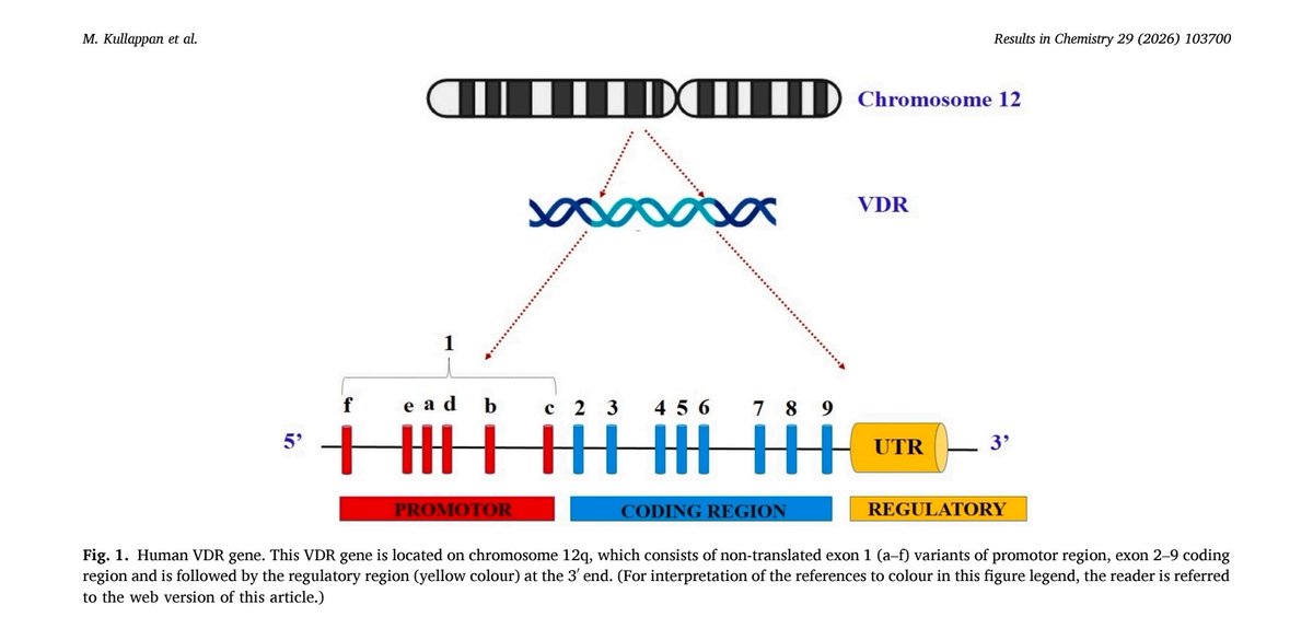
Vitamin D Receptor (VDR) R343C Polymorphism: A Potential Molecular Link Between Vitamin D Deficiency and Dementia
Based on: Malathi Kullappan, Adithya Kasagani Naga Vikram, Krishna Mohan Surapaneni. Results in Chemistry. 2026.
🔹 This in silico molecular modeling study investigated whether VDR gene polymorphisms reduce calcitriol binding and thereby contribute to progression from mild cognitive impairment (MCI) to dementia.
🔹 Among 459 missense VDR variants, R343C (rs1057521095), R391C, and R391H were identified as the most pathogenic and protein-destabilizing mutations.
🔹 Wild-type VDR showed the strongest binding to calcitriol (−11.61 kcal/mol), whereas R343C demonstrated the weakest binding (−8.3 kcal/mol), suggesting impaired vitamin D receptor activation.
🔹 Molecular dynamics simulations over 200 ns showed that the R343C mutant had greater structural instability, higher flexibility, and poorer compactness than wild-type VDR, indicating reduced receptor function.
🔹 The mutant receptor formed fewer hydrogen bonds with calcitriol (3 vs 5), further supporting weaker ligand-receptor interaction.
🔹 Vitamin D signaling through VDR is involved in neuronal survival, neurotrophin production, anti-inflammatory activity, β-amyloid clearance, and maintenance of CNS homeostasis. Impaired VDR signaling may therefore accelerate neurodegeneration.
🔹 The authors propose that R343C VDR polymorphism may predispose individuals to functional vitamin D deficiency, increasing susceptibility to progression from MCI to dementia.
Clinical Message
✅ Vitamin D deficiency in older adults with cognitive decline may not always reflect inadequate intake alone; genetic variation in the vitamin D receptor may influence biological responsiveness.
⚠️ Take-home point: This is a computational (in silico) hypothesis-generating study, not clinical proof. Routine genetic testing for VDR polymorphisms is not recommended in current clinical practice, and prospective human studies are needed before translating these findings into patient care.
https://t.co/NzF5RfsMGo






![CMEINDIA1's tweet photo. 🫀🫁 CME INDIA CLINICAL PEARLS
Liver for the Cardiologist: Is It Time to Move From CKM → CKML?
Based on: Liver for the Cardiologist: Why the Liver Belongs in the Cardiometabolic Workflow and the 2026 AHA/ACC/ADA/ASN CKM Guideline. The 2026 guideline now explicitly places MASLD assessment inside cardiovascular-kidney-metabolic care, rather than treating fatty liver as a separate hepatology issue.
🔥 The paradigm shift
The old cardiometabolic conversation was Heart–Kidney–Metabolism. The emerging concept is Heart–Kidney–Liver–Metabolism.
The reason is simple: MASLD is not merely a liver complication of obesity or diabetes. It is a systemic cardiometabolic phenotype and an independent marker of cardiovascular risk. Advanced hepatic fibrosis identifies an especially high-risk population.
🫀 Pearl 1 — In MASLD, patients often die “with the liver” rather than “from the liver”
Cardiovascular disease remains a major cause of morbidity and mortality among people with MASLD. The liver therefore provides additional information about the cardiometabolic milieu—insulin resistance, ectopic fat, inflammation, atherogenic dyslipidemia and vascular dysfunction.
Clinical message: When you see fatty liver, think beyond ALT and cirrhosis. Think ASCVD, heart failure, CKD and diabetes progression.
❤️ Pearl 2 — “The liver leads, the heart follows” is biologically plausible
The spectrum frequently progresses from:
Obesity/T2D/metabolic dysfunction → hepatic steatosis → MASH → fibrosis → systemic cardiometabolic dysfunction → subclinical cardiac disease → overt HF.
This does not imply simple one-way causality; liver, adipose tissue, heart and kidney communicate bidirectionally through inflammatory, metabolic, hormonal and hemodynamic pathways.
⚠️ Pearl 3 — Fibrosis matters much more than simply detecting steatosis
Finding liver fat identifies MASLD, but fibrosis stage is the major prognostic discriminator.
A patient with mild steatosis and low fibrosis risk is very different from a patient with MASH and F2–F4 fibrosis.
This is why contemporary guidelines emphasize fibrosis risk stratification rather than ultrasound alone.
🧮 Pearl 4 — FIB-4 should become a cardiometabolic vital sign
The 2026 CKM guideline recommends calculation of FIB-4 every 1–2 years in adults with CKM syndrome who have:
T2D OR ≥2 cardiometabolic risk factors.
In CKM stage 1 due to prediabetes, FIB-4 assessment every 2–3 years is considered reasonable.
FIB-4 =
Age × AST / [Platelets × √ALT]
It requires information already available in almost every diabetes/cardiology clinic.
No additional imaging. No additional blood draw. Minimal cost.
🚦 Pearl 5 — Think of FIB-4 as a traffic-light test
🟢 FIB-4 <1.3
Usually indicates low probability of advanced fibrosis.
Continue cardiometabolic risk modification and periodic reassessment.
🟡 FIB-4 1.3–2.67
This is the indeterminate zone.
Do not simply repeat LFTs.
Proceed to a second-line fibrosis assessment, most commonly:
VCTE/FibroScan or ELF.
🔴 FIB-4 ≥2.67
High probability of advanced fibrosis.
These patients generally warrant specialist hepatology/gastroenterology evaluation and secondary fibrosis testing.
FIB-4 thresholds need clinical interpretation, especially in older adults and situations in which AST/ALT or platelet counts are altered for reasons unrelated to MASLD.
📡 Pearl 6 — Second-line testing converts screening into staging
The 2026 guideline identifies vibration-controlled transient elastography (VCTE) and the Enhanced Liver Fibrosis (ELF) test as secondary tools.
For VCTE:
<8 kPa → low risk
8–12 kPa → intermediate fibrosis risk
>12 kPa → high risk, usually prompting hepatology referral.
The precise ELF cut-offs differ somewhat among algorithms, so they should be interpreted according to the assay and pathway being used.
🚨 NEXT .....
https://t.co/OEzOVwLQFb](https://pbs.twimg.com/media/HPILnmXbgAAZAJI.jpg)