cIMPACT-NOW Update 8: Clarifications on molecular risk parameters and recommendations for WHO grading of meningiomas #neuropath
https://t.co/ZXcRZ2e1i8
@MAHoureih@DrJPLow EBV+ SMTs usually have an evident lymphocytic infiltrate and varying amounts of fascicular regions with blunted nuclei and primitive round cell areas. This tumor has a more uniform appearance with tapered “neural” nuclei to me. Any history of immunodeficiency?
Not before your pathologist does! Important opportunity for pathologists and other diagnosticians to communicate their results directly to patients and explain results. Patients need to advocate for this as it’s not currently supported by insurance. #pathology
Glad to finally have our work on ROS1-rearranged gliomas in diverse age groups published!
#Neuropath#PathTwitter#pathX#pathology
https://t.co/IWrfJGzxuu
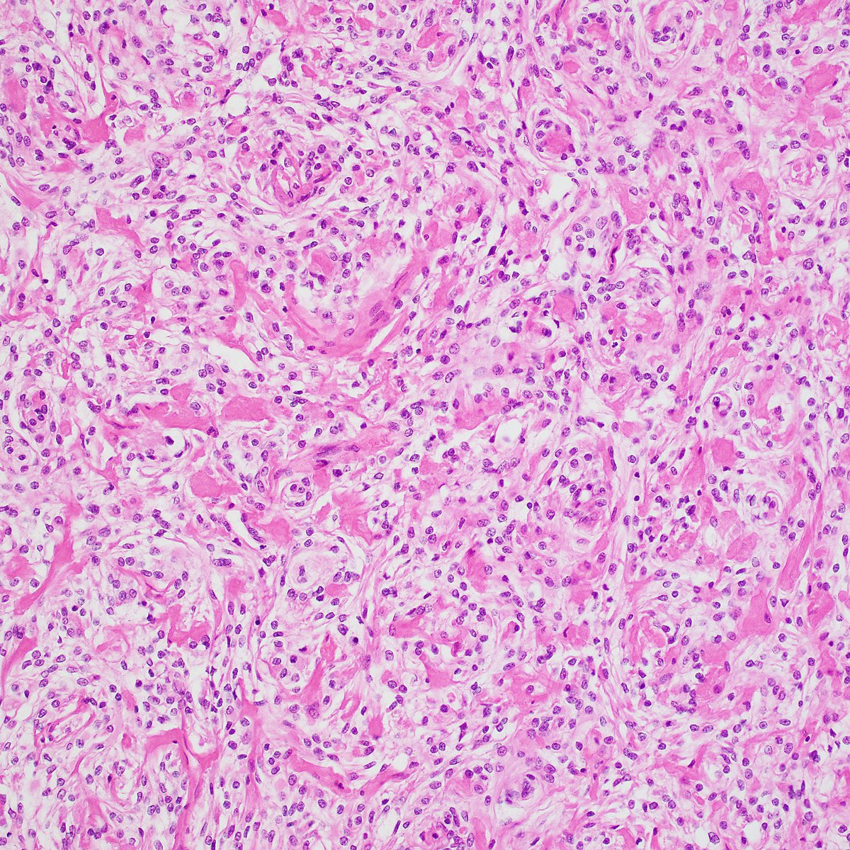
Rare example of chondroma arising in the dura (falx). Histo shows characteristic mature hyaline cartilage with increased cellularity and mild nuclear atypia.
#pathology#PathTwitter#BSTpath#Neuropath
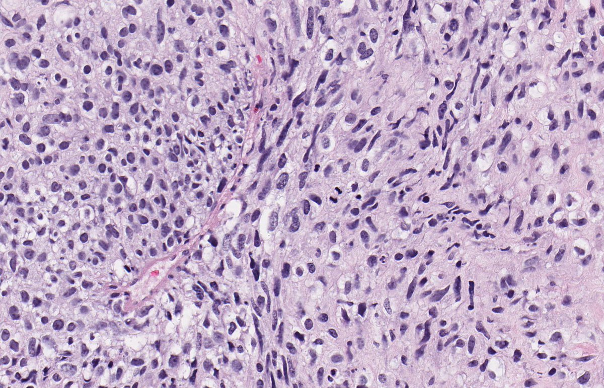
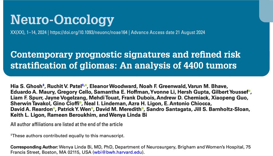
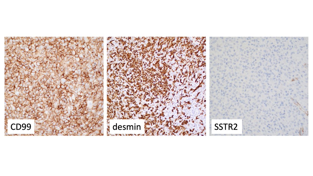
Additional IHC (CD99+/des+/SSTR2-) was highly characteristic of Intracranial Mesenchymal Tumor, FET::CREB fusion positive. EWSR1 FISH showed rearrangement. This particular case has a solid growth pattern with no myxoid stroma.
#Neuropath#pathology#PathTwitter#BSTpath
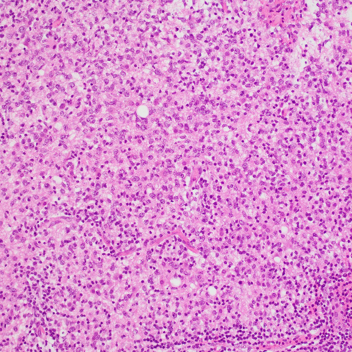
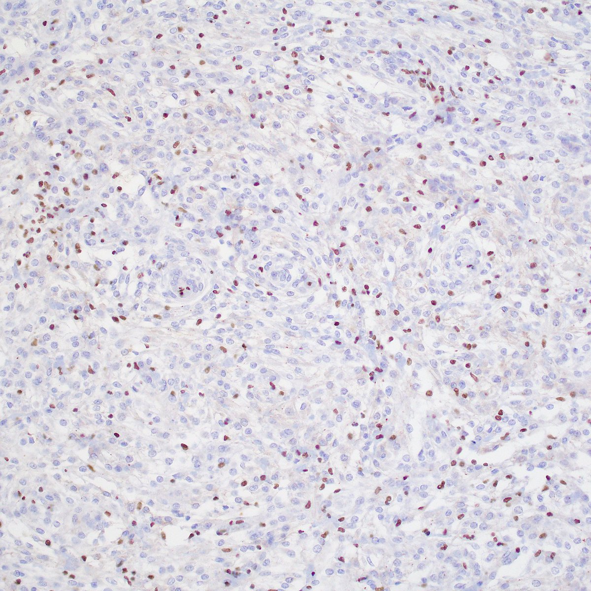
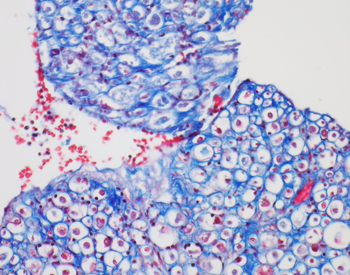
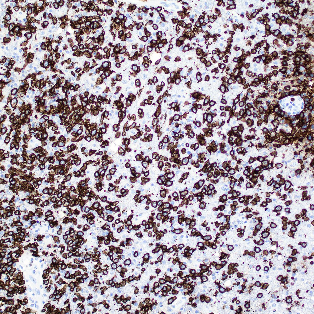
A good clue is the presence of lymphoglandular bodies in the background. Smear prep is probably the best way to diagnose CNS lymphoma at frozen section.
Here is the accompanying H&E permanent section and CD20.
#PathTwitter#pathology#Neuropath#hemepath
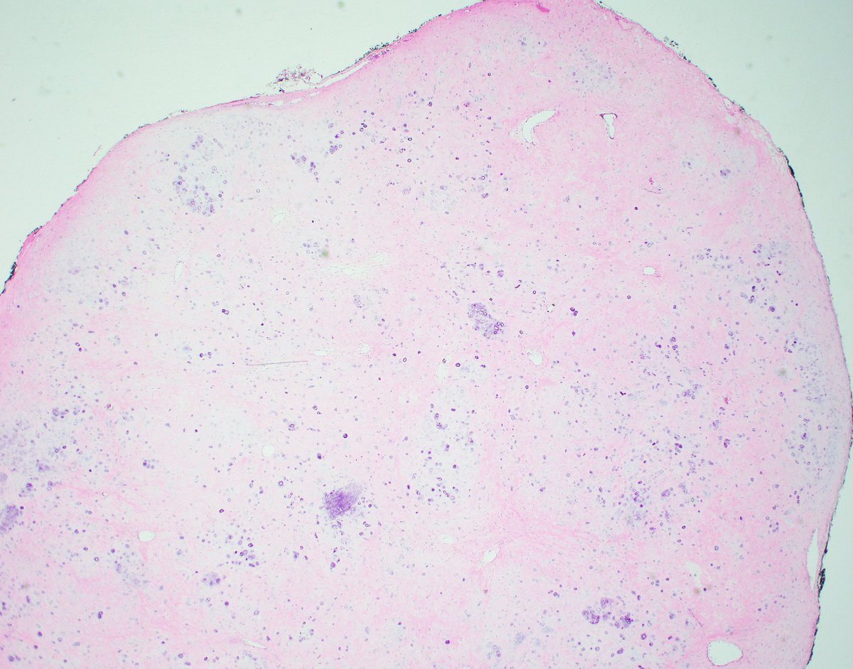
Can you diagnose in one cell?
(Smear preparation from an enhancing deep white matter frontal mass in an older patient.)
#Pathology#PathTwitter#Neuropath