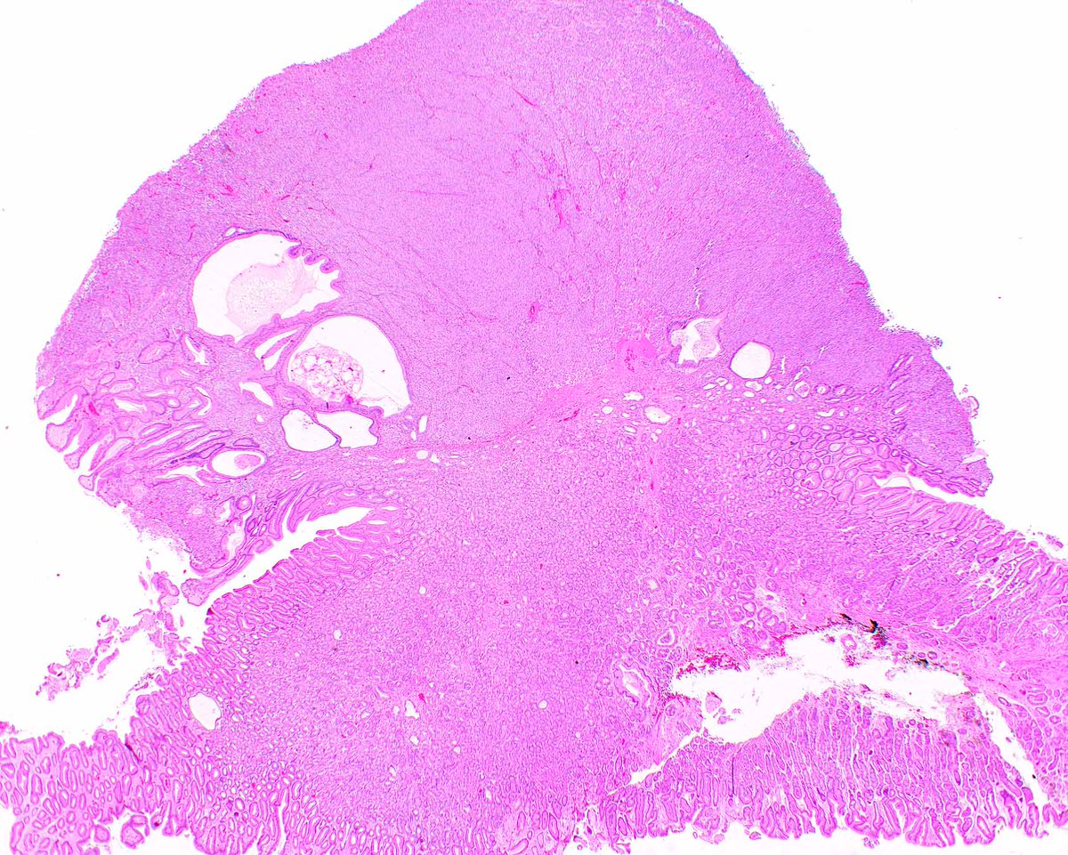
🔬Prostatic stromal tumour of uncertain malignant potential – STUMP
(with emphasis on the differential diagnosis with prostatic stromal sarcoma)
A hypercellular prostatic stromal neoplasm with large, pleomorphic nuclei is necessarily a sarcoma? No. STUMP may show quite striking cytologic atypia — and recognizing its degenerative nature is one of the main diagnostic pitfalls. 🧵
🔬 Definition
STUMP is a rare mesenchymal neoplasm arising from the specialized prostatic stroma, characterized by uncertain biological behavior and absence of the unequivocal criteria of malignancy seen in prostatic stromal sarcoma (PSS).
📊 Epidemiology
• Rare.
• Occurs predominantly in adults.
• In general, patients are slightly older than those with PSS.
📍 Sites
• Prostate, involving the transition and/or peripheral zones.
🧬 Pathogenesis
• Unknown etiology.
• Genomic alterations are heterogeneous.
• STUMP and PSS may share some chromosomal alterations and coexist within the same prostate.
• However, there is no convincing demonstration of an obligatory STUMP → PSS sequence.
🩺 Clinical Features
• Lower urinary tract obstructive symptoms.
• Hematuria or hematospermia may occur.
• It may occasionally present as a prostatic mass.
🧪 Laboratory Diagnosis
• Serum PSA is not a specific marker of the neoplasm.
• Diagnosis is fundamentally histomorphologic.
🔬 Histopathology
Five patterns may be observed, either alone or in combination:
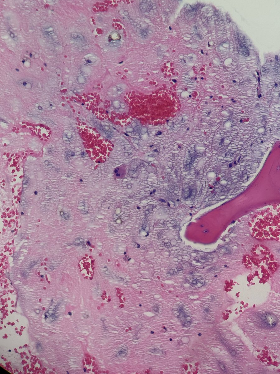
• Hypercellular stroma with atypical cells showing a degenerative appearance.
• Hypercellular spindle-cell stroma with cytologically bland features.
• Phyllodes/leaf-like pattern.
• Myxoid pattern.
• Epithelioid stromal pattern.
An important clue: very large, hyperchromatic, pleomorphic nuclei may be present, but isolated, without the overall constellation of changes expected in a sarcoma.
🧫 Immunohistochemistry
• Vimentin: positive.
• PR: frequently positive.
• CD34: variable.
• SMA/desmin: variable.
• ER: uncommon.
• KIT, S100, and STAT6: usually negative.
⚠️ None of these markers, in isolation, distinguishes STUMP from PSS. Morphology is central.
🔎 Differential Diagnosis
• Prostatic stromal sarcoma – favored by marked hypercellularity, non-degenerative pleomorphism, increased mitotic activity, atypical mitoses, and necrosis.
• Leiomyoma/leiomyosarcoma – convincing smooth muscle differentiation, with desmin/SMA/h-caldesmon.
• Solitary fibrous tumor – nuclear STAT6.
• GIST – DOG1 and KIT/CD117.
• Sarcomatoid carcinoma – look for epithelial differentiation and cytokeratin expression.
📈 Prognosis
Behavior is unpredictable, although most STUMPs do not show aggressive behavior. Local recurrence may occur, supporting the need for clinical follow-up.
💊 Treatment
There is no universal strategy due to the rarity of the disease. Localized lesions are usually treated surgically, considering size, extent, and the possibility of complete resection. Follow-up is recommended.
🎯 Take-Home Messages @Notas_Patologia
• Bizarre nucleus ≠ sarcoma: STUMP may show striking degenerative atypia.
• Look for the combination: mitoses + atypical mitoses + necrosis + non-degenerative pleomorphism to favor PSS.
• CD34 and PR may support specialized prostatic stromal differentiation, but do not resolve STUMP × PSS.
• In small samples, tumor heterogeneity is an important limitation.
• Morphology remains the key to diagnosis.
📚 Selected References
• WHO Classification of Tumours Editorial Board. Urinary and Male Genital Tumours. 5th ed. IARC.
• Bostwick DG, Egevad L. Prostatic stromal proliferations: a review. Pathology. 2021;53:12–25.
• Acosta AM et al. Re-evaluating tumors of purported specialized prostatic stromal origin. Mod Pathol. 2021;34:1763–1779.
• Gaudin PB et al. Sarcomas and related proliferative lesions of specialized prostatic stroma. Am J Surg Pathol. 1998;22:148–162.
• PathologyOutlines – Prostatic stromal tumors: https://t.co/yteSms2T6w
⚠️Disclaimer⚠️
This text is an educational summary for healthcare professionals and students. It does not replace full pathology reports, local guidelines, or individualized clinical decision-making.
#MedicalEducation #NotasDePatologia #Pathology #PathTwitter #GUPath #Uropathology
Holy Soma Batman!
You never know what you may find when you cruise our MICrONS Explorer, such as a blood vessel piercing the soma of a neuron!
👀 See this incredible find up close using this shortcut: https://t.co/QkoavsnhtS
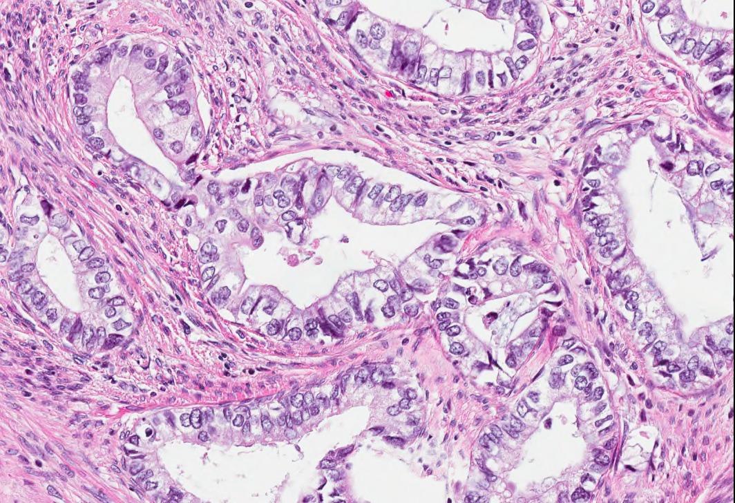
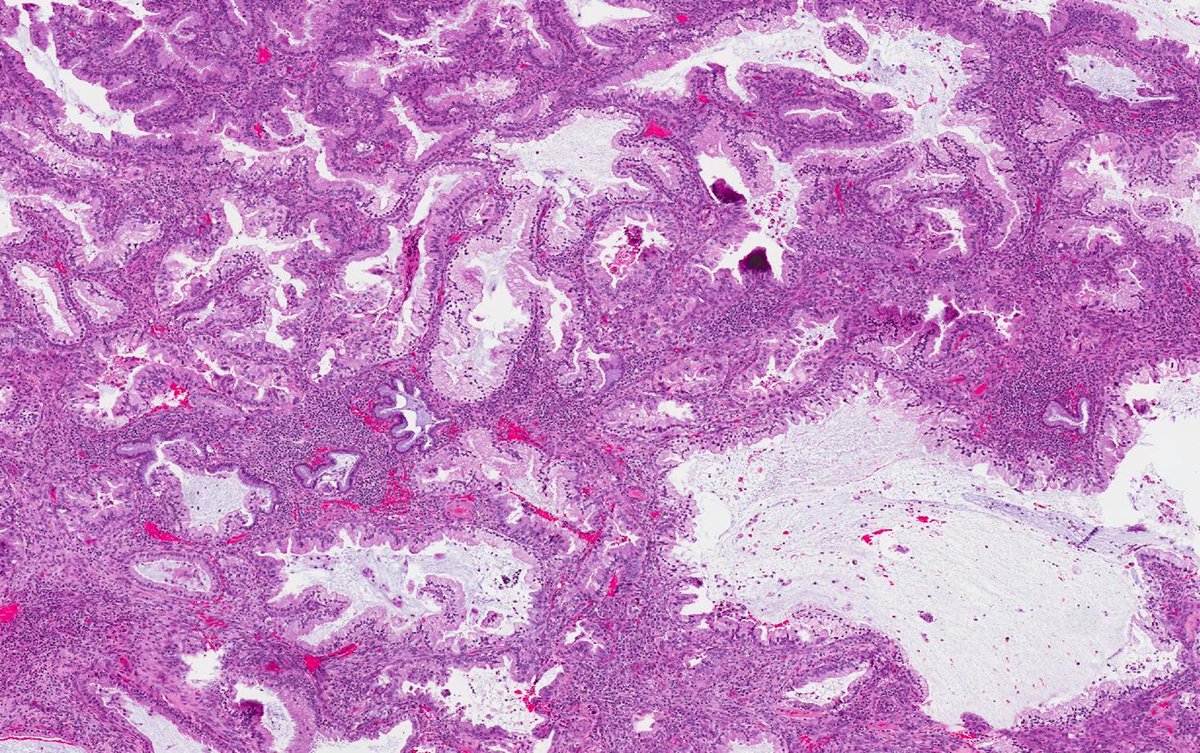
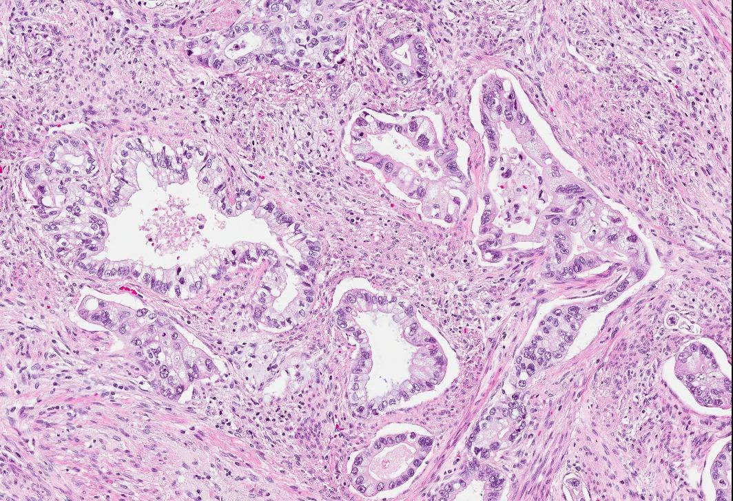
✅ Gastric-type Adenocarcinoma 🎯🧬
• Aggressive HPV-independent adenocarcinoma exhibiting gastric (pyloric) differentiation.
• Glands show voluminous clear or pale eosinophilic cytoplasm, distinct cell borders, and neutral mucins.
• Frequently associated with STK11 mutations and Peutz-Jeghers syndrome, with abnormal p53 expression.
• Importantly, p16 is typically negative or patchy, distinguishing it from HPV-associated tumors.
When cells grow together, they often align with their neighbors, forming liquid crystal-like patterns known as “nematic order.” But that order isn’t perfect, and at certain points, the alignment can break down, creating “topological defects”: small regions where cells swirl or fan out instead of lining up cleanly.
And according to research published in Science, what looks like a defect can be used strategically as built-in instructions for shaping larger structures. Instead of growing upward, cells can store and release stress in certain places along a previously flat sheetlike structure, forming a bowl, a ridge, or a fold.
Learn more: https://t.co/OSMGhNzR6d
@TungPath In my example, my first clue was they were a bit more abundant than a needle carry over, then the brush border-like condensations: normal gatric foveolar mucosa doesn’t have that.
"In cytopathology, one signet ring cell may whisper, but a cluster always shouts malignancy." Hepatic hilar node with metastatic colonic adenocarcinoma, signet ring cell type. (FNA, Smear and Cell block).
📣Excited to share our recent article on the practical approach to gastric lesions; the background mucosa & new nomenclatures!
Deeply grateful to Dr. Elizabeth Montgomery for her incredible support, mentorship & guidance.
https://t.co/tXBJEMzcLl
@LizMontgomeryMD#PathX#GIPath
ABC Of Cytology_30
2026
Carcinoid tumour
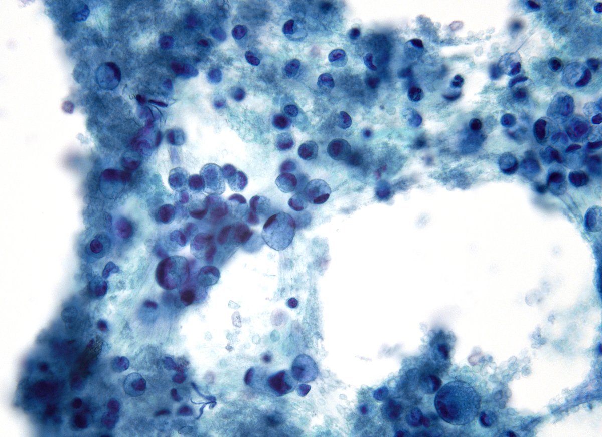
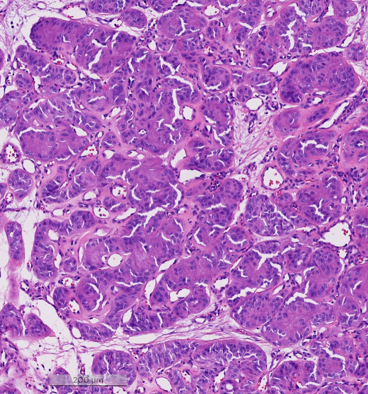
Carcinoid tumors are low-grade neuroendocrine neoplasms, most often associated with the lung or gastrointestinal tract (GIT). In cytology FNA/ EBUS-FNA, or bronchial brushings, they are observed as a uniform population
Nearly thirty years as a published writer and this is the first time I’ve used a new device that became instantly irreplaceable (no, I’m not being paid to say this.) It feels like using ink and paper and does everything I want it to do and nothing I don’t need it to do.
I know I’m late to the party, but my son gave it to me on my birthday so to me it’s a very recent revelation. It’s already joined my laptop at the top of my Things I Dread Losing list. #supernote
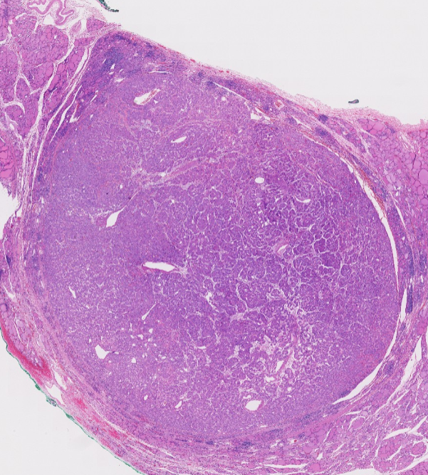
HTT (hyalinizing trabecular tumor)
Rare follicular-derived thyroid tumor with trabecular growth, hyalinized stroma, and PTC-like nuclei
Once considered a PTC variant; now regarded as benign or of low malignant potential.
Shows unusual membranous/cytoplasmic MIB-1 (Ki-67) staining, but only when the stain is done at room temperature (rarely seen in routine practice)
Dr. Scognamiglio #USCAP #pathology #PathX #PathTwitter
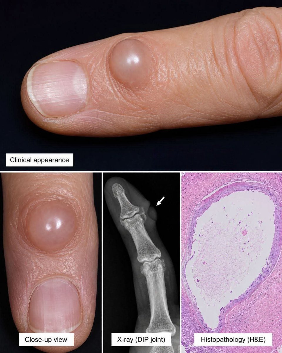
Which hand lesion is classically associated with DIP osteoarthritis and longitudinal nail grooving?
A. Glomus tumor
B. Digital myxoid (mucous) cyst
C. Pyogenic granuloma
D. Herpetic whitlow
The 6th edition of the WHO classification of tumours of the breast (2026) is now in use. Here is a comprehensive, well-illustrated, practical summary by a group of leading experts of the main changes since the last edition. #openaccess#breastpathology
https://t.co/pMWu3oIxm6
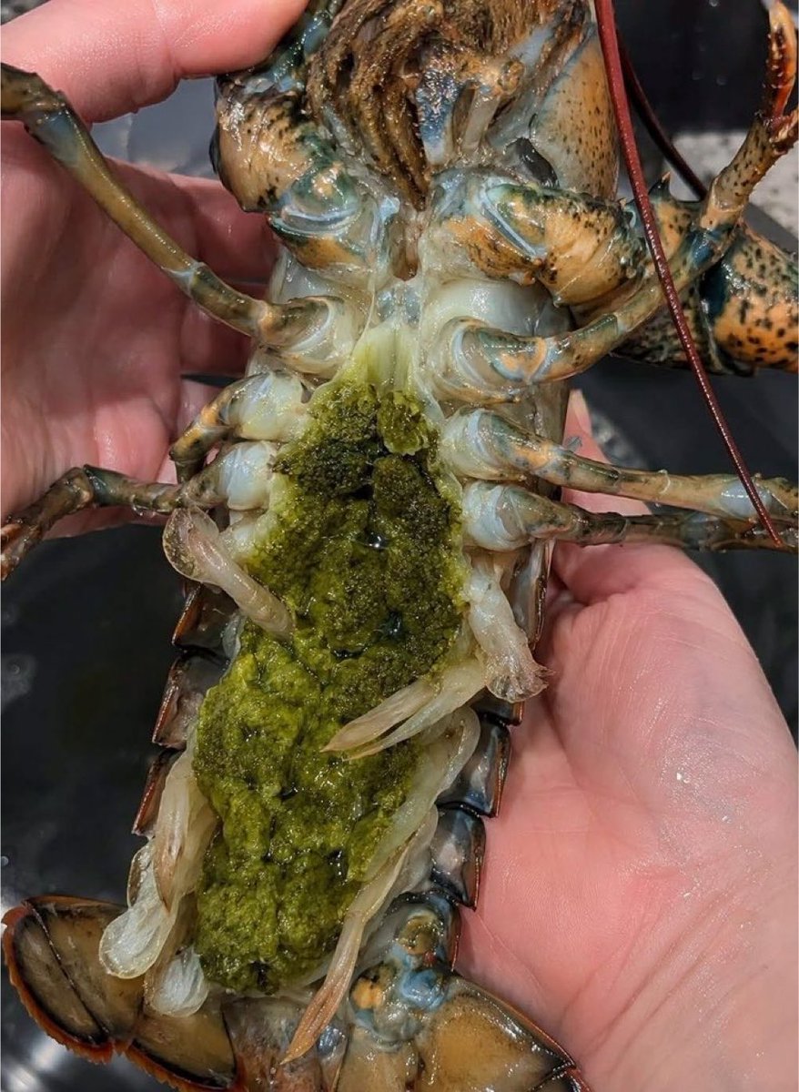
According to @grok, the green stuff on this lobster is the hepatopancreas, hence the bile-like colour. Chefs and food connoisseurs call this tomalley. Now that you know, would you still eat it?
What can pythons teach us about fat and metabolism? A Stanford Medicine researcher studies the dramatic metabolic shifts pythons undergo after eating — and what they could reveal about appetite, metabolism and weight loss in humans. https://t.co/p1UkXoXWTB
@StanfordBrain