@UWMSTP graduate and #Neurology resident @MayoNeuroRes. Dedicated to basic neuroscience research to treat neurodegenerative disease. Tweets/views are my own.
Congratulations to the Mayo Clinic Adult Neurology Residency Class of 2029! We are so delighted to have you join our Mayo Clinic Family! #MayoNeuroResidency
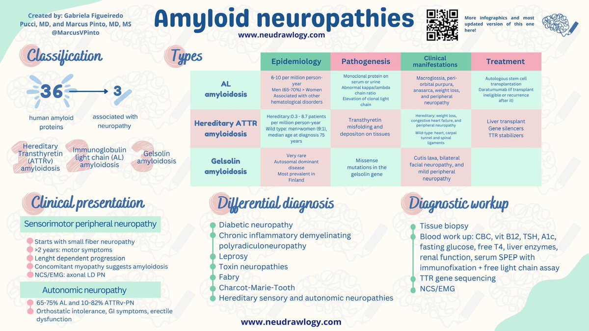
Amyloid neuropathies are treatable and should not be missed. @gabifpucci and I updated the @neudrawlogy amyloid neuropathy infographic and tweetorial that I share below.
Remember that amyloid neuropathy is one of the causes of rapidly progressive neuropathy and that's why you must order serum SPEP/immunofixation, free light chains, and fat aspirate in all these patients and consider a nerve biopsy If rest of work up is inconclusive.
1/x
Have MULTIPLE questions about MULTIPLE sclerosis?
Having trouble seeing neuromyelitis optica?
In a fog about MOG?
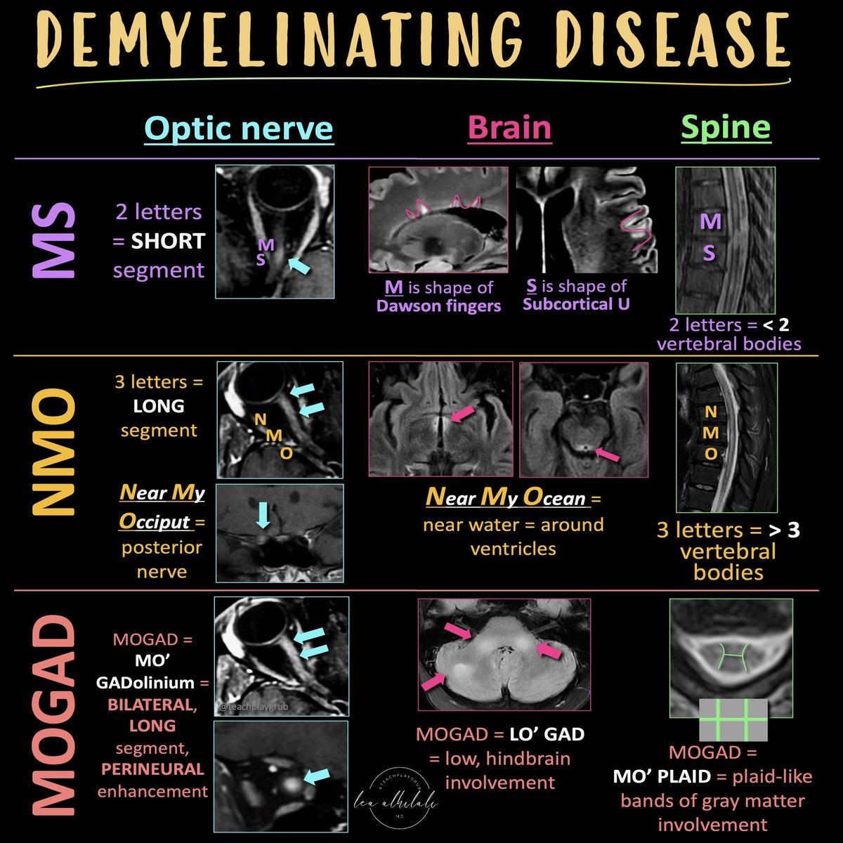
Here’s the cheat sheet you NEED to distinguish the demyelinating diseases!
Demyelinating diseases predominantly involve the optic nerves, brain, & spine.
The three main chronic demyelinating diseases are Multiple sclerosis (most common), neuromyelitis optica (NMO), & myelin oligodendrocyte glycoprotein (MOG) antibody associated disease or MOGAD
Each has its own features in the optic nerve, brain, & spine. Here’s how to remember them!
MS
Optic nerve:
MS only has 2 letters, so MS involvement of the optic nerve tends to be short segment
Brain:
Letter M makes the shape of the perivascular distribution of lesions along the ventricles (Dawson’s fingers)
Letter S makes the shape of the subcortical U fiber involvement
Spine:
MS is only 2 letters, so lesions are usually less than 2 vertebral bodies in length
NMO
Optic nerve:
NMO is a longer abbreviation, three letters, so longer involvement
NMO can stand for Near My Occiput. Occiput is posterior, so more posterior nerve involvement
Brain:
NMO can stand for Near My Ocean. What is your brain’s ocean? The ventricles. NMO lesions are all periventricular
Spine:
NMO is 3 letters, so lesions usually more than 3 vertebral bodies in length
MOGAD
Optic Nerve:
Remember MO’ GAD-olinium. So things that cause more regions of enhancement. MOGAD lesions are commonly bilateral & long segment & enhancement can extend perineural
Brain:
Remember LO’ GAD. MOGAD typically involves the lower areas of the brain
Spine:
Remember MO’ PLAID. MOGAD can give a plaid-like H shape in the cord from predominantly gray matter involvement
Hopefully, this cheat sheet will help you remember how to distinguish the demyelinating diseases! It ain’t lyin’ about diseases of myelin!
There has been a small fiber neuropathy (SFN) “epidemic” over the last 20 years, but few people know that isolated SFN (normal EMG) is rare. In Olmstead County, MN, it is rarer than CIDP. Below, I debunk a few other SFN myths 1/8 https://t.co/mWtSL0WP1V
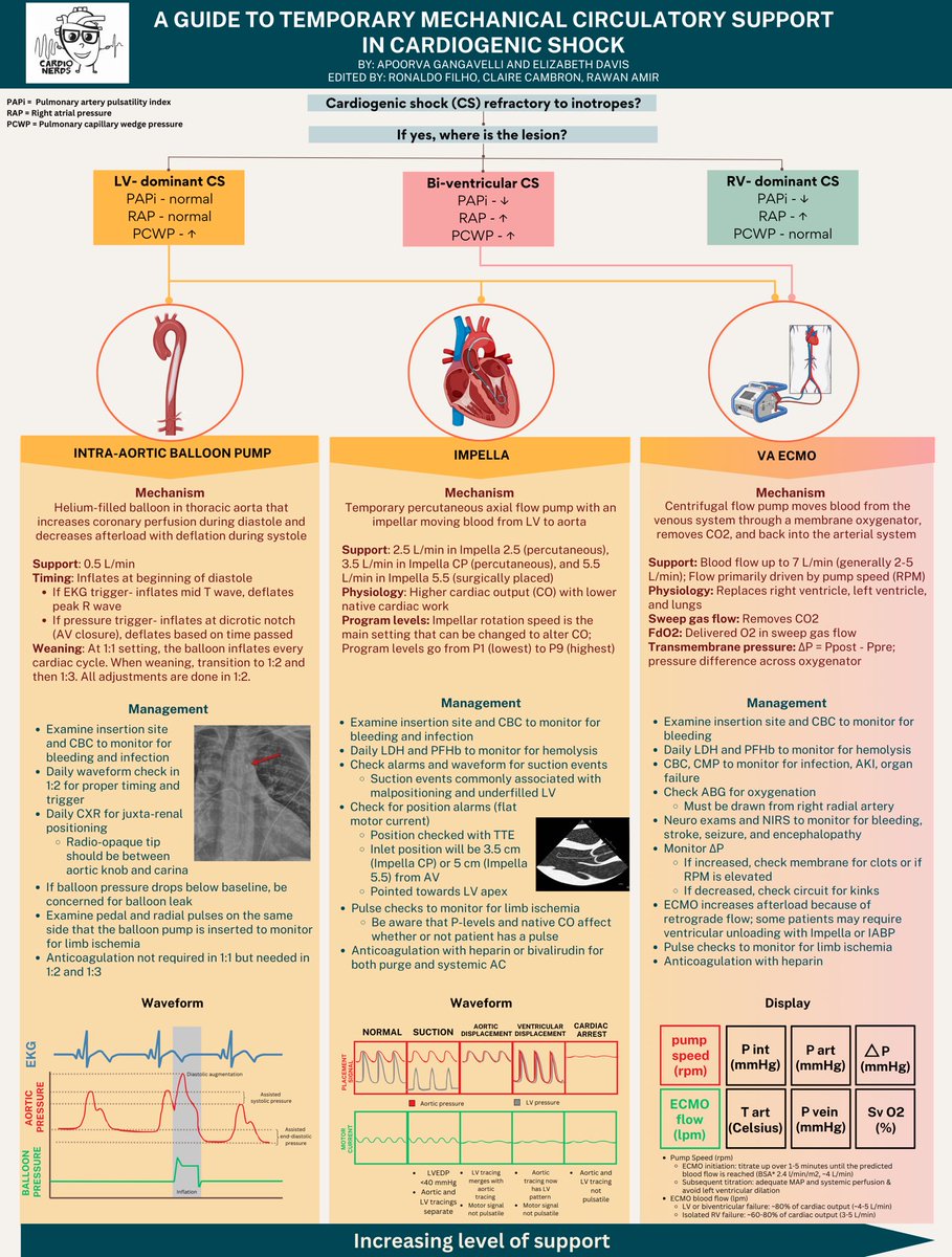
🏥New to the CCU? Not sure how to manage temporary mechanical circulatory support?
⭐️Check out our new @CardioNerds infographic on using TMCS in cardiogenic shock! Explore their mechanisms, management, and waveforms you need to know!
What a shame… so many patients exposed to an experimental treatment with questionable basic science foundation. Makes me think of the root drivers of these scandals - publish or perish culture, academic promotion - science is not immune to greed!
The @NIH's research integrity scandal involving neuroscience chief and #Alzheimer’s/#Parkinson’s scientist Eliezer Masliah is of an extraordinary scale. It might be useful to follow one thread of the investigation involving the experimental drug prasinezumab from @ProthenaCorp.
Does epilepsy imaging cause you to seize up in fear?
Everyone knows mesial temporal sclerosis (MTS)! But did you know there are different KINDS of MTS??
This month's @theAJNR SCANtastic shows YOU what you need to know in the latest in epilepsy imaging!
https://t.co/GzzekpwAJG
Here's what to remember:
Hippocampus has 4 subfields w/CA abbreviation standing for “Cornu Ammonis”:
CA1: Horizontal & ascends slightly. Number 1 on its side is horizontal w/a slight acsent!
CA2: Superior hippocampal dome. Number 2 has a curve at the top that looks like a dome!
CA3: Hippocampal curve ends & turns toward the dentate. Number 3 makes a curve heading down!
CA4: Indistinguishable from the dentate. Number 4 on its side looks like CA4 being supported by the dentate underneath!
Hippocampal sclerosis or MTS has 3 subtypes:
Type 1
Classic form w/neuronal loss in all subfields
Remember this rhyme: In type 1, hippocampus is all done!
Type 2
CA1 predominant volume loss
Remember this rhyme: Type 2 loses the glue! (CA1 glues the hippocampus to the rest of the temporal lobe)
Type 3 is CA4 & dentate predominant volume loss
Remember this rhyme: Type 3 loses the belly! CA4 & dentate are the hippocampus belly!
Next time you have an epilepsy study, hopefully you'll seize the opportunity to use this!
But this only scratches the surface! Follow @theAJNR & read the full article:
https://t.co/zW4WuKB1Ar
Met @DGlaucomflecken last night at the @mnmed annual meeting - such a hilarious yet moving discussion about using humor in medicine and how it can actually be used to inspire change in our healthcare system. #advocacy#HealthcareForAll#wellness
Where are we with biological markers and staging for Parkinson's? Have we considered the impact to persons w/ disease? Should we start using the new term neuronal synuclein disease instead of the term Parkinson's disease? Are we ready? This is a story we should all be following closely. Great viewpoint just out by Kalia and colleagues from the Movement Disorders Society in the @MDJ_Journal and I recommend it as a great read.
Key Points:
- The authors bring us up to speed on the questions regarding the link between α-synuclein and neurodegeneration in Parkinson's disease and many that 'remain unanswered.'
- The authors discuss that Lewy pathology can occur before motor symptoms and spread across the neuroaxis via a 'prion-like mechanism.'
- Two recently published novel frameworks were published in early 2024 in @TheLancetNeuro.
- The new proposals are 'not anchored to the presence of the clinical syndrome.'
- What are the MDS recommendations?
- “Parkinson's disease” and “dementia with Lewy bodies” remain the accepted clinical diagnostic terms, with their well-defined limitations, but importantly utility in guiding patient care.
- "Premature and potentially limiting to use terms such as “NSD” or “Parkinson's type synucleinopathy.” for the diagnosis of patients or classification of disease."
- Neither of the current proposals has 'universal applicability nor the robust validation.'
My take: I think this is a well thought out viewpoint and that tapping the brakes is the right path. I agree with the authors that we need to look at pathological processes other than 'α-synuclein aggregation.' I love that they discuss ethical considerations and actual effects on persons w/ disease.
https://t.co/4Eo6f8Pfp5 #parkinson #synuclein
Time to go for the jugular when it comes to carotid stenosis!
Degree of stenosis isn’t all that matters
Here’s cheat sheet you NEED to remember the risky features of carotid plaques
Read on for the newest @theAJNR SCANtatistic on carotid stenosis:
https://t.co/SieEdSWFrc
NASCET isn’t the end of what you need to know—it’s just the beginning.
In addition to degree of stenosis, there are features of plaques on imaging that suggest they are prone to rupture & cause strokes.
You can remember the high-risk features by remembering that atherosclerosis is influenced by a bad diet—so the features you get when you have a bad diet are the same features vulnerable plaques will show on imaging!
High risk features:
🔸Size
You get increased size w/outward remodeling of your belly w/a bad diet. Same w/vulnerable plaques—larger plaques with outward remodeling are riskier
🔸Irregularity
If you eat a poor diet, you will get constipated & irregular. Same w/vulnerable plaques—irregular plaques are at higher risk
🔸Ulceration
Obesity is associated with ulcers. Same w/vulnerable plaques—ulcerated plaques are more vulnerable
🔸Lipid necrotic core
If you eat a lot, you get more belly fat or lipid in your core. Same w/vulnerable plaques—plaques with fatty deposits in their core are more vulnerable
🔸Loss of fibrous cap
If you eat a lot, your belt can’t hold your fat in anymore & will break. Same w/vulnerable plaques—loss of the fibrous cap holding in the lipid core is a high-risk feature.
🔸Neovascularity
Poor diet leads to constipation & hemorrhoids, a sort of neovascularity. Same w/vulnerable plaques—increased vasculature in the plaque & enhancement is a high-risk feature.
🔸Hemorrhage
Poor diet & hemorrhoids leads to bleeding. Same w/vulnerable plaques—intraplaque hemorrhage is associated with increased chance of rupture
More and more research like the research in this month’s @theAJNR have shown that looking at only degree of stenosis & symptomatology are not enough
Now you know all the imaging features that suggest a high-risk plaque beyond the degree of stenosis. Don’t narrow your focus when it comes to carotid narrowing!
Thinking about genetic testing for #neuromuscular disorders? Bookmark this thread.
Here's a guide to next gen panels that you can get at no cost to patients.
#tweetorial
Presenting the Mayo Clinic Neurology Class of 2027!
We're excited to have you fully in neurology and look forward to seeing the amazing things you will do... it's going to be a great journey!
@mayoneurores@MayoPedNeuroRes@MayoClinicNeuro
You've probably heard "don't give lactated ringers because it raises lactate"
This statement is ~98% false, but there's one crucial practice-changing fact that you need to know.
A 🧵 all about lactic acid and lactated ringers!
1/