If you have ever heard, read or written the phrase “the patient is edematous but intravascular dry”, you have to check this review (https://t.co/6RxKeRzoqv) 👇
Fluid normally exchanges freely between the plasma and interstitial space and is returned to the “central circulation” primarily via the lymphatics. In disease states, such as sepsis, the return flow of fluid from the interstitial space to the plasma seems to be very slow, which promotes the well-known triad of "hypovolemia + hypoalbuminemia + peripheral edema"
The authors discuss studies suggesting that there two key mechanisms contributing to this triad:
(1) acute lowering of the interstitial pressure by inflammatory mediators such as TNFα, IL-1β, and IL-6 and,
(2) nitric oxide-induced inhibition of the intrinsic lymphatic pumping
As you know, fluid is filtered in the (pre-)capillary side and reabsorbed at the post-capillary end, right? Well, not really...
According to the traditional model historically taught in introductory physiology and medical school (I still remember it...), in the precapillary (arteriolar) end, the high capillary hydrostatic pressure exceeds plasma oncotic pressure, causing net filtration of fluid out of the vessel into the interstitial space. In the post-capillary (venular) end, the hydrostatic pressure drops below plasma oncotic pressure, creating a net pressure gradient that causes reabsorption of ~90% of the filtered fluid back into the vessels. The remaining ~10% was thought to be drained by the lymphatic system
According to the modern model (established via the discovery of the endothelial glycocalyx), in most tissues under normal, steady-state conditions, there is no sustained venular reabsorption. The endothelial glycocalyx layer acts as the primary semi-permeable filter. The effective oncotic gradient exists between the plasma and the subglycocalyx space, rather than the main interstitial fluid
Under normal blood pressure, capillary hydrostatic pressure exceeds the effective opposing oncotic pressure across the entire length of continuous capillaries and post-capillary venules. Consequently, virtually all fluid filtered at the capillary level is picked up by the lymphatics, which then route the fluid through lymph nodes and return it to the venous system via the lymphatic ducts.
Some exceptions exist in specialized vascular beds designed for continuous fluid absorption, such as the intestinal mucosa and renal peritubular capillaries. Also direct vascular reabsorption may occur temporarily during acute, severe drops in capillary hydrostatic pressure (e.g., severe hypovolemic shock or hemorrhage) to preserve plasma volume
#foamed #foamcc #physiology
There is a common assumption that if you exercise for an hour, you have basically taken care of the metabolic consequences of sitting for the rest of the day. This study suggests that is not quite how it works.
Sixteen sedentary adults with elevated blood lipids completed three different two-day conditions while doing their normal desk jobs. In one, they sat for most of the day. In another, they sat for most of the day but completed a 60-minute moderate cycling session. In the third, they completed the same cycling session, but also stood for 30 minutes during every hour of desk work. The exercise was identical in the last two conditions. The only difference was what they did during the rest of the workday.
When the researchers measured the response to a standardized meal, the group that combined exercise with regular standing breaks had a lower insulin response than exercise alone, while glucose remained essentially the same. That suggests they were able to handle the same glucose load with less insulin, which is consistent with better insulin sensitivity. Triglycerides did not differ between the two active conditions, so this was not an improvement across every metabolic marker. It was one significant finding in a study of only 16 people.
This was also an acute study. It tells us what happened over a couple of days, not whether standing more at work prevents diabetes or cardiovascular disease years later. Still, the design is useful because participants were doing their actual desk-based jobs rather than sitting in a tightly controlled laboratory environment, and each participant completed all three conditions.
exercise and sedentary time may not be interchangeable. An hour of exercise is clearly beneficial, but it may not completely erase what happens during another eight hours of almost uninterrupted sitting. Those may be two separate behaviors worth addressing.
Vanherle et al., Scand J Med Sci Sports. 2026;36(7):e70336. PMID 42400437
Bey & Hamilton, J Physiol. 2003;551:673. PMID 12815182
Dunstan et al., Diabetes Care. 2012;35:976. PMID 22374636
Healy et al., Diabetes Care. 2008;31:661. PMID 18252901
Less protein, more lifespan!
A comprehensive review of the merits of protein restriction for healthy aging
A flip from the proteinmaxxing craze
"https://t.co/qJ5vcgq988
🧠💪 La obesidad no solo afecta al tejido adiposo: también altera directamente el músculo esquelético
La grasa ectópica, la inflamación crónica y la disfunción mitocondrial deterioran su función metabólica y endocrina🔬🔥
https://t.co/Q768a8qaQk
SPIN Tweetorial Wednesday 🧵
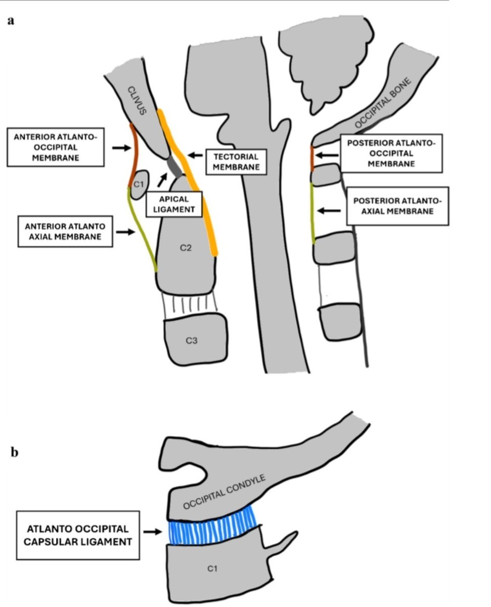
Today, we journey to the craniocervical junction—where the skull, atlas and axis form a remarkably mobile yet highly vulnerable osteoligamentous unit.
Let’s meet the ligaments that keep the skull attached to the spine.
A patient with atrial fibrillation is taking apixaban regularly.
No missed doses.
Correct dose.
Yet he develops an ischemic stroke.
Our first reaction is often:
The DOAC has failed.
Not so fast.
The first question is NOT
Which anticoagulant should I switch to?
The first question is
Why did the stroke happen despite anticoagulation?
That single change in thinking can completely change management.
I now use a simple bedside approach.
DOAC BREAKTHROUGH STROKE CHECKLIST
1️⃣ Was the patient really taking the DOAC?
• Missed doses
• Stopped before a procedure
• Couldn't obtain the medicine
2️⃣ Was the dose actually correct?
Many elderly patients receive reduced-dose DOACs without fulfilling guideline criteria.
Underdosing can become undertreatment.
3️⃣ Any hidden drug interaction?
Think especially of enzyme-inducing antiseizure drugs.
Carbamazepine.
Phenytoin.
Phenobarbital.
Primidone.
These can significantly reduce DOAC levels.
4️⃣ Any absorption problem?
One practical pearl many people miss:
Rivaroxaban 15 mg and 20 mg should be taken with food.
Also ask about bariatric surgery or inflammatory bowel disease.
5️⃣ Was AF really the cause?
Not every stroke in an AF patient comes from AF.
Always look for:
• Carotid stenosis
• Intracranial atherosclerosis
• Carotid web
• Dissection
• Small vessel disease
6️⃣ Is there another prothrombotic state?
Think of:
• Occult malignancy
• Antiphospholipid syndrome
• Left atrial appendage thrombus
Only after excluding all of these should we call it a true breakthrough stroke.
The biggest practical message from the 2026 BMJ review?
There is no good evidence that routinely switching from one DOAC to another reduces recurrent stroke.
There is also no evidence to routinely add aspirin, but there is clear evidence that it increases bleeding risk.
The answer is usually not a new drug.
The answer is finding the missed mechanism.
In stroke medicine, changing the prescription is easy.
Finding the real reason the patient stroked is what changes outcomes.
#NeuroX #MedX #Anticoagulation #AF
Reference
Lun R, et al.
Investigation and management of breakthrough ischemic stroke in anticoagulated patients with atrial fibrillation.
BMJ. 2026;394:e084905.
1/2
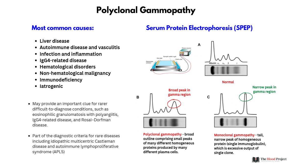
POLYCLONAL GAMMOPATHY
A broad gamma-region peak on SPEP reflects increased production of many different immunoglobulins by many different plasma-cell clones.
Think inflammation, infection, autoimmune disease, liver disease, IgG4-related disease, and other reactive states.
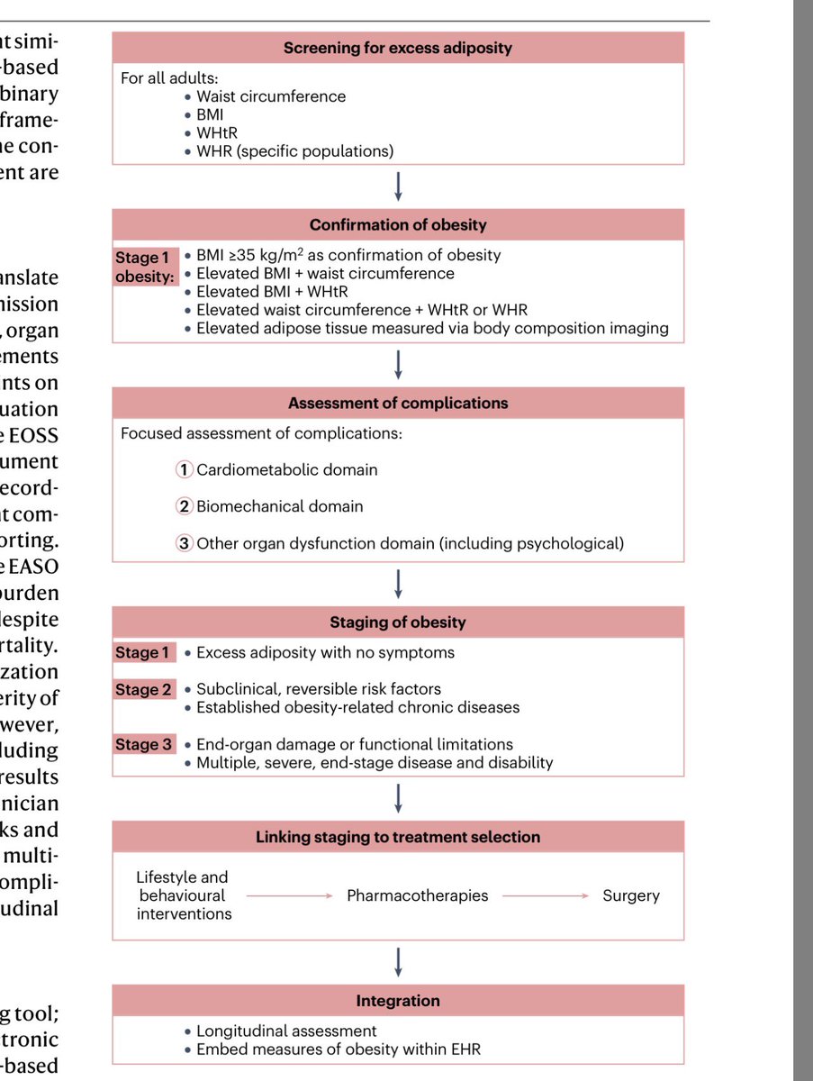
Este paper es probablemente la mejor síntesis hasta ahora en obesidad. No es una nueva guía. Compara las más recientes EASO (2024), (2025), ACC (2025) y AACE (2025) en un paper.
Obesidad deja de ser definida por IMC y se define ahora por exceso de adiposidad que genera daño.
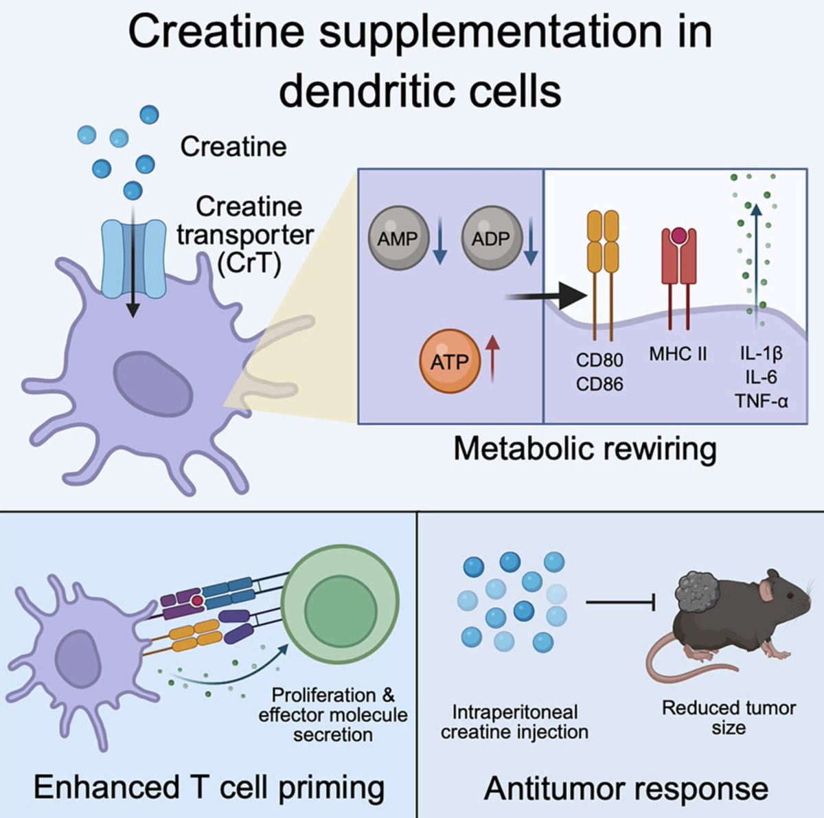
Creatine has been used by bodybuilders for decades to gain muscle. A new study shows it does something entirely different to the immune cells that detect and fight cancer.
Researchers found that creatine supplementation enhances dendritic cell function, the specialized immune cells responsible for activating T cells against tumors. The effect worked in mice and human cells, and it slowed melanoma tumor growth through a mechanism that preserves cellular energy.
This matters because dendritic cells are central to cancer immunotherapy. They're the alarm system of the immune response. These cells patrol tissues, capture tumor antigens, and present them to T cells in lymph nodes, activating tumor-specific immune responses. When dendritic cells can't activate properly, T cells don't get the signal to attack cancer.
The problem is energy. Dendritic cell activation requires substantial ATP to support inflammatory signaling, surface molecule expression, and cytokine production. In tumors, cancer cells compete aggressively for glucose and other nutrients, creating a metabolically hostile environment that starves immune cells of the energy they need to function.
Creatine addresses this through an ATP-buffering system. Here's how it works:
Creatine enters cells through a transporter called CrT (SLC6A8). Inside the cell, an enzyme called creatine kinase adds a phosphate group, converting creatine to phosphocreatine. Phosphocreatine acts as an energy reserve. When ATP gets consumed during cellular work, phosphocreatine donates its phosphate back to ADP, rapidly regenerating ATP without requiring glucose metabolism.
This buffering system is well known in muscle and brain tissue, which have high energy demands. What this study reveals is that dendritic cells use the same system when they activate.
The researchers started by measuring creatine transporter expression in dendritic cells isolated from mouse tumors. They found CrT was markedly upregulated in tumor-infiltrating dendritic cells compared to splenic dendritic cells. The same upregulation occurred when they cultured bone marrow-derived dendritic cells and stimulated them with LPS, a molecule that mimics bacterial infection and triggers immune activation.
To test whether creatine uptake was functionally important, they compared dendritic cells from normal mice versus mice genetically engineered to lack the creatine transporter (CrT knockout).
Key findings when dendritic cells couldn't import creatine:
• Cell survival dropped dramatically after LPS stimulation
• Surface activation markers (CD86 and I-Ab) were significantly reduced
• Production of inflammatory cytokines (TNF-α, IL-6) decreased
• Ability to activate antigen-specific T cells was impaired
When they co-cultured CrT knockout dendritic cells with OVA-specific T cells, those T cells showed reduced proliferation, lower viability, decreased cytokine production (IL-2, IFN-γ), and diminished expression of activation markers (CD25, CD69, CD44).
The opposite intervention produced opposite results. When they supplemented normal dendritic cells with 0.5 mM creatine during activation:
• Cell survival increased significantly
• CD86 and I-Ab expression enhanced
• IL-1β, IL-6, and TNF-α production increased
• Capacity to activate T cells improved
They tested this in a melanoma tumor model. Mice received daily creatine supplementation (2% in drinking water, roughly equivalent to standard athletic supplementation doses adjusted for body weight). Creatine treatment resulted in:
• Slower tumor growth compared to controls
• Increased frequencies of activated dendritic cells in tumors
• Enhanced expression of costimulatory molecules on tumor dendritic cells
• Higher numbers of tumor-infiltrating T cells
• Increased IFN-γ production by tumor T cells
Crucially, the antitumor effect required functional T cells. When they depleted CD8 T cells using antibodies, creatine supplementation no longer slowed tumor growth. This confirms the effect works through enhanced dendritic cell activation of T cell responses, not through direct effects on cancer cells.
The mechanism comes down to ATP preservation. Using metabolic profiling, the researchers showed that creatine supplementation maintained higher intracellular ATP levels in activated dendritic cells. This sustained energy supply supported:
• NF-κB signaling, a master regulator of inflammatory gene expression
• MAPK pathway activation, which controls cytokine production
• mTOR activity, which supports protein synthesis needed for activation
All of these pathways are energy-dependent. When ATP drops, these signaling cascades falter, and dendritic cells can't complete their activation program.
The findings extended to human cells. When researchers isolated monocytes from human blood and differentiated them into dendritic cells, creatine supplementation similarly enhanced their activation, increased costimulatory molecule expression, and improved their ability to stimulate T cell proliferation and cytokine production.
Limitations worth noting: This was tested primarily in one tumor model (melanoma) with one dosing protocol. The human data comes from in vitro experiments with monocyte-derived dendritic cells, not from actual cancer patients. Whether creatine supplementation enhances dendritic cell function sufficiently to improve clinical outcomes in human cancer immunotherapy remains untested.
The tumor microenvironment presents additional complexity. Creatine needs to reach dendritic cells within tumors, which means crossing barriers created by abnormal vasculature, dense extracellular matrix, and immunosuppressive factors that may limit both creatine delivery and dendritic cell responsiveness.
Still, the mechanistic clarity is notable. A widely available, well-tolerated supplement preserves ATP in immune cells operating in energy-depleted environments. This enhances their capacity to activate antitumor T cell responses through well-defined signaling pathways.
The broader context: dendritic cell metabolism has emerged as a target for improving cancer immunotherapy. Multiple studies have shown that metabolic interventions can restore dendritic cell function in tumors. This study adds creatine to that toolkit as a specific intervention that addresses the ATP depletion problem.
For context, creatine supplementation in humans typically involves loading doses of 20g per day for 5-7 days, followed by maintenance doses of 3-5g per day. The supplement has an extensive safety profile from decades of use in athletic populations. Serious adverse effects are rare at standard doses.
Right now, we know that creatine supplementation enhances dendritic cell activation and antitumor immunity in preclinical models through ATP preservation. Whether this translates to improved outcomes when combined with existing cancer immunotherapies in patients is the next question that needs clinical testing.
🧵 TERAPIA ANTIRETROVIRAL en VIH 2026: los 12 conceptos que TODO especialista debe dominar 🚀🦠
Basado en la revisión de NEJM 2026 y las guías DHHS/EACS/BHIVA más recientes.
The Gut-Brain-Muscle Axis: Microbial Regulation of Neuromuscular Aging and Cognitive Frailty
"Collectively, the gut-brain-muscle axis provides a novel systems biology framework for understanding cognitive frailty and developing integrated therapeutic strategies for healthy longevity."
https://t.co/U1ltKP2tUR
People are moral hypocrites
The tendency for people to consider themselves morally good while behaving selfishly is known as moral hypocrisy
We found that people engaging in moral hypocrisy with regard to their own behavior as well as the groups the belong to
Specifically, we observed evidence of in-group favoritism in minimal groups and out-group derogation in political groups: https://t.co/sfYtGQeHdU
Does this ring true?
At the cellular level, your body has about forty different ages (according to one of the largest aging studied ever run and published this week)
A new study in Nature Medicine took blood from 60,542 people, measured over 7,000 proteins, and traced them back to the specific cell types that made them: brain cells, muscle cells, lung, gut, bone marrow. Then they built a separate "aging clock" for each one.
The finding: your cell types don't age in sync. Your muscle can be a decade older than your liver. Your brain's support cells can be racing ahead while your immune cells stay young. One to three percent of people have ten or more cell types aging fast all at once.
Fast-aging brain support cells (astrocytes) flagged future Alzheimer's about as strongly as the highest-risk Alzheimer's gene. Fast-aging muscle cells flagged ALS more than three years before diagnosis. Aging airway cells stacked on top of smoking to push lung cancer risk higher still.
But the one to sit with is muscle. Across all forty-plus cell types, accelerated muscle aging was the single strongest predictor of dying from any cause. Not brain, not heart. Muscle.
muscle is the most modifiable tissue you have. This study can't prove training rewinds the clock, but everything we already know points the same way. Muscle loss tracks death in every population it's been measured in, and the protein signatures behind this clock are the same ones tied to how your muscle is built and how well it makes energy. The strongest death-predictor in the body is also the one you have the most power over.
Normal cellular aging: 9 in 10 alive at fifteen years. More than twenty cell types aging fast: about 1 in 3.
this isn't a test you can buy. It's a research finding from banked blood, in a group skewing older and mostly white. Younger, more diverse validation comes next.
Cette photo mérite absolument tous les prix. Elle a même sa place au Louvre.
Elle relate précisément l’inertie de l’individu face à l’effondrement.
Regardez la composition. Un chef-d’œuvre involontaire.
Au premier plan, le sable doré, les tongs abandonnées, la glacière. Le décor ordinaire du bonheur estival. Deux personnages assis, de dos, comme dans un tableau de Caspar David Friedrich. Sauf que Friedrich peignait des hommes contemplant le sublime. Ici, ils contemplent la catastrophe. Et ils ne bougent pas.
Au centre, le parasol. Publicitaire, criard, dérisoire. Un pastis face à l’apocalypse. C’est presque trop parfait : la France qui prend l’apéro pendant que le monde brûle. Aucun scénariste n’aurait osé.
Et puis le ciel. Les deux tiers de l’image. Cette muraille de fumée qui avale la lumière, ce dégradé d’ocre et de noir digne d’un Turner. La nature qui reprend la palette des maîtres pour peindre notre propre fin.
La femme photographie l’incendie avec son téléphone. Mise en abyme totale : nous documentons notre disparition au lieu de l’empêcher.
Cette image dit tout. Le déni, le confort, l’habitude. Nous sommes tous sur cette plage. Nous voyons tous le nuage. Et nous restons assis, parce que l’eau est bonne et qu’on a posé nos congés.
Les historiens de l’art parleront un jour de cette photo comme on parle du Radeau de la Méduse.
Sauf que cette fois, le naufrage, c’est nous qui le regardons depuis la plage.
💔🌍
💊 Are mineralocorticoid receptor antagonists still underused in heart failure?
Despite being one of the four foundational therapies for heart failure with reduced ejection fraction (HFrEF), mineralocorticoid receptor antagonists (MRAs) remain underutilized, largely because of concerns regarding hyperkalaemia and worsening renal function. This comprehensive 2026 review reminds us that, when used appropriately, their benefits clearly outweigh their risks.
The evidence is strongest in HFrEF, where spironolactone and eplerenone consistently reduce mortality and heart failure hospitalizations, confirming why they remain a Class I recommendation in international guidelines.
The therapeutic landscape has now expanded. Finerenone, a nonsteroidal MRA, has demonstrated clinically meaningful reductions in worsening heart failure events in patients with HF with mildly reduced (HFmrEF) and preserved ejection fraction (HFpEF), offering an important option where traditional therapies have shown more modest benefits.
An equally important message concerns patients with chronic kidney disease (CKD). Historically, clinicians have been reluctant to prescribe MRAs because of concerns about renal dysfunction. However, the review shows that the cardiovascular benefits remain consistent across different levels of kidney function. Although a modest decline in eGFR is common after initiation, this has not translated into loss of clinical benefit. Finerenone also provides additional renal protection in patients with diabetic CKD while reducing cardiovascular events.
Hyperkalaemia remains the major adverse effect, but the review emphasizes that serious hyperkalaemia is uncommon when patients are appropriately selected, dosed according to kidney function, and monitored regularly. Importantly, MRAs also reduce hypokalaemia, which itself is associated with adverse cardiovascular outcomes. The authors propose a practical strategy based on baseline eGFR, serum potassium, diabetes status, and scheduled laboratory monitoring rather than avoiding these drugs altogether.
The practical message is straightforward: fear of hyperkalaemia should not become a reason to withhold a life-saving therapy. Appropriate patient selection, dose adjustment, and careful monitoring allow most eligible patients to receive the proven mortality and morbidity benefits of MRAs.
Reference 📚
Van Spall, H. G. C., & Vardeny, O. (2026). Mineralocorticoid receptor antagonists for the treatment of heart failure and kidney disease: A state-of-the-art review. Heart Failure Reviews, 31, 68. https://t.co/XNKdS9a6il
During lecture on bradycardia, Med student asked:
“If atropine speeds up heart rate, why can a low dose slow it down even further?”
We all memorize board doses, but real clinical mastery comes from seeing which receptor gets hit first.
Critical Physiology Series #14
Unstressed and Stressed Blood Volume in Critical Care: Preload Is Not Simply How Much Fluid the Patient Has
Unstressed volume is the blood required to fill the vessels without stretching their walls significantly. It occupies the vascular space but generates little pressure. It behaves like blood stored in a highly compliant reservoir.
Stressed volume is the blood that stretches the vascular walls and creates elastic recoil pressure. This pressure contributes to mean systemic filling pressure and helps drive blood back toward the right atrium.
Venous return depends on the pressure difference between mean systemic filling pressure and right atrial pressure. Increasing stressed volume raises mean systemic filling pressure and may increase venous return, provided that right atrial pressure does not rise by the same amount and the heart can accept and eject the additional blood.
This explains the effect of intravenous fluids.
A fluid bolus adds blood volume. Part of that volume fills the unstressed compartment, while part increases stressed volume. If the resulting rise in venous return increases stroke volume, the patient is preload responsive.
However, when the heart is already operating on the flat portion of the Frank Starling relationship, additional stressed volume mainly increases cardiac filling pressure and venous congestion. More preload then means more pressure, not more flow.
Norepinephrine can recruit preload without administering additional fluid. By constricting venous capacitance vessels, norepinephrine shifts blood from the unstressed compartment into the stressed compartment. Mean systemic filling pressure rises, the pressure gradient for venous return may increase, and cardiac output may improve.
This is sometimes described as an internal or functional autotransfusion.
Sympathetic activation produces a similar response naturally during acute circulatory stress. Loss of sympathetic tone, as may occur during induction of anesthesia or severe vasoplegia, can move blood in the opposite direction. Vascular capacitance increases, stressed volume falls and venous return may decrease even though total blood volume has not changed.
Positive pressure ventilation can also interfere with preload recruitment. Increased intrathoracic pressure raises right atrial pressure and may reduce the gradient driving venous return. A patient may therefore possess adequate stressed volume but still have impaired venous return because the downstream pressure has increased.
How can this be monitored at the bedside?
Passive leg raising temporarily transfers venous blood toward the central circulation. It acts as a reversible preload challenge without administering fluid.
PPV, SVV, EEOT, TV challenge may also be useful, but only when their physiological requirements are satisfied.
References 📚
Magder, 2016, https://t.co/zAGYN4knaQ
Persichini, 2022, https://t.co/RaR7Ff7mYZ
Monnet, 2011, https://t.co/wQXqtpQaYt
SEDENTARISMO: UNA HUELLA MITOCONDRIAL DETECTABLE. El sedentarismo se asocia con menor capacidad mitocondrial, reducción del transporte de piruvato y ácidos grasos, mayor acumulación de lactato y pérdida de flexibilidad metabólica. Las pruebas de ejercicio podrían detectar precozmente este deterioro bioenergético antes de que aparezca enfermedad. (lee el artículo completo en BLOG JL Chicharro en https://t.co/ghlyA0rsYU) https://t.co/irUD64Wwp8