@jinzhanglab@nchembio Congratulations!! I was wondering whether your lab is developing a similar chemigenetic sensor for endogenous RhoA activity, or whether the HaloRasAR design could potentially be adapted to detect RhoA-GTP?
We are arranging an international islet and transcale imaging meeting in Aug, with a good combination of imaging technology, fluorescent probes, mathematical modeling, and of course, the islet researcher from the world! Here is the preliminary notice. We welcome all of you😀
Excited to announce this new paper from the Kirchhausen and Hatzakis labs on DeepSPT, a deep learning framework for single-particle/vesicle tracking. https://t.co/JjU5w94sbb
Three years on, our #sparsedeconvolution has reached 300 citations, and the codes have been downloaded by over 2,200 times.
So happy to see our developed method being widely used!
https://t.co/JzlyH3ad6Z
New preprint countdown!
Mitochondria are cells within our cells. They need the same core activities - replication, transcription, translation. How do cells enable these diverse activities in both compartments? We uncover an unexpected strategy with ancient origins. Stay tuned!
Tetanus on demand! (not for bioweapons, but for neuroscience research)
Tetanus toxin induces paralysis by using a protease to cleave VAMP2, a protein required for synaptic vesicle fusion and neurotransmitter release.
To harness this toxicity and use it as a beneficial tool for neuroscience research, we fused a light-sensitive LOV domain from oat plants into tetanus’ protease domain and made its activity controllable by light.
LATeNT (for light-activated tetanus neurotoxin) can be expressed in specific cell populations, and then turned on with a brief pulse of blue light. Synaptic inhibition is potently inhibited only in those cells expressing LATeNT while neighboring cells are unaffected.
We used LATeNT in the mouse brain to discover a new cell population (SST+ interneurons in the hippocampus) that modulates anxiety. 24 hours after shutting off LATeNT by taking away the light, anxiety levels returned to normal.
We are excited for neuroscientists to try this new tool! The inhibitory effect is stronger and more sustained than with other tools like halorhodopsin and DREADDs, but the timing and location of the effect are completely under your control.
Preprint here: https://t.co/ire30ouvXA
Very proud of amazing protein engineer @heegwangroh, our fantastic neuroscience collaborator Ji Won Um, co-first author Dongwook Kim from the Um lab, and our other wonderful co-authors.
Image credit: painting by Sir Charles Bell depicting opisthotonos, which can be caused by tetanus (1809)
I am super excited to share our article addressing a longstanding question in neurobiology: how Ca²⁺ signaling cascades initiated at dendritic spines operate at a distance. Check it out here: https://t.co/t7zqOdKvFt @CellCellPress
Our latest work on #actin assembly is now online @ScienceMagazine!
We show how #formins bind and move with the growing barbed end of F-actin.
Awesome collab. between @Intein and Bieling labs @mpimoph, and big thanks to co-first author @maikaboiero
https://t.co/I6A17Jcz8a
Excited to share the first paper from the lab! https://t.co/8ZypMSclii
Limited by photobleaching in single-molecule imaging? We present multicolor long-term single-particle tracking in live cells for 10 min. The probes are actually stable for more than several hours!
#FluorescenceFriday#cellbiology#bioart@wonderofscience 4D organelle remodeling in dividing HeLa cells. Please expand to see greater detail.
Left: 3D volumes of chromosomes, mitochondria, and the endoplasmic reticulum (ER) in whole cells.
Middle: Inner details revealed in successive 2 micron thick slabs computationally cut though the cells.
Right: Expanded view in one slab. As the cells progress through division, mitos fragment, ER forms sheets, and mito fragments nestle into ER pockets as both organelles partition into the two daughter cells.
As seen by 4D lattice light sheet microscopy. https://t.co/vpdwTXqrdm
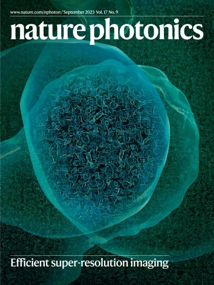
It's a great pleasure for our recent work #SACDimaging to be selected as the cover article of Volume 17 Issue 9, September 2023
@NaturePhotonics
https://t.co/42yq7EXhVa
Some of you may have seen an edited version of our video circulating recently, here is the original! Filopodia extension in a migrating fibroblast. #cell#biology#education#animation@rainmaker1973