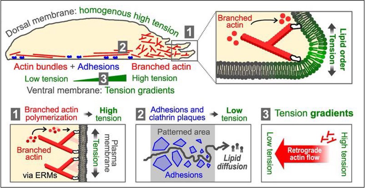
How does nano-scale plasma membrane structure underpin tissue development?
Let’s find out!
First opportunity to do a PhD with me, co-supervised by Darius Koester, at Warwick
Fully funded studentship by #MIBTP and University of Warwick
More info here:
https://t.co/lctwKQ93zq
How can you take your data one important step closer to #FAIRdata 🤔? Check out #EuroBioImaging's FAIR image data decision tree🌳- designed to guide you in the selection of appropriate repositories.
Separate trees exist for different image data types!
Check it out⤵️
https://t.co/jYz8pdVDDR
Shellard, A., & Mayor, R. (2021). Collective durotaxis along a self-generated stiffness gradient in vivo. Nature, 600(7890), 690–694. #EpithelialMechanics
https://t.co/lLiq4OX8O8
The @NIH's research integrity scandal involving neuroscience chief and #Alzheimer’s/#Parkinson’s scientist Eliezer Masliah is of an extraordinary scale. It might be useful to follow one thread of the investigation involving the experimental drug prasinezumab from @ProthenaCorp.
Brooks, J. W., Tillu, V., Eckert, J., Verma, S., ..., Parton, R. G., & Yap, A. S. (2023). Caveola mechanotransduction reinforces the cortical cytoskeleton to promote epithelial resilience. Molecular biology of the cell, 34(12), ar120. #EpithelialMechanics
https://t.co/dBN4LMXszu
WHaloCaMP is a chemigenetic calcium indicator based on HaloTag that can be combined with different rhodamine dyes for multiplexed or FLIM imaging in vivo, as demonstrated in neuronal cultures, brain slices, Drosophila, zebrafish larvae and mouse brain.
https://t.co/bL751a2Mg7
Introducing #NovaFLIM: our new #fluorescence lifetime imaging analysis software. It is designed for an efficient and multi-faceted analysis of #FLIM, FLIM-FRET, and #anisotropy. ➡ Learn more: https://t.co/xGpOrnyDYX
Are you interested in the proteins that enable macrophages to eat, kill, and digest bacteria, apoptotic cells, or other debris? Here, we present PhagoID, phagosome proximity labeling we used to profile human and mouse phagosomes with quantitative mass spec! (1/ 11)
I added image-generation capabilities to the ipy-llm-kernel so you can use #DALLE3 within Jupyter. I hope it's useful, e.g. for crafting prompt-engineering training materials. 🌴🏝️
https://t.co/FKnWR4Srgl
Mathieu, M., Isomursu, A., & Ivaska, J. (2024). Positive and negative durotaxis – mechanisms and emerging concepts. Journal of Cell Science, 137(8), jcs261919. #EpithelialMechanicsReview https://t.co/yzlDLqqUju
📢 Do cells migrating on a confined environment have a mechanical memory of their previous state ? We tried to answer this question in our new preprint 👇🏼
https://t.co/UZPyg5fhrB
an outstanding work by @KYohalie and very great collab with @d_brueckner
A paper in @NatureNeuro presents a suite of genetically encoded opioid peptide sensors for probing opioid drugs and brain-region/circuit-specific opioid release in behaving animals. https://t.co/Dt5Cm9Rkvy
It took more than a DECADE... but finally it’s published!!! It started as a CRAZY idea but after tons of hard work we built it: We hijacked the endogenous secretion systems of Toxoplasma gondii, a brain parasite, to deliver active proteins to neurons 👇
https://t.co/I2j2pAel69
"Flipper-TR reveals the secrets behind membrane tension gradients."... Check out my latest
@preLights post 👇covering the latest @biorxivpreprint
from the team of @RouxLab with the first author's response @juanmagararc. https://t.co/1s6IXMfqEz
.
Since long time we tried to achieve “permanent” labelling of plasma membranes. Now it become possible with MemGraft probes, which exploit a new principle of lipid-directed covalent fluorescent labeling of membrane proteins. The MemGraft probes are compatible with fixation, permeabilization, trypsinization, presence of serum in the medium and enable long-term cell imaging, co-culture of cells and their imaging in multiple colors, cell barcoding and cell manipulation. Congrats and thanks to co-authors: Nathan Aknine @n_aknine & Remi Pelletier. @unistra@CNRS@CNRS_Alsace@LBP_Illkirch
Just out in biorXiv:
https://t.co/tKxnUhvFxT