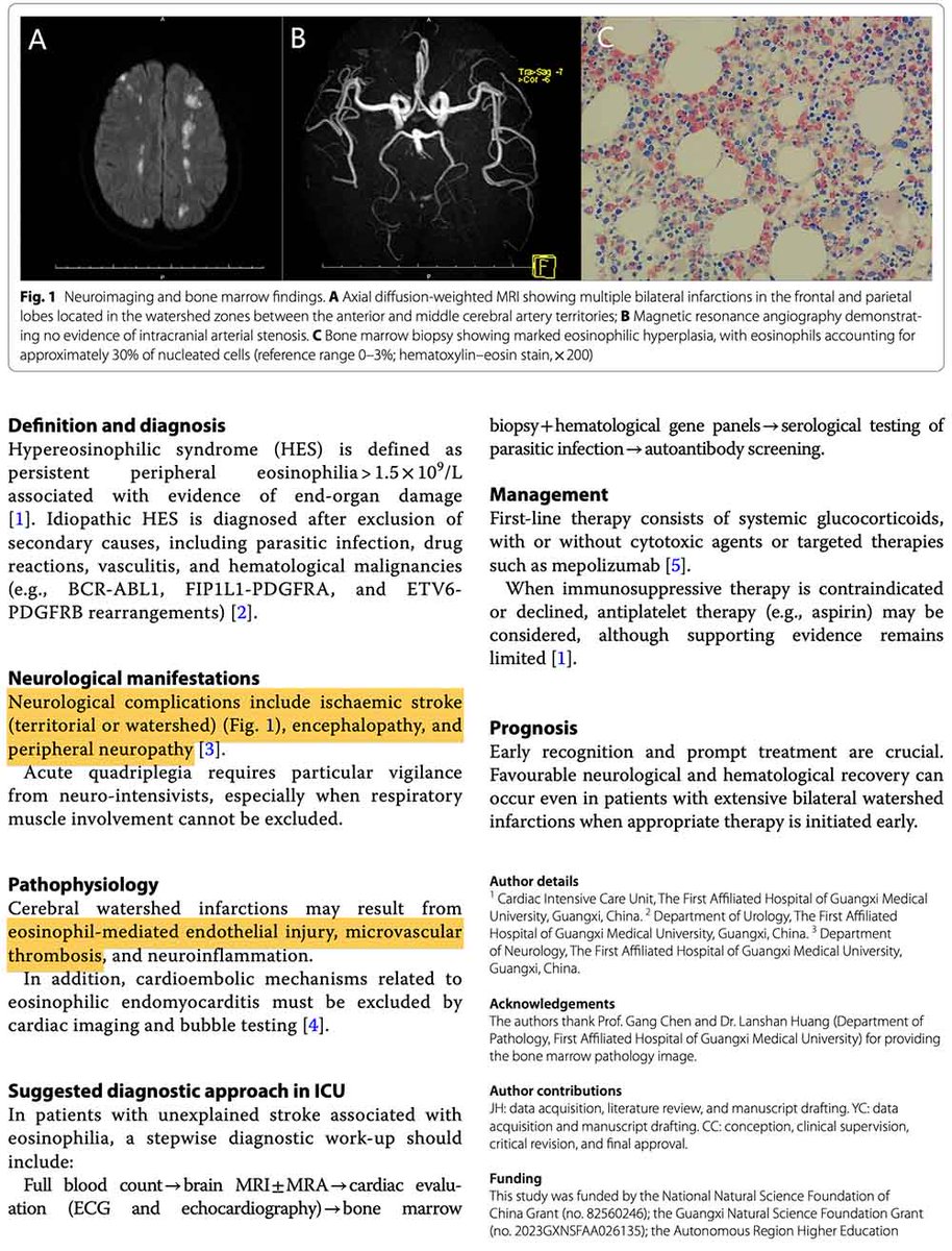
Hypereosinophilia can cause a variety of different stroke syndromes.
#1 Most common = Watershed infarcts (case below)
1⃣ Not due to hypotension or carotid disease.
1⃣ Mechanism unclear, but might relate to some combination of vascular inflammation & eosinophil-platelet aggregates.
#2 Cardioembolic strokes
2⃣ Due to endocardial thrombi (Loeffler endocarditis).
2⃣ Widely distributed in a typical cardioembolic pattern.
Other stroke patterns are less common:
- Single large territorial infarct
- Lacunar & deep perforator infarcts
- Cerebral venous sinus thrombosis
Take-home messages:
🥡 Eosinophilia >1.5 k/uL is defined as hypereosinophilia. This is unlikely to be due to allergy and requires investigation.
🥡 Always get a CBC *with* differential (otherwise you will miss this). It's more effective to be systematic than smart.
🥡 Eosinophils are angry cells that can wreak havoc on the cerebral vasculature through a variety of mechanisms. Hypereosinophilia of any etiology can cause end-organ damage, including stroke.
(Further discussion: IBCC chapter on eosinophilic lung diseases)
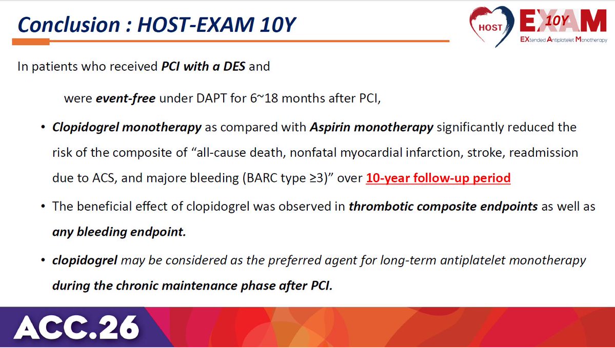
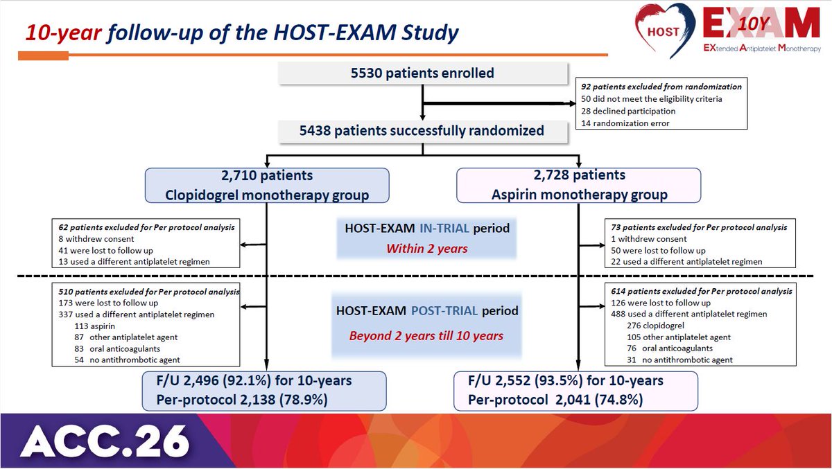
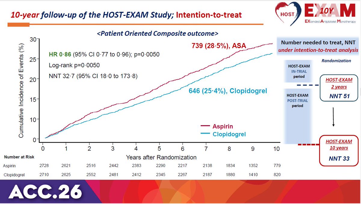
HOST-EXAM trial 10-year results: Clopidogrel monotherapy is superior to aspirin for chronic maintenance after PCI, significantly reducing both thrombotic and bleeding events. #ACC26 View slides here: https://t.co/ihoudzlxIi
Also thrilled to share the future leaders of the Osler Resideny—our 2027-2028 ACSs! @maxmcgredy@jackbuyske@mahamkaratela@Liam_P_Coyne@helminthhenry . Our ACSs are the heart of the program & embody our mission and values. We are in great hands with these 5 amazing people!
South Asian adults in the US develop cardiovascular disease risk factors earlier than other groups, according to new research from the MASALA cohort study. Here’s what that means for prevention. https://t.co/SweVoZgOhS
From @JAMA_current:
📊 This Rational Clinical Examination evaluates the diagnostic accuracy of clinical examination, radiographic imaging, and laboratory testing in assessing volume overload in nonintubated patients.
https://t.co/ATTOSfiAcG
Here’s a situation many of us have seen in the ICU or ED: “It looked like there was ST elevation on the monitor but when I took a 12 lead it was gone?!”
A STEMI went MIA? Here’s a #tweetorial all about why ST segments look different on monitors.
#FOAMed#FOAMcc
1/
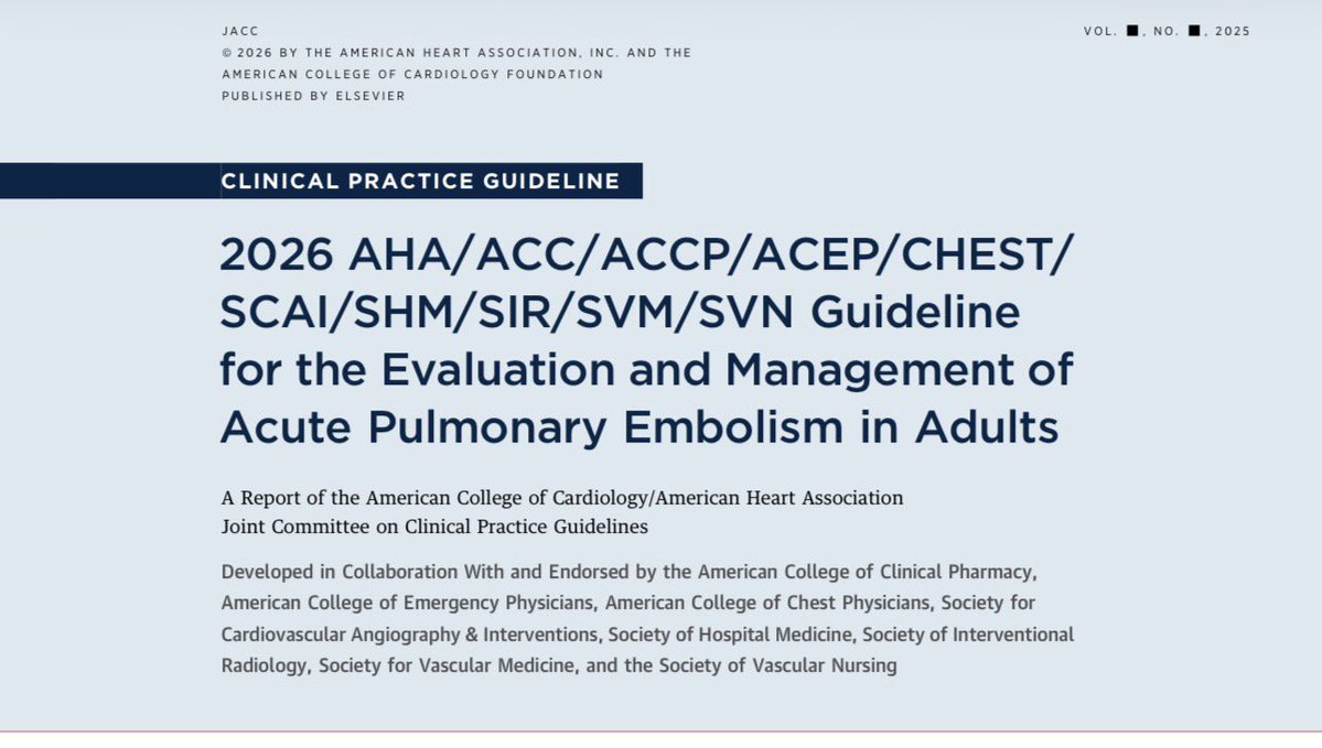
🧵 1/ First ever AHA/ACC/multi-society guidelines re: diagnosis & management of acute PE released today!
2 year effort with 38 authors from 10 specialties.
Link attached & summary in this thread:
https://t.co/uUUyUvz3pR
Excited to share our work examining CAC distribution patterns in older adults. Thank you to Siyu Zou, @KuniMatsushita and @MichaelJBlaha for their collaboration and mentorship.
https://t.co/HM4lINwdLK


![AHAScience's tweet photo. Key updates to this guideline include:
➡️ The use of the American Heart Association PREVENT-ASCVD equations to guide primary-prevention and lipid-lowering therapy decisions.
➡️ Testing Lp(a) at least once in a lifetime and selective apolipoprotein B measurement to improve risk assessment and guide treatment
➡️ The return of LDL-C and non-high-density lipoprotein cholesterol treatment goals (with lower targets for higher-risk groups)
➡️ Expanded use of coronary artery calcium scoring to reclassify risk[ME1.1]
✍🏼 @rblument1 @tygluckman @RonBlankstein @PamelaBMorris @pnatarajanmd @AnnMarieNavar @SethShayMartin @APRN_CNS @nyulangone @DrMichaelShapir @kgradneyrd @eugeniagianos @virani_md @KellieMcLain1 @ijeomaheartdoc @SamiaMoraMD @DrHeatherJohn @dmljmd](https://pbs.twimg.com/media/HDPXv0SXsAIEPMD.png)