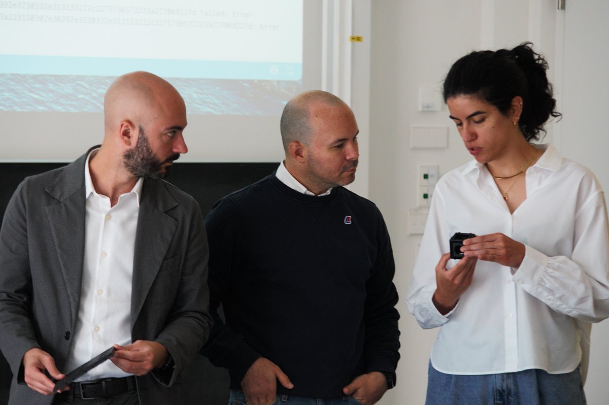
🏆STEDYCON 2 has received the 2026 Microscopy Today Innovation Award!
STEDYCON 2 was recognized as “the first modular add-on system combining multicolor STED and phasor-based FLIM (TIMEBOW) in an alignment-free, browser-controlled platform.”
https://t.co/W9MfYKXSlW
We have been working for a while on effect of crowding on enzyme activity and it is finally out 🤩. Incredible collaboration with @RGolestanian@Sugita_Riken https://t.co/702IBOJRIN
Some RFD4-Proteina tool calling examples using GFP as a template.
Insert a motif: ALFA tag insertion
Iterative redesign: fix an atom clash by increasing the tag linker length
Loop redesign: replace a 3-aa loop with a longer one
Binder design: generate a 60-aa computational binder against GFP
Circular permutation: join the original N and C termini and introduce new termini
pi-stacking: aromatic side-chain stacking against a SAM molecule
Motif scaffolding: retain GFP’s chromophore and adjacent residues, then generate the rest of the protein
Agents ran these on Modal cloud compute and made this demo video.
Excited to share our latest @biorxivpreprint, led by Dr. Chelsea Fries @assemblingfries 🥳
Here, we establish multistate interface design as a strategy for engineering protein assemblies that break symmetry and switch between distinct geometries.
https://t.co/TEjb7MpGow
Two Golgi markers. One MINFLUX acquisition. The new abberior DNA-PAINT FLASH 2color kit resolves Giantin and GM130 simultaneously in 3D. Vero cells · MINFLUX · 3D
#MINFLUX#DNAPAINT#Golgi#FLASHPAINT
De novo design of light-controlled conformational protein switches
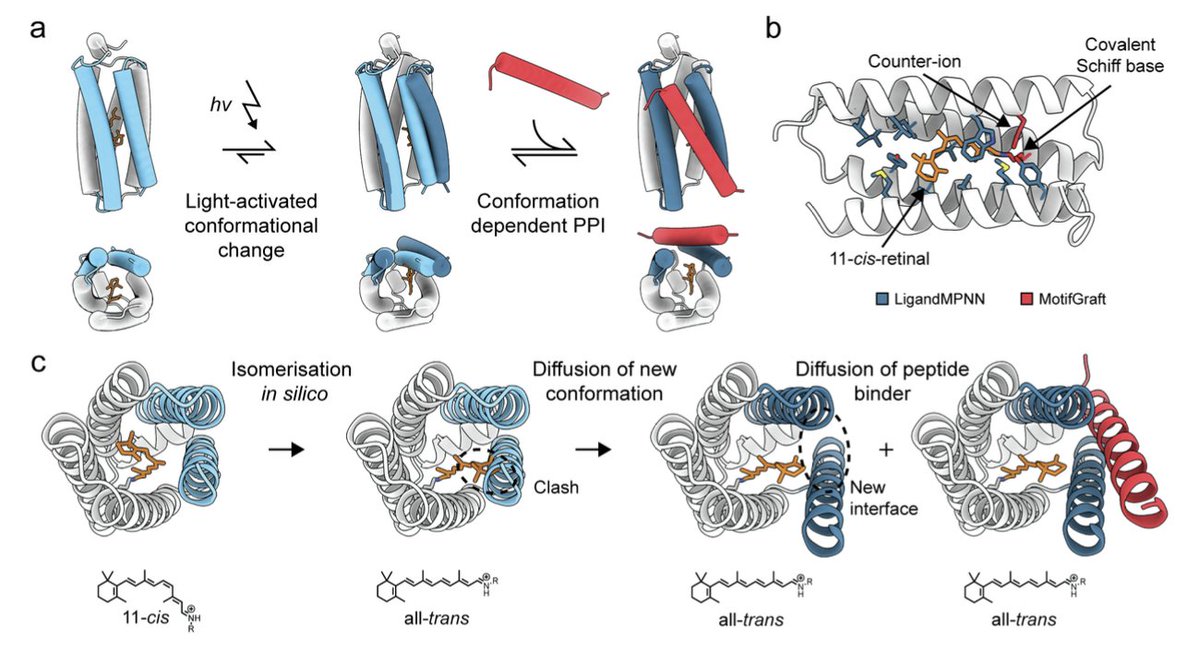
1. The study reports fully bottom-up computational design of new-to-nature light-responsive proteins: de novo retinal binding, a retinal-isomerisation-coupled conformational switch, and a conformation-specific protein–peptide interaction (PPI) that turns on upon illumination.
2. A key result is light-activated heterodimerisation with large dynamic range: optimized SPARC designs reach >30-fold increased binding signal upon blue-light illumination (split-luciferase readout), with further engineering pushing performance (e.g., combinations reported to reach much higher maximal signals while maintaining strong fold-change).
3. The chromophore choice is retinal (11-cis to all-trans), enabling access to broad visible-spectrum photochemistry associated with rhodopsins, but implemented here in soluble, minimal, designable scaffolds rather than membrane proteins.
4. They first design de novo retinal-binding proteins (RBPs) across multiple folds (4/5/6-helix bundles and a beta-barrel). Retinal is covalently attached via a designed Schiff base to a core lysine with a counter-ion and H-bond network to favor a protonated Schiff base, producing strong red-shifts (absorbance maxima ~440–530 nm vs ~380 nm free retinal).
5. Experimental validation of retinal binding includes native MS showing the expected +266 Da adduct consistent with Schiff base formation, top-down MS localizing the modified residue, and CD confirming well-folded, highly thermostable proteins.
6. Sequence novelty is assessed using ESM2 embeddings and UMAP: the designed RBPs/SPARCs form a distinct cluster separated from both microbial and animal rhodopsins, suggesting exploration of new sequence space despite convergent chromophore chemistry.
7. The core design innovation for switching is SPARC (Synthetic Photo-Activated Retinal-based Coupling): in silico retinal isomerisation is introduced, steric clashes identify regions that must move, and RFDiffusionAllAtom is used to generate an alternative conformation compatible with all-trans retinal while simultaneously diffusing a 25–30 aa helical peptide binder to fit the newly opened site.
8. Multistate sequence design then ties residues across dark and light conformations (ProteinMPNN/LigandMPNN), with AlphaFold2/AlphaFold3 used to filter designs that adopt the intended apo/dark vs peptide-bound/light structures and the correct retinal isomer context.
9. Screening 92 initial heterodimers yields a first functional hit (SPARC-39), and a second computational optimization round produces multiple improved switches; the best reported (e.g., SPARC-111) shows >30-fold activation. Mutating the Schiff-base lysine abolishes response, and the parent single-state RBP does not switch, supporting the multistate-mechanism requirement.
10. Structural and mechanistic support includes a dark-state crystal structure (SPARC-117prot) matching the design model closely (~0.92 Å Cα RMSD). Ultrafast spectroscopy indicates single-state RBPs can hinder isomerisation (long-lived excited-state features), whereas multistate SPARCs show signatures consistent with successful isomerisation and productive switching.
11. The platform is engineered post hoc with relatively simple interventions: (i) rational charge-swapping yields an orthogonal heterodimer pair (SPARC-111ortho) with preserved light response, (ii) pocket point mutations shift absorbance up to ~30 nm red, and (iii) interface tweaks improve sensitivity/maximum signal; the peptide partner can also be scaffolded into small globular miniproteins via diffusion + sequence design, with many scaffolds retaining light-activated binding.
12. Cellular proof-of-concept: in HEK-293T cells, SPARC-111 fused to split luciferase shows light-dependent activation when retinal is supplied. For receptor control, SPARC-based dimerisation is used to cluster FGFR1 via fused minibinders; adding a SpyTag/SpyCatcher “covalent lock” helps overcome dissociation and enables light-dependent calcium signaling responses.
13. Real-time light-activated NMR (in situ illumination inside the spectrometer) directly observes switching: spectra distinguish at least four conformational states (apo, 11-cis-loaded dark, all-trans-loaded light, and all-trans + peptide complex). Light-induced chemical shift perturbations match all-trans retinal loading, and peptide binding is only evident in the light-activated state, supporting the designed conformational-selection landscape.
14. Limitations noted include incomplete wavelength separation for multi-color orthogonal activation, absorption still often shorter-wavelength than many natural rhodopsins, and cellular efficacy not yet matching mature optogenetic tools (e.g., receptor activation benefiting from covalent locking). Retinal availability/solubility in soluble contexts is also discussed, with potential mitigation via retinal derivatives.
📜Paper:
https://t.co/ijWPyRcBek
#ComputationalProteinDesign #Optogenetics #DeNovoProteinDesign #ProteinEngineering #Retinal #RFDiffusion #ProteinMPNN #LigandMPNN #AlphaFold3 #NMR #SyntheticBiology
Thrilled to share our paper in @NatureComms
RNA-binding proteins can enhance transcription largely independently of condensation
Nascent RNA recruits FUS to transcription site, boosting RNAP loading and preventing R-loops to enhance elongation.
https://t.co/6KC1knQYkR
How does the mitotic spindle adapt to genomes that differ in size by more than 10,000-fold? Our new paper in @NatureCellBio reveals a simple scaling law and the physics behind it: chromosome crowding! ��
https://t.co/DsGvz8ulME
We've just updated our 2027 EMBL Annual Poster with even more events 🙌
Have a look and tell us which course or conference you're excited to join:
https://t.co/Ond0DeiQQ0
@embl@emblebi@EBItraining@EMBO
BindCraft2 v1.0.4 has been released: with significantly improved VHH, scFv, ARP, and induced fit targeting. WE also improved speed and code stability. Try your difficult campaigns again. Great work by Erik Mäeots, Leonardo Tredese and Elizaveta Chernova.
Three years of intensive work and overcoming both smaller and larger experimental hurdles have now revealed the practical accuracy of MINFLUX in a real, widely used system. The answer definitely makes one eager to exploit the performance of MINFLUX even further….See KB Chapman et al
Localization accuracy in single-molecule fluorescence microscopy https://t.co/Sdx59Xc7bu
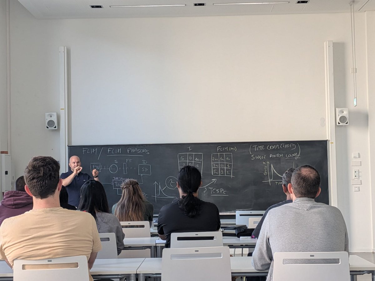
#Jena, that’s a wrap!
We’re happy to close the #FLIM MicroSpectroscopy workshop with such a positive feeling.
These two days were a great opportunity to share ideas, test workflows, and continue strengthening our collaboration with the amazing @OpenUc2 team.
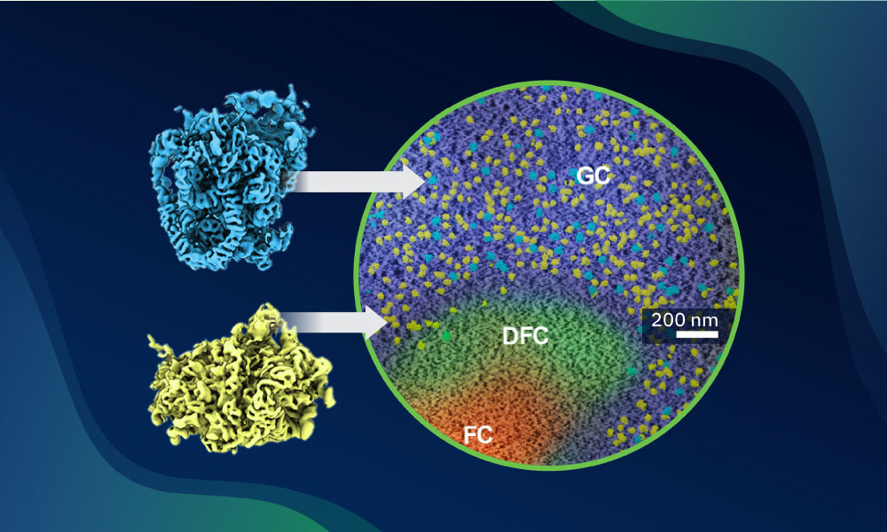
🔬A new study by EMBL researchers, published in Science, about the pre-ribosome assembly pathways inside intact human cells, reveals — at molecular resolution — in-cell structural snapshots and novel configurations.
https://t.co/fDugyaicSp
#lipidtime: It's a lipid universe. Today in Nature we publish the Lipid Brain Atlas, the first map of membrane lipid composition across the entire mouse brain, a layer of organisation that cell-type and transcript maps had left out. #lipidomics#neuroscience#BrainAtlas
ʙɪɴᴅᴄʀᴀꜰᴛ2 is out, and we're not waiting for the paper. The full code drops today, free for academic and industry use.
We're releasing it early so you can start designing right now, and bring its full power to the current Adaptyv competition.
https://t.co/O8pr6jH99t
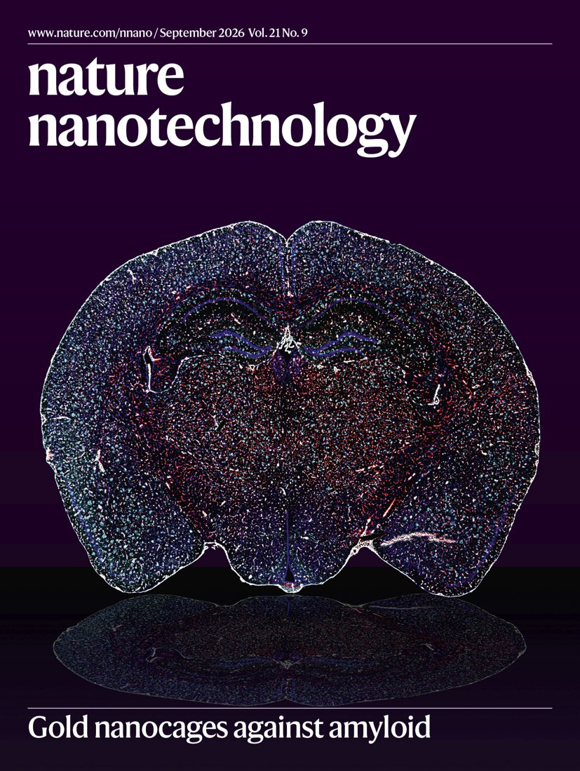
In our September 2026 issue -
Plasmonic nanocavities,
Upconverting nanoparticles for imaging,
Blue-emitting QD/SiNx lasers,
Self-assembled contacts,
Li+ separation,
Spectral biophysical cytometry,
Gold nanocages against amyloids, & much more.
https://t.co/wNlyKYOUfR
our 5th publication of the year is now out, and it comes with our shortest title yet: ONE microscopy. https://t.co/7AgYOqBuS3
ONE pushes conventional fluorescence microscopy to the molecular scale. It allows us to directly visualize the shapes of individual proteins, resolve conformational changes of around a nanometer, and achieve nanometer to subnanometer imaging precision, while extending these capabilities from purified molecules to complex cellular and tissue samples.
This Nature Protocols paper provides the complete workflow for doing this, from sample preparation and expansion to imaging and fluctuation analysis. And now there is ONEpy, our new software package that brings the analysis pipeline together and makes it substantially easier to use. Faster to set up, easier to use, and gives us a much better base for what we want to add next.
and this is not all from the team this year. We currently have 3 more papers in revision, and we are preparing a 4th manuscript to submit by the end of the year.
we are only getting started, and I could not ask for a more ambitious, creative and relentless team to build this with.
what started with ONE as something that seemed almost impossible is now becoming a field: optical structural biology.