Hi! #PathTwitter My name is Alice. I’m currently an AP resident in Thailand but I hope to pursue my career path on Path in the land of possibilities one day. I’m interested in #Gynpath#Thyroidpath and #Cytopath . I hope to share my cases and learn from #PathTwitter as well!
Hi #Pathtwitter, my name is Kanokkan, and an IMG from Thailand applying for #PathMatch2024. My interests include cytopath, gyne, and heme. I love swimming, yoga, & watching movies. Hope to be a cytopathologist one day in the land of opportunity. Also swift13 fan, and 🐶 person💕
Friday staging mini-Tweetorial!
Staging in breast cancer is a subject dear to my heart that I feel is often confusing and misunderstood. The reason is simple. On a slide, we see things in two dimensions, and therefore we are taught in two dimensions and we come to remember and understand things in two dimensions. Yet, as we all know, reality is three-dimensional, and staging ends up being regarded as a linear measurement while in its essence it must be a volumetric measurement.
Let me illustrate with an example. Assuming a spherical shape, what is the difference in volume between a tumor that is 1 cm in diameter and one that is 2 cm in diameter? This should be a relatively straightforward geometry problem but the answer is counterintuitive and surprises most. The ratio of the volumes of these two spheres is 8 times. In other words you need eight (!) 1 cm tumors to equal the volume of one 2 cm tumor. The implications of this are the following:
1. You don't add the diameters of multifocal tumors to get the final T stage, you use the diameter of the largest focus, otherwise you will overstage. One possible exception to this is numerous small foci of invasion in extensive high-grade DCIS. Because these foci are occult and we don't know how many there are, a largest focus of 2 mm might end up being an underestimate of the actual stage of the tumor and the outcome is often worse than what is expected from a T1a tumor.
2. The 5 mm separation rule between two masses (if they are less than 5 mm apart, you should add their diameters for staging purposes) is a useful one given our inadequate representation of the entirety of the involved tissue. However if you know for a fact by imaging for instance and/or by gross examination that these are two contiguous stellate "spherical" masses, then adding the diameters for staging purposes leads to overestimation of the volume of tumor.
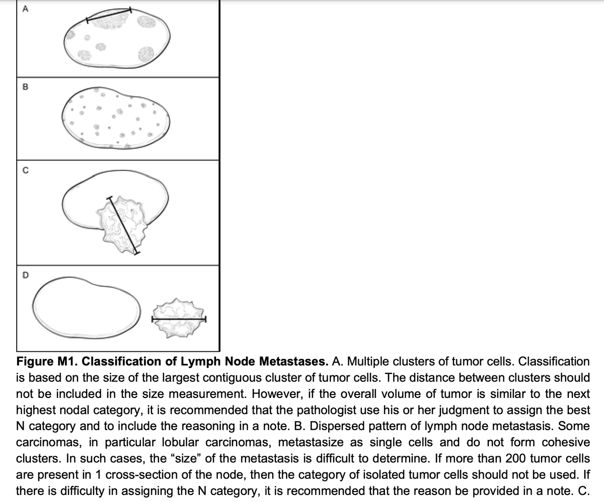
3. In a lymph node, we often struggle with the size of the metastasis, especially if there happens to be multiple foci. Is it multiple micrometastases? is it a macrometastasis. The CAP does provide some guidance that harkens back to the volume idea, but it relies on the pathologists using their judgmenet in unclear cases (see image below). The key idea again is to adopt a volumetric mindset, and, if you can, get a number of levels to allow you a better three-dimensional survey of the metastatic deposit. But do not add diameters if the foci are spherical and separate, and do not call it ITC if more than 200 cells are present.
Like I said, one of the greatest ills of breast pathology is that it is a volumetric disease, and while much of what we know about precancerous lesions has to do with probability (the greater the volume the higher the probability of an adverse outcome) we use confusing ans inadequate linear cutoffs to diagnose lesions. So, short of being armed with breakthrough technology that allows us to visualize breat diseases in 3D, and short of being able to count the cells, because, ultimately, it's all about how many bad cells we find and how bad they really are, we have to arm ourselves with the next best thing, an understanding of the fundamental principles that guide staging and cancer risk assignment, and common sense.
Finally, this is a very brief account of this topic. Please feel free to comment and ask. I have but scratched the surface, but I hope this helps.
@washupathedu @wusm_pathology #breastpath #PathTwitter #breastcancer
I'm close to done turning all* of @KurtSchaberg Kurt's Notes into one Anki deck. Nearly 6 months in the making. 8 chapters left: cyto and derm.
*I'm leaving out neuro only bc the folks at @SynaptiqHQ already did the CNS chapters!
Training is done and you’re about to start making $225,000.
This isn’t a discussion on tax detail so let’s just say you’re out 35% for taxes.
If you want to protect 20% for retirement savings, (10% in pretax 401(k) contributions and the other 10% taxed and put in Backdoor Roth and a taxable investment account) how would you organize the remaining 45% or $101,250?
That’s $8,400/month.
Do you have credit card debt to pay off?
How will you structure your student loan payoff?
Do you have emergency savings?
Do you have kids - how much is childcare? Relative to retirement savings, where does college savings fit in for you?
What kind of home can you afford?
Are your cars reliable for the foreseeable future or no?
What refreshment spending gives you real joy? And what are some old behaviors that cost money are sticking around but aren’t doing it for you anymore?
^^^^^^^^^^^^^^^
These questions are good to ask because you want to step very carefully in these early high income years.
Check out these beautiful CD20+ and CD20- aggressive lymphoma classification algorithms from Dr. Pan being presented at our @CULymphoma tumor board. Saving these for future reference! #lymsm#hemepath
I never celebrate #Thanksgiving in Thailand, but I am thankful I found #PathTwitter. I learned so many things from here, and I hope I have a chance to give it back to #PathTwitter too!
I have my copy of @AceMyPath Surgical Pathology Reimagined! Do you have yours? A must-have for trainees and practitioners alike, with high-yield facts, annotated digital slides @pathpresenter, and fully updated Kurt’s notes! Videos and MCQs in progress. #PathTwitter#MedTwitter