Skeletal muscle is remarkably flexible: what it burns depends on what we eat—and what we ask it to do.
In fasting or on a high-fat diet, muscle favors fat uptake and β-oxidation. After a carbohydrate-rich meal, insulin drives GLUT4-mediated glucose uptake, fueling glycogen synthesis and glycolysis.
Exercise changes the equation: contraction activates pathways that increase uptake of both glucose and fatty acids. Intensity pushes metabolism toward glycolysis, while prolonged exercise increasingly relies on fat oxidation.
Over time, exercise also builds a more oxidative muscle through PGC-1α-driven mitochondrial biogenesis.
https://t.co/L3z19tmmZr
@WuTsaiAlliance
#Exercise #MuscleMetabolism #Metabolism #Mitochondria #GLUT4 #HealthyAging
The fix: Start all 4 pillars (ARNI, BB, MRA, SGLT2i) simultaneously, Day 1.
STRONG-HF: rapid up-titration cut death/HF readmission 23.3%→15.2%
SHORT: cut time to optimization 112→29 days, CV death/worsening HF 5→0
Days, not months.
The window is narrow — act now
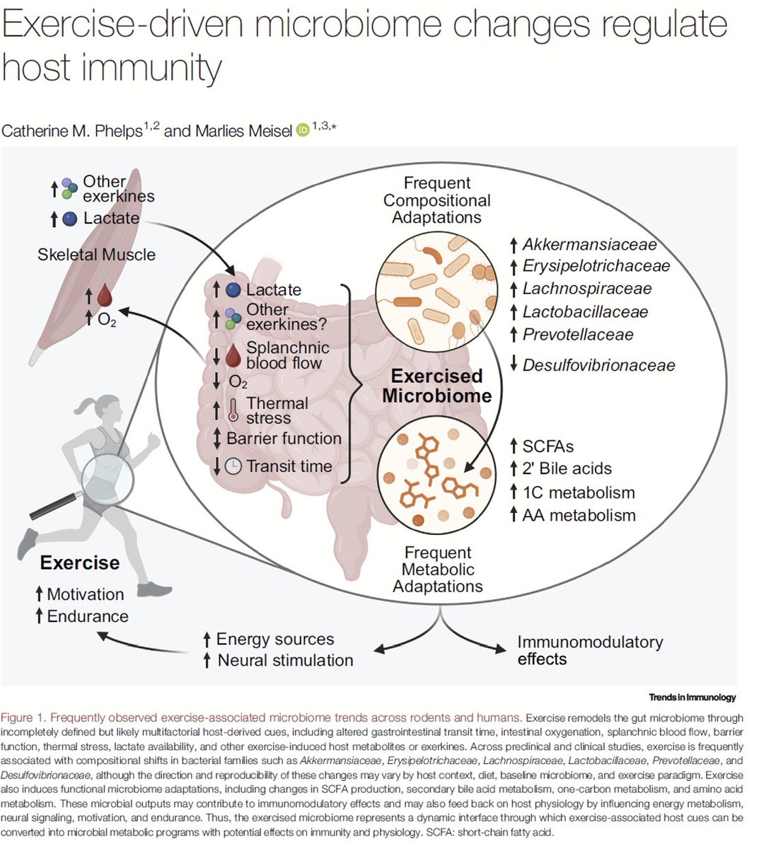
Exercise may reshape our microbiome and immune system in ways that influence disease risk.
This timely review explores how exercise-responsive gut microbes and their metabolites, including short-chain fatty acids, secondary bile acids, and formate, may influence systemic inflammation, antitumor CD8 T-cell immunity, autoimmunity, and metabolic disease.
The exciting challenge now is to move from association to mechanism: Which microbes matter? What metabolites do they produce? And how does the host sense and respond to them?
#Exercise #Microbiome #Immunity #Metabolism #GutHealth #CancerImmunology #PrecisionMedicine
@TrendsImmuno@WuTsaiAlliance
https://t.co/qz4sCRuAGU
Body Composition in Heart Failure: A Magnetic Resonance Imaging and Dual X-Ray Absorptiometry Assessment in the UK Biobank Study
Participants with HF showed higher VAT and adverse muscle changes, with weaker grip strength
https://t.co/dNt044OjeQ
A study in The Lancet Healthy Longevity found that stopping statins in adults over 75 with no history of heart disease or stroke does not increase the risk of death within three years.
Authors say further trials are needed to confirm these findings.
🔗 https://t.co/UzACLAgT2d
# Cardiac CT Is No Longer a Niche Test. It's Becoming Everyday Cardiology.
For years, Cardiac CT has been viewed primarily as a test for ruling out obstructive coronary artery disease. Yes it has been focused on the triage of obstructive CAD.
This new EACVI Clinical Consensus tells a very different story.
Cardiac CT is now presented as a comprehensive imaging platform, extending well beyond coronary stenosis to include valvular disease, cardiomyopathies, cardiac masses, congenital heart disease, pericardial disease, and large-vessel pathology.
The message is clear:
**Cardiac CT has moved from a specialized examination to an integral part of routine cardiovascular care.**
## The Critical Point
Perhaps the most important shift is conceptual.
The document no longer frames CCTA as a tool that simply detects stenosis.
It emphasizes plaque characterization, plaque burden, high-risk plaque features, calcium scoring, functional assessment, and patient-specific clinical pathways.
This reflects the broader evolution of cardiovascular imaging:
**from identifying anatomical narrowing...**
to **understanding cardiovascular disease biology.**
## My Take
The question is no longer *"Can Cardiac CT do this?"*
It is:
**"How do we integrate Cardiac CT into everyday clinical decision-making?"**
That is exactly where the field needs to go.
Technology only changes medicine when it changes clinical pathways.
And there is where the document is weak IMHO.
I believe the weak point of this document is that it does not actually provide sufficient insight into the Anatomical (and Functional and soon Tissue) imaging standard in Cardiology which is Photon Counting CT.
## Where PCCT Fits
The consensus already recognizes **Photon-Counting CT** as the next technological step, highlighting its **0.2 mm spatial resolution** and its potential in challenging scenarios such as heavily calcified plaques, in-stent restenosis, and myocardial injury—while correctly acknowledging that randomized clinical evidence is still limited. However, there are already 600+ Photon Counting CT scanners installed in the world... and the technology is available since 2021.
To me, as I said several times many years ago, the key message is:
- PCCT is not simply another scanner.
- it represents a NEW imaging modality.
If conventional CCT has now entered everyday practice, the next challenge is ensuring that **Photon-Counting CT translates its technical advantages into measurable improvements in diagnosis, clinical decision-making, and ultimately patient outcomes.**
That—not higher resolution alone—will define its success.
#CardiacCT #PhotonCountingCT #PCCT #CardiovascularImaging #CCTA #PreventiveCardiology #PrecisionMedicine #Radiology
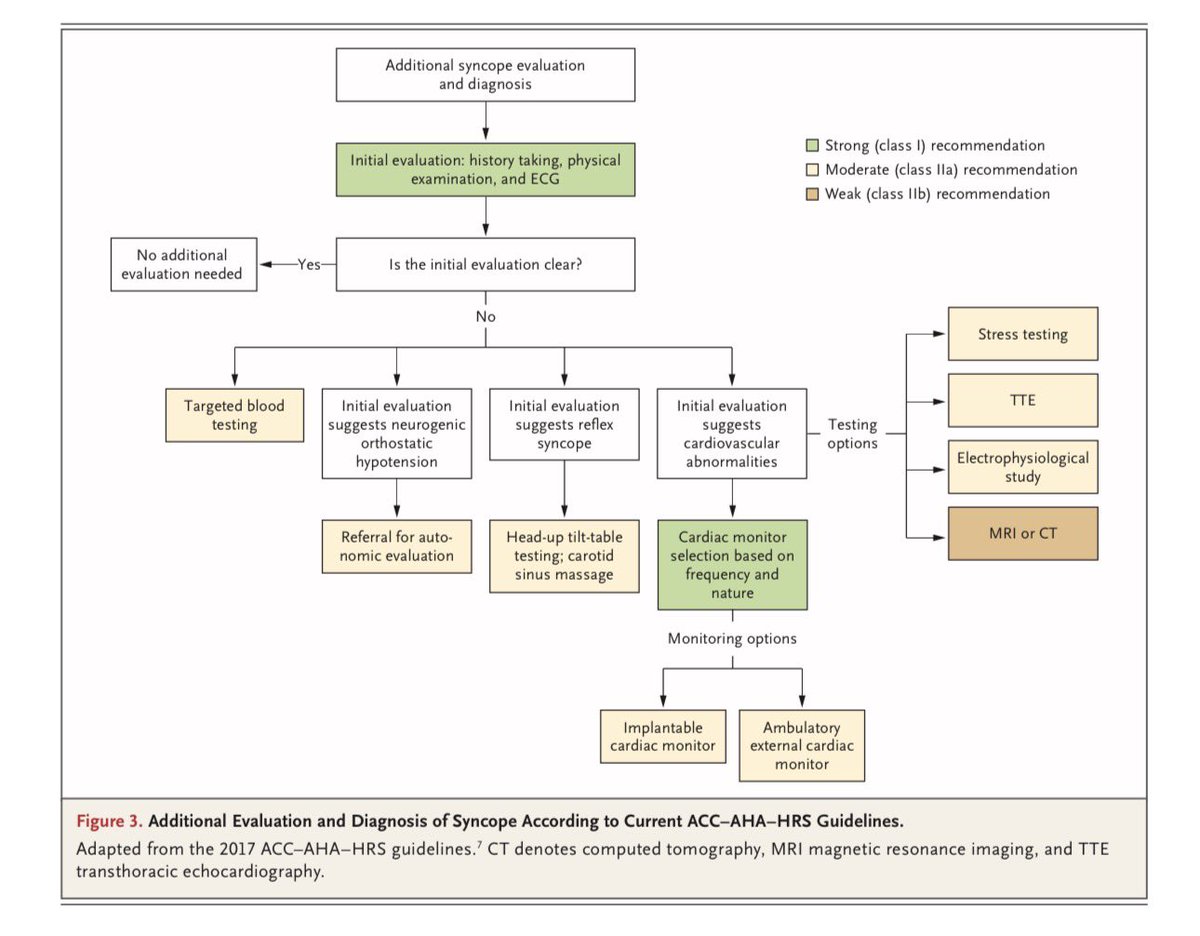
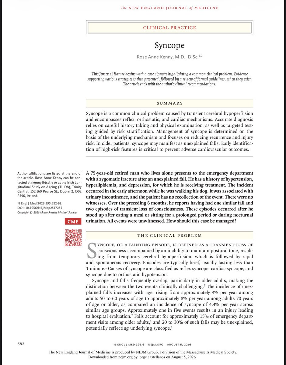
🫀 SÍNCOPE: EL DIAGNÓSTICO COMIENZA CON LA HISTORIA CLÍNICA, NO CON LOS EXÁMENES ⚡
El nuevo artículo del NEJM 2026 nos recuerda que el abordaje inicial del paciente con síncope debe ser simple, sistemático y basado en la evidencia: toda pérdida transitoria de la conciencia con sospecha de síncope requiere una historia clínica detallada, relato de testigos, medición de la presión arterial en decúbito y de pie, examen físico completo y electrocardiograma (ECG). 🩺📋 Estas cuatro herramientas permiten identificar la causa en la mayoría de los pacientes y, cuando no es posible, orientan la estratificación del riesgo para decidir quién necesita estudios adicionales, observación u hospitalización. 🚑 El mensaje clave es claro: no todos los pacientes requieren múltiples pruebas, pero ninguno debe ser evaluado sin una adecuada historia clínica y un ECG. Una evaluación inicial correcta puede detectar causas potencialmente fatales, reducir hospitalizaciones innecesarias y prevenir nuevas caídas o episodios de síncope. 📈❤️ Basado en: Rose Anne Kenny. Syncope. N Engl J Med. 2026;395:582–591.
✅ https://t.co/cYnnL8WtDt
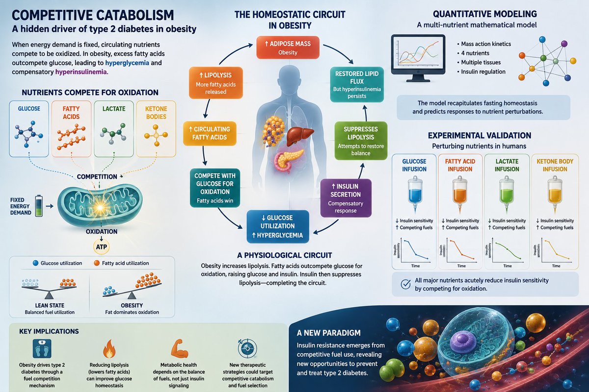
🚨¿Y si la resistencia a la insulina no comienza en el receptor de insulina?
Estudio propone un nuevo mecanismo: el catabolismo competitivo.
En obesidad, el exceso de ácidos grasos compite con la glucosa, reduciendo su utilización y favoreciendo hiperglucemia e hiperinsulinemia.
🚨¿El futuro del tratamiento de la obesidad está en la microbiota intestinal?
Revisión de 217 ensayos clínicos muestra que la investigación sobre moduladores de la microbiota está creciendo rápidamente.
🦠Los probióticos lideran la evidencia clínica.
📈Los postbióticos emergen.
👉Moderate-Intensity Statin + Ezetimibe: The Sweet Spot?
☝️A new network meta-analysis including 38 randomized controlled trials and 7,340 patients with coronary heart disease suggests that adding ezetimibe to a moderate-intensity statin may provide lipid lowering comparable to high-intensity statin-based strategies across most lipid parameters.
☝️Key findings
1️⃣ Moderate-intensity statin (MIS) + ezetimibe ranked highest for LDL-C reduction.
2️⃣ LDL-C lowering with MIS + ezetimibe was comparable to high-intensity statin (HIS) + ezetimibe.
3️⃣ MIS + ezetimibe also showed the best overall performance for HDL-C, total cholesterol, non-HDL-C, and ApoB.
4️⃣ HIS + ezetimibe ranked highest for triglyceride reduction, but without a statistically significant advantage over MIS + ezetimibe.
5️⃣ Among moderate-intensity regimens, rosuvastatin + ezetimibe appeared to provide the greatest lipid-lowering efficacy. pdf.pdf
☝️Clinical message
1️⃣ Before automatically escalating to high-intensity statins, consider early combination therapy with ezetimibe.
2️⃣ This strategy may maximize LDL-C lowering while potentially improving tolerability and long-term adherence.
3️⃣ These findings reinforce a principle increasingly supported by contemporary guidelines: “add rather than double.”
👉 Combination therapy beats dose escalation.
Adding ezetimibe to a moderate-intensity statin achieves lipid lowering comparable to high-intensity statin strategies across most parameters, reinforcing the principle of “add early, don’t just intensify.”
🔗https://t.co/iJjMtmVF0r
@LipidJournal@nationallipid@society_eas
OPTIMA-AF published: In AF patients undergoing PCI, 1 month of DOAC + P2Y12 inhibitor followed by DOAC alone was non-inferior to 12 months for death/thromboembolism (5.4% vs 4.3%; HR 1.25) and halved major/CRNM bleeding (4.5% vs 8.8%; HR 0.50, p=0.004). Therefore, shorter may be safer without compromising efficacy. However, efficacy findings should be interpreted with caution in light of the lower-than-anticipated event rates and fixed absolute non-inferiority margin. https://t.co/EpiCGxUmjk
This narrative review summarizes the latest evidence of how exercise promotes cardiovascular health, focusing on how incorporating small changes in lifestyle can lead to an overall change in the trajectory of developing cardiovascular disease and maintaining a healthy life.
👉The “low cholesterol increases mortality” argument just took another hit.
☝️A new Danish population study spanning nearly 30 years found:
📍Median LDL-C fell from 142 to 124 mg/dL
📍Median non-HDL-C fell from 174 to 150 mg/dL
📍The LDL-C level associated with the lowest mortality shifted downward from 155 to 135 mg/dL
📍The non-HDL-C level associated with the lowest mortality shifted downward from 186 to 162 mg/dL
📍At the same time, overall mortality declined
📍Cardiovascular mortality declined substantially
👉Key takeaways
1️⃣ Lower population LDL-C did not lead to higher mortality.
2️⃣ The “optimal” LDL-C observed in epidemiological mortality curves is not a fixed biological constant.
3️⃣ Mortality nadirs move as populations become healthier, cardiovascular prevention improves, and LDL-C levels decline.
4️⃣ The well-known U-shaped association between LDL-C and mortality is likely driven, at least in part, by reverse causation and residual confounding at the low end.
5️⃣ These findings are fully consistent with the extensive genetic, epidemiological, and randomized trial evidence supporting LDL-C lowering for ASCVD prevention.
👉Bottom line
📍The question is not whether lowering LDL-C increases mortality.
📍This study suggests the opposite concern may have been misplaced all along:
📍As population LDL-C levels fell, mortality did not rise—it fell too.
🔓🔗 https://t.co/q3oniarrxR
@society_eas@ATHjournal@BNordestgaard
Llevamos años obsesionados con bajar los triglicéridos. Nueva revisión ➡️los TG no son directamente aterogénicos. Son el marcador de las lipoproteínas remanentes ricas en TG, que sí entran en la pared arterial. 🫀
Lo importante no es la cifra, es la carga de partículas apo B
@ESC_Journals
🔗 https://t.co/PfieuYrQSa
¿Por qué el Alzheimer es más frecuente en mujeres? Parte de la respuesta podría estar en la grasa abdominal.
Nuevo trabajo en @DiabetologiaJnl (n=260): mayor cintura, grasa visceral e hígado graso predicen peor respuesta a insulina en el hipocampo con la edad… pero solo en mujeres, no en hombres. Y en postmenopausia, mayor vulnerabilidad.
Lo llamativo: no se explica por atrofia. Apunta a la resistencia insulínica cerebral como marcador precoz.
🔗 https://t.co/tVCbPVfiRo
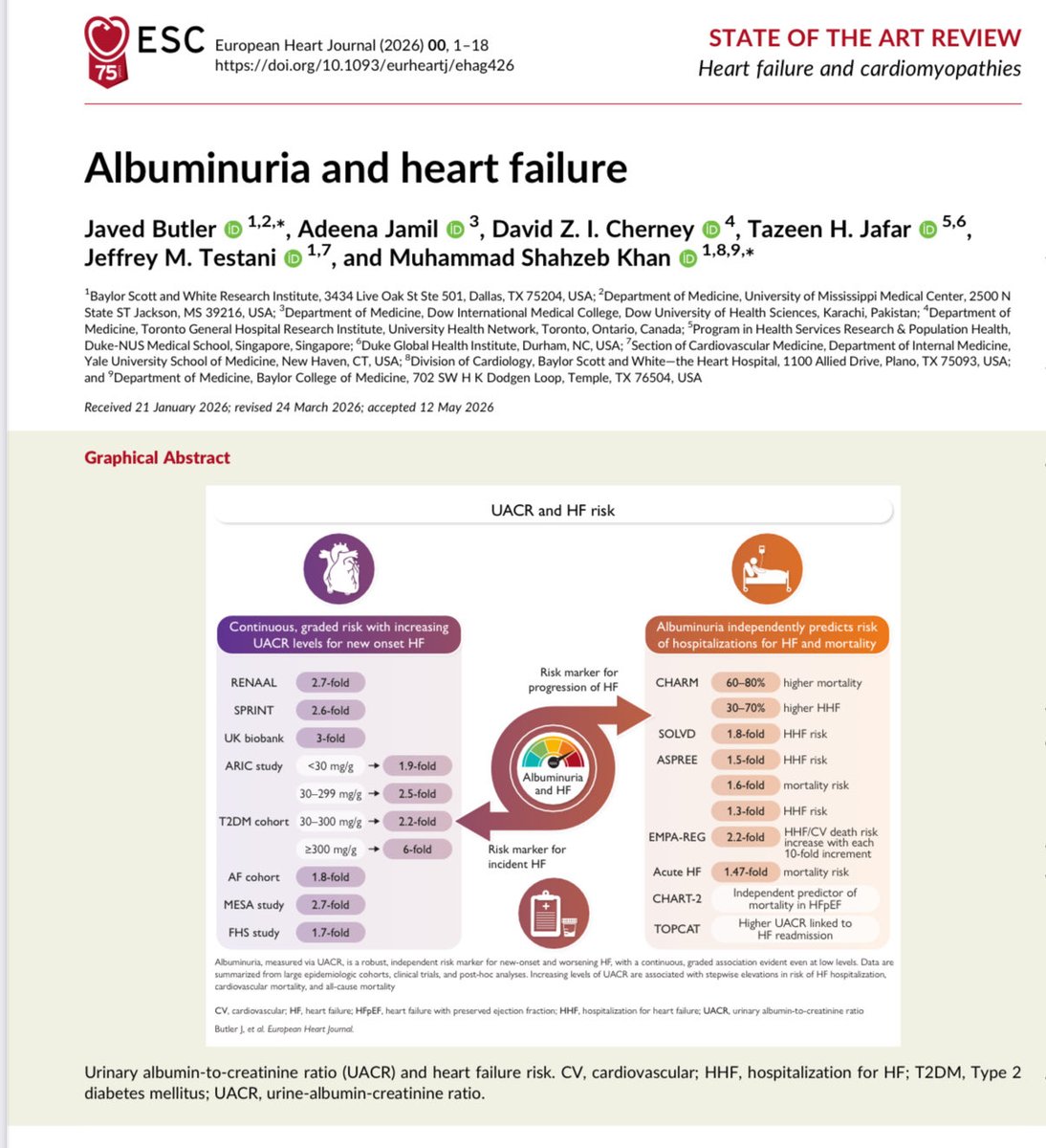
✅Albuminuria and Heart Failure- published in @ESC_Journals
📌UACR screening is important for early detection, risk stratification and intervention to improve outcomes
@JavedButler1
https://t.co/0Y2NmaTSNS
🚨 Eggs were blamed for heart attacks for decades. The American Heart Association told millions of Americans to avoid them. But the data says otherwise.
And no, eggs are not just "dietary cholesterol bombs" waiting to kill you.
🩺 Here is what actually happened. In the 1960s, scientists linked dietary cholesterol to serum cholesterol and assumed eggs were dangerous. That assumption drove 40 years of public health guidance. Millions of Americans switched to egg whites, egg substitutes, and low-fat breakfast options. Heart disease kept climbing anyway.
🔬 The mechanism matters here. When you eat dietary cholesterol, your liver compensates by producing less of its own. For roughly 70% of the population, eggs cause minimal change in LDL. The remaining 30% are called hyper-responders. In those individuals, eggs raise both LDL and HDL simultaneously, leaving the LDL to HDL ratio largely unchanged.
💓 The trial data tells a clearer story.
✅ PREDIMED (olive oil and Mediterranean diet): populations eating whole eggs as part of a whole-food diet showed no increase in cardiovascular events.
✅ The Nurses Health Study and Health Professionals Follow-Up Study tracked over 200,000 people for decades. Eating up to one egg per day showed no significant association with cardiovascular disease in healthy adults.
✅ A 2020 meta-analysis published in the BMJ covering 1,720,108 participants found no significant link between moderate egg consumption and coronary artery disease or stroke.
🫀 What eggs actually contain matters. One whole egg delivers 6 grams of complete protein, 147 milligrams of choline critical for brain and liver function, lutein and zeaxanthin for eye health, vitamin D, B12, and selenium. The yolk is where the nutrition lives. Throwing it away to avoid cholesterol is trading real nutrients for a fear that the data never fully supported.
⚠️ Context is everything though. The problem was never the egg. The problem was the dietary pattern surrounding it. Eggs eaten alongside processed meats, refined carbohydrates, and excess sugar create a metabolic environment that drives cardiovascular risk. Eggs eaten inside a whole-food, fiber-rich, low-processed diet do not carry the same signal.
🔸 If you have type 2 diabetes, the data is more nuanced. Some studies show a modestly higher cardiovascular signal with very high egg intake in diabetic populations. I counsel those patients individually based on their full lipid panel, ApoB, and insulin resistance status.
🩺 I am a board-certified cardiologist practicing preventive cardiology.
I have reviewed lipid responses to dietary changes in hundreds of patients. When patients cut processed food and keep whole eggs, I watch their metabolic markers improve, not worsen.
A patient who replaces a processed breakfast cereal with two whole eggs, vegetables, and avocado can see measurable improvements in fasting insulin and triglycerides within 8 to 12 weeks. That is the difference between a breakfast that spikes blood sugar and crashes energy by 10am and one that keeps glucose stable and supports satiety for hours.
❤️ Bottom line:
Eggs are not the villain. The dietary cholesterol hypothesis that buried them was built on incomplete science.
The evidence now covers millions of patients across decades of follow-up. Moderate egg consumption in healthy adults does not drive heart disease.
Eat the whole egg. Prioritize what surrounds it on your plate. Fix the processed food first. Track your ApoB and particle number, not just total cholesterol.
The question is no longer whether eggs raise cholesterol. The question is what your overall dietary pattern is doing to your cardiovascular risk.
This is why patients need to stop taking 1960s nutrition advice as gospel.
#Cardiology #HeartHealth #HeartDisease #CardiovascularHealth #DietaryCholoesterol #EggNutrition #LipidHealth #Nutrition #PreventiveCardiology #MetabolicHealth
🚨 A new study found that people doing time-restricted eating had a 91% higher risk of cardiovascular death compared to those eating across a normal window.
Intermittent fasting is trending as the ultimate biohack.
But the data says otherwise.
💓 Here is what the science actually says.
The American Heart Association presented preliminary data from a study of over 20,000 adults. People who compressed their eating into fewer than 8 hours per day had a 91% higher risk of dying from cardiovascular disease compared to those eating across 12 to 16 hours daily.
That is not a small signal. That is a red flag.
⚠️ And it gets worse when you look at the full picture.
🔬 What the data shows beyond that one headline number:
✅ Muscle loss is real. Time-restricted eating without adequate protein and resistance training accelerates lean mass loss. Sarcopenia is an independent cardiovascular risk factor.
❌ Fat loss from fasting is not superior to standard caloric restriction. When calories are matched, the window does not matter. The CALERIE trial proved this.
❌ Cortisol spikes from prolonged fasting increase vascular inflammation. Chronic cortisol elevation drives endothelial dysfunction.
❌ Skipping breakfast specifically is associated with higher LDL, higher blood pressure, and worse glycemic control in multiple observational datasets.
🔸 People with existing cardiovascular disease, diabetes, or metabolic syndrome face disproportionate risk from aggressive fasting protocols.
🔸 Electrolyte dysregulation during extended fasting windows triggers arrhythmias in susceptible patients.
🔸 Disordered eating patterns frequently emerge from rigid fasting schedules, and psychological stress is a direct cardiovascular risk factor.
🫀 I am a cardiologist. I see patients daily who arrive convinced that fasting is their path to heart health. They are losing muscle, spiking cortisol, skipping medications because they fall outside the eating window, and calling it optimization.
It is not optimization. It is risk.
❤️ Bottom line:
Intermittent fasting is not the cardiovascular miracle the wellness industry sold you.
The 91% increased risk of cardiovascular death is preliminary data, but it is consistent with the mechanistic concerns I have raised with patients for years.
The tools with the strongest data are unsexy, free, and require your participation.
Move your body. Eat enough protein. Sleep 7 to 9 hours. Control your LDL, blood pressure, and blood sugar. Take your medications.
A patient who builds those four habits consistently can cut their 10-year cardiovascular event risk by more than 50% without fasting a single day.
That is the real story.
The question is no longer whether fasting is trendy. The question is whether you are optimizing for optics or optimizing for outcomes.
#Cardiology #HeartDisease #HeartHealth #CardiovascularHealth #IntermittentFasting #TimeRestrictedEating #MetabolicHealth #HeartRisk #PreventiveCardiology #LifestyleMedicine
El tamaño de tu abdomen y estatura
SI IMPORTAN
📢 El diagnóstico de #Obesidad se amplía, con base en el nuevo capítulo de Tamizaje y Diagnóstico de los Standards of Care de la American Diabetes Association (ADA)
⚖️ IMC > 30 = Obesidad (sin cambios)
📏 IMC > 25 + relación cintura/estatura > 0.5 = Obesidad
🌎 IMC > 27.5 en personas de origen asiático = Obesidad
🌎📏 IMC > 23 en personas de origen asiático + relación cintura/estatura > 0.5 = Obesidad
@StratCons@doctormacias@DrMauinforma@docramiro@Obesidades_mx@WorldObesity
https://t.co/KQE4k4MKNf