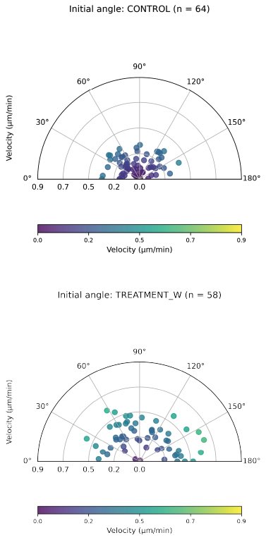
How can we analyze and visualize axonal growth dynamics and guidance?
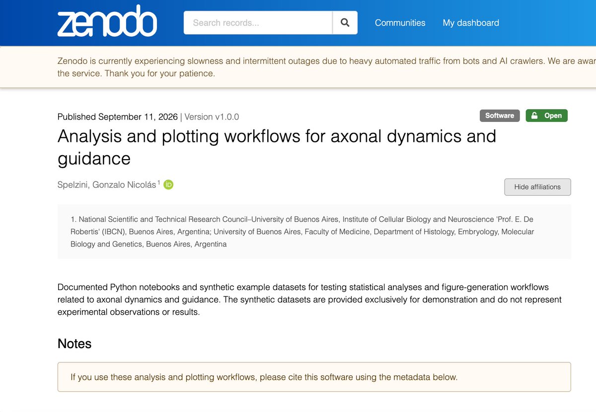
I’ve shared a collection of documented Python notebooks on GitHub for statistical analysis and figure generation related to axonal dynamics and guidance.
Archived release on Zenodo: https://t.co/upqEJ0hqea
I am very happy to share that in March 2026 I successfully defended my PhD thesis in Basic Medical Sciences at the University of Buenos Aires, receiving honors. This achievement closes an important chapter and strengthens my commitment to research and teaching in neuroscience.
A speck in a dish—that’s all the nervous system of the fruit fly Drosophila melanogaster looks like to the naked eye.
But look closely enough, and it resolves into a network of 166,700 nerve cells connected through 124.2 million synapses.
The intricate map—called a connectome—has now been published, marking the end of an ambitious 20-year research project. Learn more: https://t.co/EKsDRQOgAS @NewsfromScience
New issue is out👉https://t.co/uP2kI6DjtL
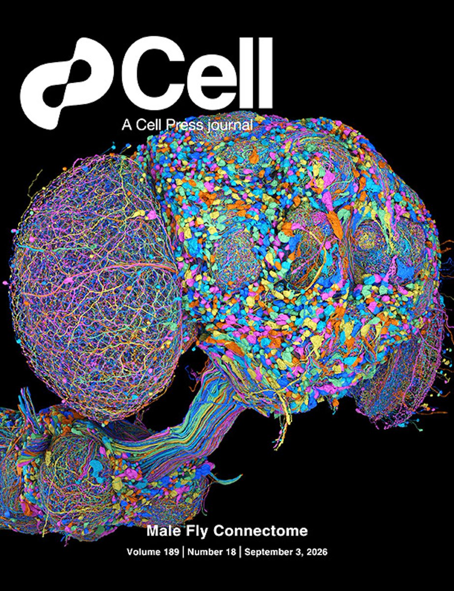
On the cover: the complete connectome of the male adult D. melanogaster central nervous system. The cover depicts a single representative cell from each of the 11,710 neuron types in the dataset, which contains 166,700 neurons in total.
Inspired by the cell network of the retina, a new photonic computing system described in @ScienceAdvances can rapidly learn to solve complex visual tasks—from classification to image segmentation—from very restricted datasets. https://t.co/enRyRVgzlI
Click anywhere on the brain and see that region's projections.
Fibers are diffusion tractography (dMRI) from Human Connectome Project
https://t.co/Fl4O6YnbiB
How do our brain cells interact with each other to pull off the symphony of activity needed to solve problems, create or learn? The Simons Collaboration on the Global Brain has investigated these questions for over a decade: https://t.co/qveGzrrXuT
#science#neuroscience
#MedNews: A major push to complement and replace animal testing with “human-based” models like organoids is underway.
These tiny organ-like structures grown from human cells are being used to model disease and study human development, identify and screen new therapeutics, test chemical or drug toxicity, and evaluate drug response.
And in what some call a “watershed moment,” the biomedical field is pairing these laboratory specimens with increasingly powerful computational tools.
Learn more: https://t.co/qIxcrH0q9e
The brain may compare its predictions to actual sensory input by having different brainwave frequencies interact with each other (fast waves nested in slow waves). It acts like a built-in error-detector.
https://t.co/YAsM4Jog9s
#neuroscience
Nearly 400 Argentine researchers have been left jobless and uncertain about their future in science after a delay in the process for securing permanent research positions in the country.
On 12 August, researchers staged protests around the country, including in downtown Buenos Aires, calling on the National Scientific and Technical Research Council, Argentina’s main science agency, for support.
The case is one of the most visible consequences of the budget cuts implemented by President Javier Milei’s government, which have caused the loss of thousands of jobs in the science sector. Public investment in science and technology is at its lowest level in 54 years.
Learn more: https://t.co/fCHvXacabG @ScienceCareers
Today's AI systems learn mostly by passively absorbing data, but the brain builds intelligence in the reverse order.
Grounded world models in biological organisms and future embodied AI
https://t.co/jIj7bfeoN7
#neuroscience
Video of myosin proteins "walking" along actin. Insane that we can image this!
It was made by Toshio Ando's group using atomic force microscopy. All of their papers are a goldmine; they've also recorded a Cas9 protein cutting DNA and ATP synthase spinning.
Our paper "Highly attenuated dendritic propagation of isolated synaptic potentials in vivo", demonstrating that individual synaptic potentials in vivo do not reach the soma, is published in Science Advances: https://t.co/8w0Bz8cUEb
High-speed microscopy reveals electrical activity across the brain. New technique, demonstrated in zebrafish, could help scientists learn how the entire brain works as a whole. https://t.co/gapyEigGja #neuroscience
Engineers in Ed Boyden's lab have developed a microscope that captures electrical activity across an entire zebrafish brain at millisecond speed—revealing how neurons coordinate as an emergent whole. https://t.co/zPvWfhnRs7
An emerging body of work shows that glial cells act as leading architects of the neural extracellular matrix, which serves as a critical signaling hub for nerve synapses and neuronal circuits, according to a new Focus in @SciSignal. https://t.co/jY3vIDmEP0
Memories can survive even after the brain temporarily loses more than half of its synaptic connections, according to a new mouse study in Science, which challenges the long-held view that long-term memories depend on stable individual synapses.
Learn more: https://t.co/6PbiaMd1F8