Today in @Nature, we report MouseMapper: foundation-model AI to map disease perturbations across the entire mouse body cell-by-cell.
In obesity, it revealed body-wide inflammation & unexpected facial nerve damage. 🧵👇🔉
https://t.co/BERf5GQ10Z led by @Dorie00 & @yingchen733
Excited to share our preprint, reviving @kristyredhorse's interest in the placenta from her PhD ~20 years ago! We find using 3D imaging that the mouse placenta is far more invasive than previously thought. Furthermore, we find a surprise from our favorite chemokine CXCL12...🧵:
A century ago, “vitamin hunters” discovered micronutrients. Today, vitamins are taken adhoc. We revisited this with modern genetics: CRISPR screens -> new NAXD disease mouse -> over 40× lifespan increase w/ vitamin B3. Huge credit to Ankur & Skyler! https://t.co/0cRmrng7ux
🧵👇
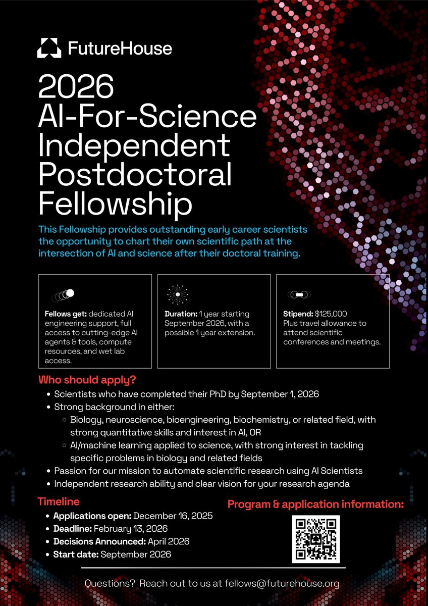
We are opening applications for our 2026 cohort of FutureHouse AI-for-Science Independent Postdoctoral Fellows! Apply our AI tools to specific problems in biology and biochemistry, in collaboration with world-leading academic labs:
--$125,000 annual stipend.
--Access to all tools developed by FutureHouse and Edison Scientific at scale, including Kosmos and several as-of-yet unreleased agents, with under-the-hood access to them to specialize them for your workflows.
--Receive dedicated software engineering support.
--1 year with possible 1 year extension.
Even more exceptional co-advisors than last year. Deadline for applications is February 13th, 2026. Link in next post.
Announcing our new preprint! We built SPICE, a framework that combines large-scale experiments and generative AI to design RNA sequences that control cell type-specific gene expression using alternative splicing - a powerful new modality! (1/10)
Preprint: https://t.co/rlSdgBXGZ9
When and how do different tissue physical structures deteriorate during aging (structural aging)? What molecular changes occur in tissues during periods of major changes?
Are there tissue-specific periods of accelerated structural aging? Which organs age early vs. late?
Is there cross-organ coordination in structural aging? And, finally, how lifestyle, diseases and genetics impact these organ-specific trajectories?
While important, these questions haven’t been answered yet because we lack structural and molecular data from normal aging tissues at scale.
We present a framework taking the first stab at scale at these questions using high resolution histology images and omics (+more) from 25,000 post-mortem tissues (public data: GTex).
We reason that structure determines function and learning how tissue structure changes with age can help us understand the process of aging in different tissues - a central question with yet little understanding. An example is to visualize these two ovaries histology: young vs. old ovary- young ovaries cortex is intact, with follicles, no fibrosis - basis of its function (partially).
First, we extract tissue structure from these organs using their high-res histology images using a pre-trained digpath foundation model (UNI) and asked how much they change with age (Structural Aging Rate)?
As an example, the ovary has two peaks around the late 30s and then around 55. First aligns with accelerated follicle loss and second is just after post-menopause.
This shows that change in morphology of the ovary captures its functional milestones during aging with no training.
Bonus Puzzle: Does anyone know how these two functional milestones of ovary were originally found in the last century?
What if we repeat the same analysis on bulk-omics, transcriptomics and methylation, from the same samples? Can they capture this bimodal functional decline? No. (read the paper of our explanation)
Can molecular clocks trained on chronological age track this? No. They assume aging is linear.
Note: Unlike molecular clocks trained on chronological age, PathStAR learns with no age labels. We simply ask: When and how does tissue morphology change most rapidly during life?
🚨 New preprints from our lab!
First, we introduce Cryo-mtscATAC-seq, led by Maren (@ms-maren.bsky.social), enabling high-throughput clonal tracing from frozen human samples by isolating nuclei with their mitochondria (“CryoCells”).
👉 https://t.co/0mD7JaS30T
Check out this cool work led by @nithyabhasker@mweber_PU where we develop Contrastive Poincaré Maps (CPM), a self-supervised hyperbolic encoder for scRNA-seq that preserves hierarchies & scales efficiently!
Single-cell data can reveal hierarchical patterns in organismic development but popular embedding approaches often distort them. We introduce Contrastive Poincaré Maps, a self-supervised hyperbolic encoder that preserves hierarchies, scales efficiently, and uncovers developmental lineages across diverse datasets.
Led by @nithyabhasker. Preprint here: https://t.co/Q3w5skFvxN
Building on the legacy of many, I'm incredibly excited that we have successfully translated our polygenic risk scores into a validated clinical assay @MassGenBrigham@broadinstitute, orderable by any clinician in the US today.
🧬To order: https://t.co/pivEDisCZR
🧬Announcement: https://t.co/V3GJaa4SHf
The CVD PRS panel is already expanding, and panels in other disease areas are in development. Single sequencing for multiple disease areas.
Methods:
https://t.co/YkPzcgSC8A @CellGenomics
https://t.co/hezzuwNTvZ @NatureComms
[1/4]


![pnatarajanmd's tweet photo. Building on the legacy of many, I'm incredibly excited that we have successfully translated our polygenic risk scores into a validated clinical assay @MassGenBrigham @broadinstitute, orderable by any clinician in the US today.
🧬To order: https://t.co/pivEDisCZR
🧬Announcement: https://t.co/V3GJaa4SHf
The CVD PRS panel is already expanding, and panels in other disease areas are in development. Single sequencing for multiple disease areas.
Methods:
https://t.co/YkPzcgSC8A @CellGenomics
https://t.co/hezzuwNTvZ @NatureComms
[1/4]](https://pbs.twimg.com/media/Gz8K76JXgAAAXjh.jpg)
![pnatarajanmd's tweet photo. Building on the legacy of many, I'm incredibly excited that we have successfully translated our polygenic risk scores into a validated clinical assay @MassGenBrigham @broadinstitute, orderable by any clinician in the US today.
🧬To order: https://t.co/pivEDisCZR
🧬Announcement: https://t.co/V3GJaa4SHf
The CVD PRS panel is already expanding, and panels in other disease areas are in development. Single sequencing for multiple disease areas.
Methods:
https://t.co/YkPzcgSC8A @CellGenomics
https://t.co/hezzuwNTvZ @NatureComms
[1/4]](https://pbs.twimg.com/media/Gz8IpFsXUAEzfsG.jpg)


![pnatarajanmd's tweet photo. Building on the legacy of many, I'm incredibly excited that we have successfully translated our polygenic risk scores into a validated clinical assay @MassGenBrigham @broadinstitute, orderable by any clinician in the US today.
🧬To order: https://t.co/pivEDisCZR
🧬Announcement: https://t.co/V3GJaa4SHf
The CVD PRS panel is already expanding, and panels in other disease areas are in development. Single sequencing for multiple disease areas.
Methods:
https://t.co/YkPzcgSC8A @CellGenomics
https://t.co/hezzuwNTvZ @NatureComms
[1/4]](https://pbs.twimg.com/media/Gz8LEYOWoAUIEjL.jpg)


