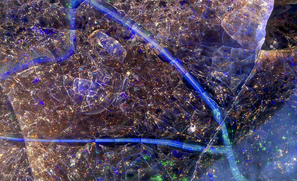
MIT research scientist and science photographer Felice Frankel has released a book, Phenomenal Moments, showcasing striking images of natural phenomena that invite viewers to uncover the scientific processes at work. Read more in @PopSci and @NextAvenue: https://t.co/7vHUxH8RXp
Our PLAMseq paper is out today in @ScienceAdvances . We developed a TurboID-based protein-genomic approach to map proteins and protein interactions in the genome. Validated with CTCF and RNApolII. Applied to map Histone H1 SUMOylation genomic loci.
https://t.co/7wDxkMLt2E
A team of researchers at the University of Victoria (UVic) have achieved a major breakthrough in electron microscopy that will allow scientists to visualize atomic-scale structures with unprecedented clarity using lower-cost and lower-energy microscopes than ever before.
Led by Arthur Blackburn, co-director of UVic’s Advanced Microscopy Facility, the team developed a novel imaging technique that allowed them to achieve sub-Ångström resolution (less than one ten-billionth of a meter) using a compact, low-energy scanning electron microscope (SEM)—a feat previously possible only with a large, high-cost transmission electron microscope (TEM).
https://t.co/gxdHUaAKxT
🌱 Excited to share my new personal academic website! 👉 https://t.co/jjIyiOFxEO
Inspired by the Nature Index article "Build-your-own website for scientists", I wanted to create a hub to present my research & foster collaborations.
Following on from their successful meeting in 2023, @J_Cell_Sci are delighted to announce a second iteration of the Imaging Cell Dynamics meeting to be held in 2026. Find out more and sign up for registration and abstract updates at
https://t.co/JOemNCgNQF
#JCSimaging
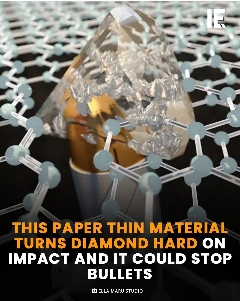
Researchers at CUNY have developed diamene, a groundbreaking material formed by stacking two graphene layers. Under sudden pressure, diamene transforms into a diamond-like structure, offering exceptional hardness while remaining incredibly thin and flexible. This innovation holds potential for revolutionizing protective gear, providing lightweight yet robust defense against high-impact forces.
For more content like this, please visit: https://t.co/rX1UfTwie0
#Diamene #GrapheneInnovation #MaterialsScience #BulletproofTechnology #CUNYResearch
So cool - Yang et al. used microfabrication techniques to make microscale and nanoscale patterns on frozen tardigrades before successfully reviving them!
https://t.co/tgkiLJP4J0
Experiments and simulations of DNA nanostar hydrogels reveal that microscopic topology determines macroscale elasticity in amorphous networks. @Dmichiel1
https://t.co/SvPKuRQuPw
Recent data suggests microbes form nanotubes with neighbors. This occurs not only between members of the same species, but also between different species.
Other research, though, suggests that nanotubes only emerge from DYING cells. So who's right, and how might we find out? 🔻
Happy #Newtonmas! Isaac Newton was born Christmas Day, 1642. The apple tree in MIT's President's Garden is a direct descendant of the actual tree believed to have inspired Newton's theory of gravity.
Here’s a great one-tube, four-step, 5–15 min DNA extraction method for on-bead PCR using alkaline polyethylene glycol (PEG) and paramagnetic beads. It could be very useful for point-of-need testing or other rapid field research.
The method, by Lee et al. (2024), is called ASAP (Abridged Solid-phase extraction with Alkaline Polyethylene glycol lysis). It was developed for the detection of bacteria in saliva, sweat, and urine, and it simultaneously lyses cells and binds DNA to magnetic beads.
Once the DNA is bound the reagent can be removed (leaving the magnetic beads) and a PCR mix can be added directly to the tube without washing. It’s reportedly capable of detecting single copies of E. coli DNA in 20 µL biological fluid when using qPCR.
ASAP works by exploiting the fact that PEG is the main component of the alkaline PEG DNA extraction method, while also being a major component of the binding buffer in magnetic bead cleanup suspensions.
PEG’s role in the alkaline PEG lysis method is to induce a molecular crowding effect that increases the pH (improving cell lysis), but it allows the pH to drop significantly when the reagent is diluted, making it suitable for direct PCR.
Similarly, PEG and NaCl are used together as a crowding agent that reversibly binds DNA to the coatings of paramagnetic beads.
By using the same PEG for both extraction and binding (15% PEG-8000), and by including an alkali (3.5 mM KOH) and paramagnetic beads in the same reagent, cells can be lysed and DNA bound during a single room-temperature incubation. Sensitivity can be increased by extending the incubation time from 5 to 15 minutes.
The authors found that no paramagnetic bead washing step was necessary due to the compatibility of the reagent with qPCR, simplifying the procedure, saving time, and reducing opportunities for DNA loss.
In their tests, the authors found ASAP to have 10x the sensitivity over commercial kits and 100x sensitivity over the original alkaline PEG extraction method, detecting as few as 15 colony-forming units in 50 uL of saliva, sweat, and urine.
The ASAP reagent consists of 15% PEG-8000, 0.5 M NaCl, and 3.5 mM KOH, with 1.5 µg paramagnetic beads per 20 µL PCR for optimised sensitivity. KOH is added separately to the reaction tubes to avoid storage issues. So it’s easy to source the reagents, and it’s easy to make.
Its ease of use, low cost, and quick extraction also compare favourably with other methods based on the World Health Organisation’s REASSURED criteria for point-of-need nucleic acid amplification testing.
The researchers are now investigating how the starch-based nanofibers could be used for medical purposes such as wound dressing, for scaffolds in tissue regrowth and even in drug delivery https://t.co/8yynWc3qXS