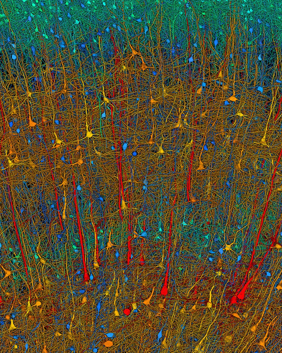
Using more than 1.4 petabytes of electron microscopy (EM) imaging data, researchers generated a nanoscale-resolution reconstruction of a millimeter-scale fragment of human cerebral cortex, providing an unprecedented view into the structural organization of brain tissue at the supracellular, cellular, and subcellular levels.
The human brain is a vastly complex organ and, to date, little is known about its cellular microstructure, including the synaptic and neural circuits it supports. Disruption of these circuits is known to play a role in myriad brain disorders. Yet studying human brain samples in such great detail comes with a host of challenges, ranging from technological limitations to the availability and preservation of tissue samples from healthy individuals.
In a 2024 Science study, researchers performed a high-resolution EM reconstruction of the ultrastructure of a cubic millimeter of human temporal cortex. According to the authors, the reconstruction contains roughly 57,000 cells, about 230 millimeters of blood vessels, and nearly 150 million synapses, comprising 1400 terabytes of data. The authors generated a three-dimensional reconstruction of nearly every cell and process in the cubic millimeter sample and developed a freely available tool for visualizing and analyzing the vast dataset.
Learn more on #WorldBrainDay: https://t.co/ye8JncNkrJ
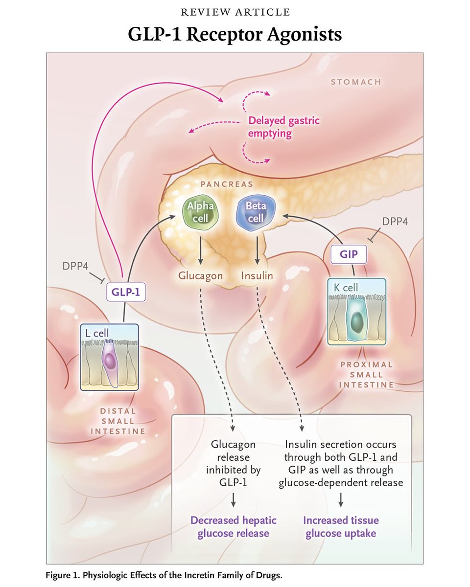
GLP-1 receptor agonists are increasingly used to treat type 2 diabetes and obesity, and trials have shown reductions in cardiovascular risk and slowing of kidney failure. Adverse events are mostly gastrointestinal.
Read the Review Article “GLP-1 Receptor Agonists” by Clifford J. Rosen, MD, and Julie R. Ingelfinger, MD, from @tuftsmedschool and the Maine Medical Center Institute for Research: https://t.co/D0XSLv7U8n
@NTFabiano I apologize, but being a scientist requires not only motivation but also knowledge and technical skills to provide quality information. My degree is more than a fancy document.
Two 2025 studies confirm it:
In some people, COVID leaves parts of the brain functionally switched off.
PET and EEG both show disruptions in regions that control attention, motivation, and emotion.
What that actually means🧵
Metabolic brain changes in post-acute COVID-19: systematic review and meta-analysis of [18F]-FDG-PET findings | Brain Structure and Function https://t.co/UsUKn1lcYP
Belo Horizonte researchers reviewed 584 brain scans and found consistent glucose hypometabolism in the frontal cortex of long COVID patients, linking it to cognitive symptoms and urging more longitudinal PET studies
https://t.co/Q8mpfDAv2r