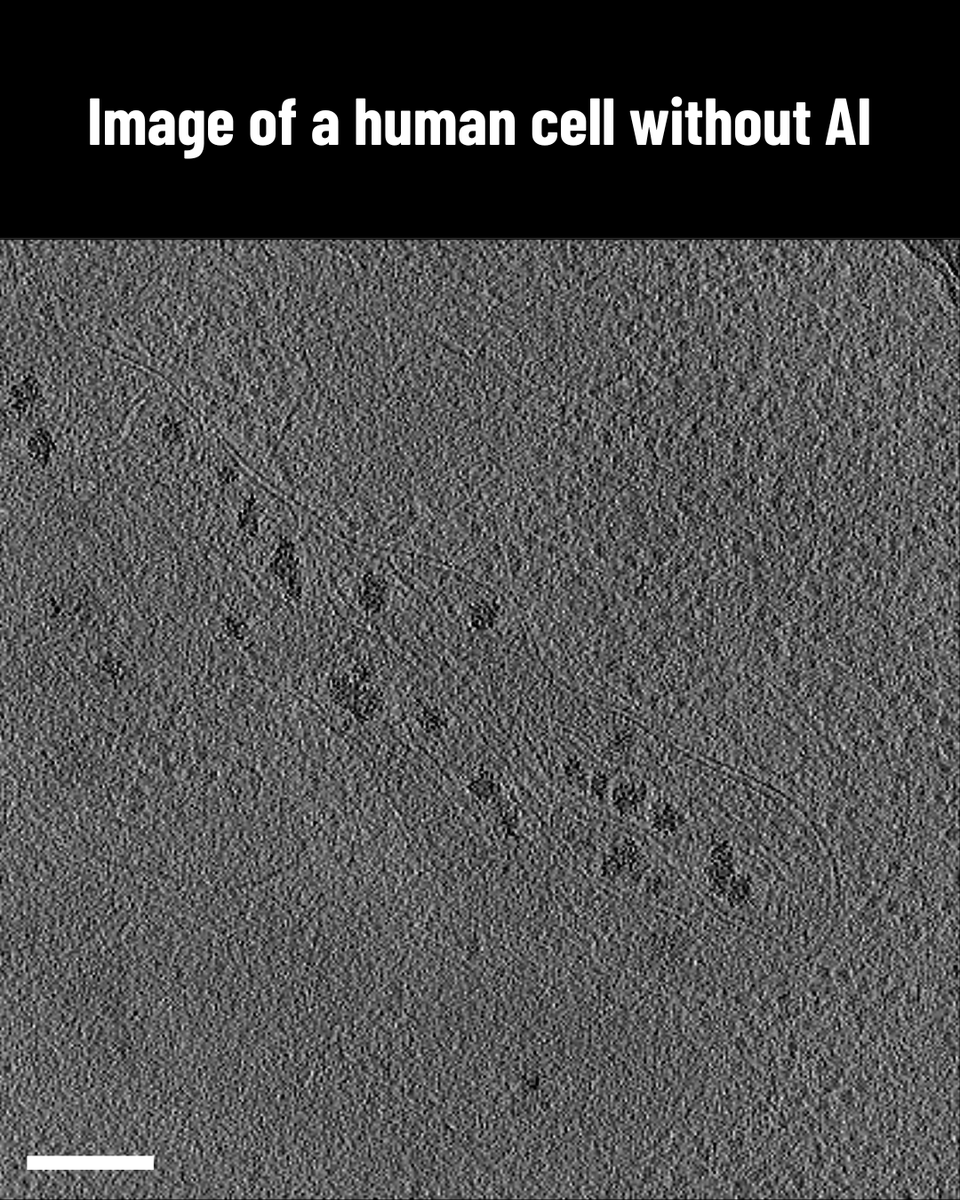
„My finger hurts from clicking all those membrane proteins! 😩”
– A sentence you will never have to say again.
Check out the preprint about MemBrain 💻🧠: Our #DeepLearning -aided tool for membrane protein detection in cryo-ET.
https://t.co/Jhpmvx8fM3
🧵How it works: 🧵 (1/N)
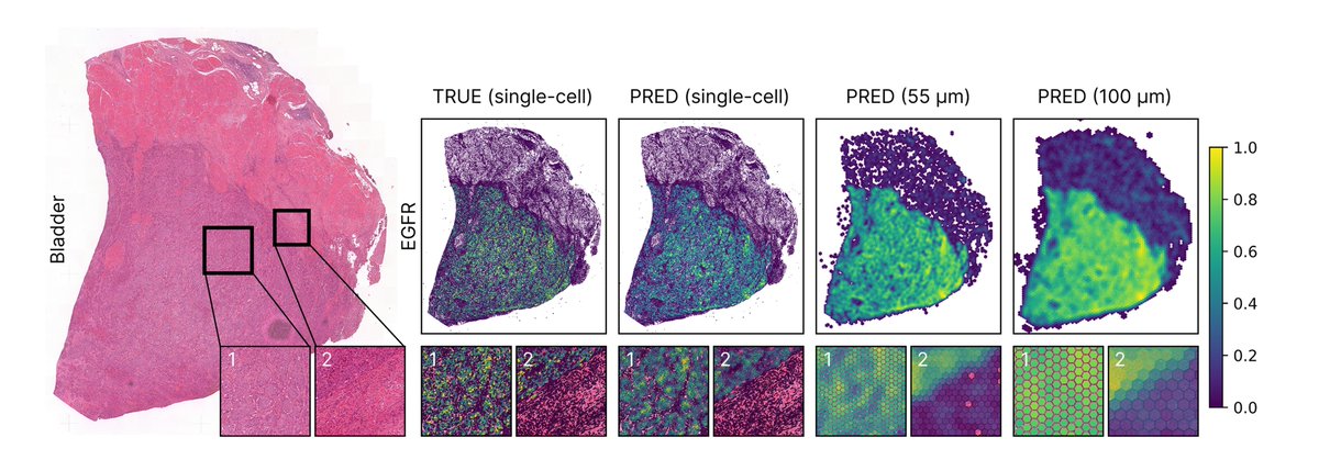
1/7🎉 Introducing Phoenix: generative AI predicting spatially resolved gene expression from routine H&E slides-enabling virtual spatial-omics. Zero-shot. Across cancers, organs & species.
Huge thanks to my co-first @TranManuel & all collaborators! 🙌
📜: https://t.co/MtmnyHfWZn
You want to start tomography? Solve structures inside cells? Reach Nyquist😳?
We have a website for you! https://t.co/ulvbbhEY6d
You'll find a tutorial on how to reconstruct tomograms, pick particles and do subtomo averaging, using different software!
Hope it will be useful !
Excited to share nnInteractive: The nnU-Net moment for 3D Interactive Segmentation! Supports points, scribbles, boxes, & a new lasso prompt for full 3D masks from 2D inputs. Check it out: https://t.co/MyNQyUDd4x
https://t.co/waDcltB9dG
https://t.co/uWoqHI3Ruf
A new pre-print for LSFM lovers in the bioimaging community: https://t.co/GJb6u7C46a w. python + napari. We would love to hear your feedbacks. Thanks for the Peng Lab in Helmholtz Munich and the Huisken Lab in Göttingen for having us along the journey in this great collaboration.
Excited to share TITAN, a multimodal foundation model for pathology trained on >330k WSIs, synthetic captions, and reports🔬⚡
co-lead w @TongDing99@GreatAndrew90@richardjchen
Thanks to @AI4Pathology@p_tingying!
Preprint: https://t.co/LpgmNlbvMa
Model: https://t.co/gYJxLcjKiS
🥳New cryo tomography Preprint ☀️🌱🔬Molecular architecture of thylakoid membranes within intact spinach chloroplasts.
https://t.co/ePKx2jhRYD
Proud to be part of this great work with @bengeliscious@woj_wie@LorenzLamm@DrLornaMalone @whjwood @PPS_UoS
We present MultiOrg: an #organoid dataset tailored for #object_detection tasks with #uncertainty quantification. Over 400 high-resolution 2D #microscopy images and multi-expert annotations of more than 60,000 organoids.
Paper: https://t.co/39WjGkSVF4
Data: https://t.co/64vBOSvPGi
SpotMAX, our software tool for analysing multi-dimensional microscopy data, is finally out! 🎉 And conveniently, I just presented it at the #I2K conference 😀
A multi-year-long effort of many great collaborators and users. But what can SpotMAX do? A small thread
Tingying Peng (@p_tingying) of @HelmholtzMunich presented MemBrain v2, “an end-to-end tool for analysis of membranes in cryo-electron tomography” #FEMPL3
I’m thrilled to introduce MemBrain V2(https://t.co/YLuu6oKEqt), our latest research tool developed by @LorenzLamm and co-supervised by @bengeliscious, right here at the beautiful Princeton campus. It’s exciting to see how widely it’s being adopted by the community!
1/ 🧵Introducing UNICORN,🦄 the first #AI model for integrating multi-stain #histopathology images for classification, designed to tackle missing data during both training and inference. Great colab between @HelmholtzMunich & @TU_Muenchen German Heart Centre! the architecture ⬇️
Really happy to share that the last chapter of my submitted phd thesis is already on biorxiv!🥳
Using a tophat transform, I made template matching more precise, allowing automated annotation with little false positives! #teamtomo
https://t.co/Y7K8l3fSCk
Super excited to share our latest work on the native in-cell organization of the mitochondrial respiratory chain 🥳
Using cryo-electron tomography🔬, we show how the respiratory complexes (and other complexes) are organized inside native mitochondria!
https://t.co/cxHl1j20CJ
⚡️ Excited to announce that CoLIE has been accepted to #ECCV2024!
CoLIE uses a neural implicit representation to enhance low-light images according to the Retinex theory.
@p_tingying@ja_schnabel@eccvconf
Paper: https://t.co/EHwJEJpavg
Code: https://t.co/K2ioykLMKL
The future is bright! We're going to make amazing discoveries about how cells work, and that means better understanding of health, disease, and even how we age!
Thanks to @ErmelUtz , @LorenzLamm , @bengeliscious for their amazing work.
Paper: https://t.co/O7ZSIRDlMd
It can take months to annotate a set of tomograms captured by #CryoET manually.
@ErmelUtz adapted @lorenzlamm + @bengeliscious’ deep-learning algorithm MemBrain to annotate cell membranes in over 13k tomograms in just 3.5 days
https://t.co/on1WyjDsPt
These are 3D images of the inner workings of human cells. Because they are so detailed, each set can take months to analyze.
To accelerate discovery, #CZImagingInstitute scientists applied @lorenzlamm + @bengeliscious’ AI technique that could analyze 13k+ images in just 3.5 days