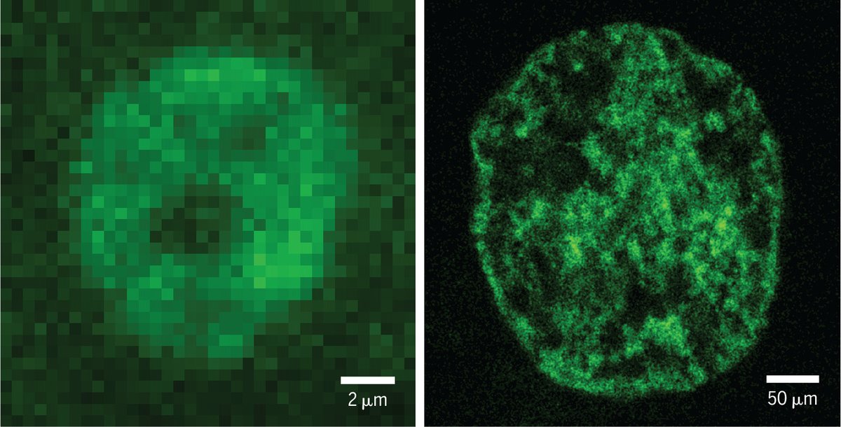
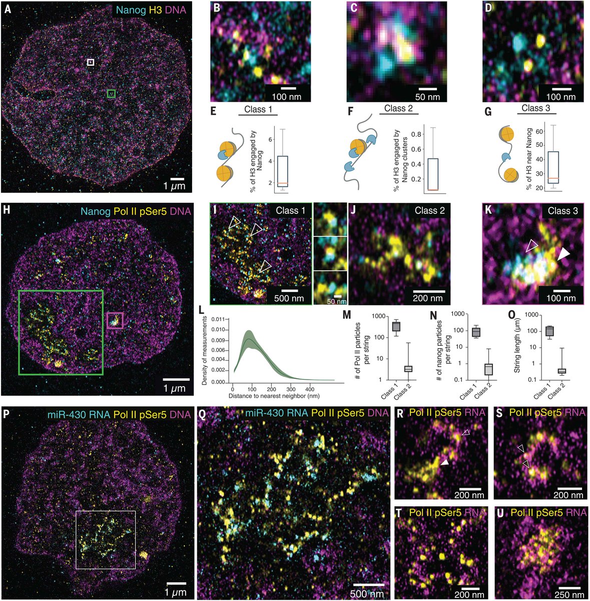
Our work describing Chromatin Expansion Microscopy is now out in @ScienceMagazine https://t.co/ysL8Q1xmqA Check out how we were able to visualize transcription, chromatin, and TFs with molecular resolution in developing zebrafish embryos to understand gene regulation!
First preprint from the lab 🚨
How can enhancers regulate target promoters across vast genomic distances, and what is the role of cohesin loop extrusion in the process?
https://t.co/l7bwPsTSkd
(1)
📣 Congrats to @GalJaschek, who was recently awarded a highly competitive PhD Fellowship from Boehringer Ingelheim Fonds. Gal is a @YaleGenetics graduate student in the @GiraldezLab. 👏 #TraineeTuesday [1/4]
We have open @ERC_Research-funded post-doc positions to study #3Dgenome organization and #generegulation. We are looking for talented and motivated candidates, but prior experience in the field is not required. Please RT!
https://t.co/Mms2UwDVVP
Breaking news! We're announcing the names of the six laureates of the International Birnstiel Award for Doctoral Studies in Molecular Life Sciences 2023! 🏆
Congratulations to all awardees and honourable mentions! 🎉 1/
➡️ https://t.co/7gjnvpbpT8
This week’s #FluorescenceFriday user is Mark Pownall! #HCRimaging allows @MarkPownall5 to better understand how individual molecules of transcription factors are organized in the context of a specific gene. Check out his recent publication here: https://t.co/kyNp1yDPMw
It took Sergey Ulyanov and I a while to shape fountains, a novel type of chromatin architecture. Here is our story of pioneering factors binding (@onichtchouk) and the loop extrusion (@leonidmirny) working together. (can you tell that I’ve been into fishkeeping since I was six?)
Technologies to model early mammalian embryo development in vitro have advanced rapidly, providing new insights into self-organization and environmental control. @BernaSozen_ and I wrote a review as part of a special issue on #earlyembryonicmodels: https://t.co/F8o9WaxTcJ (1/3)
A new form of expansion microscopy called ChromExM can be used to expand chromatin to visualize transcription at high resolution, according to new research in Science.
Learn more ⬇️
📄: https://t.co/fIb7aFv4AJ
#SciencePerspective: https://t.co/K6HSlLl3Me
Researchers apply #SuperResolution expansion #Microscopy#ExM towards imaging of chromatin in a developing embryo during zygotic genome activation, illuminating how Nanog interacts with nucleosomes and RNA polymerase II. Learn more: https://t.co/s6dxxUL2IT
🔬🧬 Excited to share our latest study https://t.co/kb3GfMrK32 @ScienceMagazine developing ChromExM which uses expansion microscopy to visualize the chromatin,
transcription, RNA POL II and pioneer factor Nanog during genome activation.
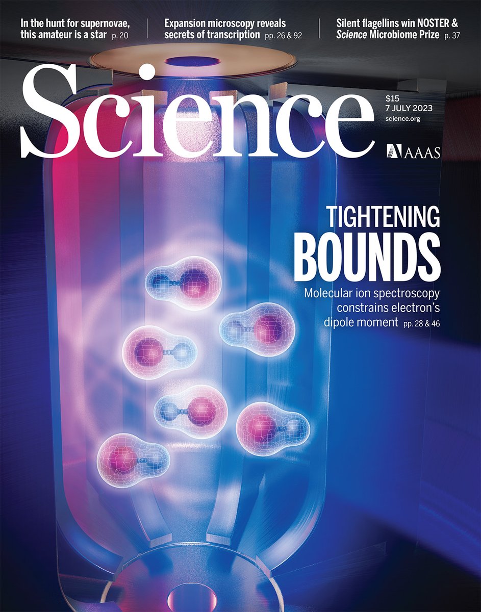
This week in Science, a study reports “the most precise measurement yet” of the size of the electron’s permanent electric dipole moment.
The results have implications for an important unresolved question in physics—the imbalance of matter and antimatter. https://t.co/GyuCXRk6Zl


![YaleGenetics's tweet photo. 📣 Congrats to @GalJaschek, who was recently awarded a highly competitive PhD Fellowship from Boehringer Ingelheim Fonds. Gal is a @YaleGenetics graduate student in the @GiraldezLab. 👏 #TraineeTuesday [1/4] https://t.co/4AmriLEY4X](https://pbs.twimg.com/media/GQ7CdbjWcAAwo2s.jpg)