⚡ Every neuronal signal has a story to tell.
From uncovering disease-related changes in neuronal activity to exploring the functional complexity of biological samples, high-resolution electrophysiology (HD-MEA) is opening new possibilities for neuroscience research.
The MaxWell Biosystems team is heading to Washington, DC, for Neuroscience 2026 (SfN 2026)! Join us to discover how our HD-MEA technology enables multiscale functional insights for disease modeling and drug discovery. Meet our scientific experts, explore the latest MaxLab Live analysis capabilities, and experience live demonstrations of our platforms and innovative accessories.
📅 November 15–18, 2026
📍 Booth #636 | Washington, DC, USA
Here is what you won't want to miss:
🔬 Organoids in Action | November 15–17, Daily at 2 PM | Booth #636
Three days, three exciting presentations showcasing new possibilities in brain organoid research! Discover innovative solutions for organoid plating, explore high-throughput functional pharmacology in 3D models, and watch with us "Organoid Sumi", where neuroscience meets creativity through art powered by brain organoids.
Also, do not miss the two poster sessions highlighting how HD-MEA technology is advancing functional phenotyping in brain organoids and human disease models.
📄 Dr. Laura D’Ignazio – Head of Commercial Excellence and Engagement
November 16, 1-5 PM | Poster LL60
A scalable HD-MEA workflow for multiscale functional phenotyping of human midbrain organoids
📄 Dr. Marian Hruska-Plochan – Scientific Business Developer
November 17, 8 AM - 12 PM | Poster V2
Multiscale HD-MEA profiling reveals early hyperexcitability and axonal dysfunction in a human iPSC-derived GRN-FTD model
🎉 And there's more! Try your luck at our spinning wheel, join us for a Run for ALS on November 18, and connect with fellow researchers over our signature cocktail at the SfN Mixer!
👋 Come meet our team to learn more or send us a message:
• Dr. Laura D'Ignazio – Head of Commercial Excellence & Engagement
• Dr. Tom Dufor – Field Application Scientist
• Dr. Urs Frey @u_frey CEO
• Elvira Guella – Account Manager
• Dr. Marian Hruska-Plochan @m_hruskaplochan Scientific Business Developer
• Dr. Marie Obien @engeleneo – Chief Commercial Officer
• Dr. Silvia Oldani @silvia_oldani Head of NAM
• Dr. Carlos Sanchez Priego – Field Application Scientist
See you in Washington, DC! 🇺🇸
🔗 https://t.co/mQfRKnE8s3
#SfN26 #Neuroscience #Organoids #Electrophysiology #HDMEA #MxWConnect
🧠 What if you could continuously record neuronal networks for five weeks without repeatedly disturbing the culture? Longitudinal recordings reveal how networks mature, but evaporation and medium exchanges can influence the very activity being measured.
In a new Advanced Science @AdvPortfolio paper, Maurer et al. demonstrate 35 days of continuous electrophysiological recordings with only one half-medium exchange, using human iPSC-derived neuronal networks patterned in PDMS microstructures on MaxWell Biosystems' MaxOne+ HD-MEA chips.
But how?!
The team developed a custom recording environment around the MaxOne High-Density Microelectrode Array (HD-MEA) System, combining an innovative water-compartment culture lid that effectively eliminates evaporation while preserving gas exchange with a temperature-controlled incubator.
Using the MaxLab Live Python API, they followed spontaneous neuronal activity over five weeks, revealing how firing patterns emerge and evolve during network maturation.
A great example of how HD-MEA technology and innovative bioengineering can push the boundaries of longitudinal neuronal recordings!
👉 Read the full publication here: https://t.co/V7sMVV0eYK
👏Congratulations to Benedikt Maurer, Fabio Fischer, Giulia Amos @amos_giulia, Vaiva Vasiliauskaitė @VVasiliauskaite , and János Vörös from the Laboratory of Biosensors and Bioelectronics, Institute for Biomedical Engineering, ETH Zürich! @ETH_en
#HDMEA #Neuroscience #iPSC #Neuroengineering #Electrophysiology
🧠 How do neuronal networks mature? It is not just about how often neurons fire together, but how they do it.
In our new joint application note with Thermo Fisher Scientific @thermofisher , we explored how culture medium influences neuronal network development over time.
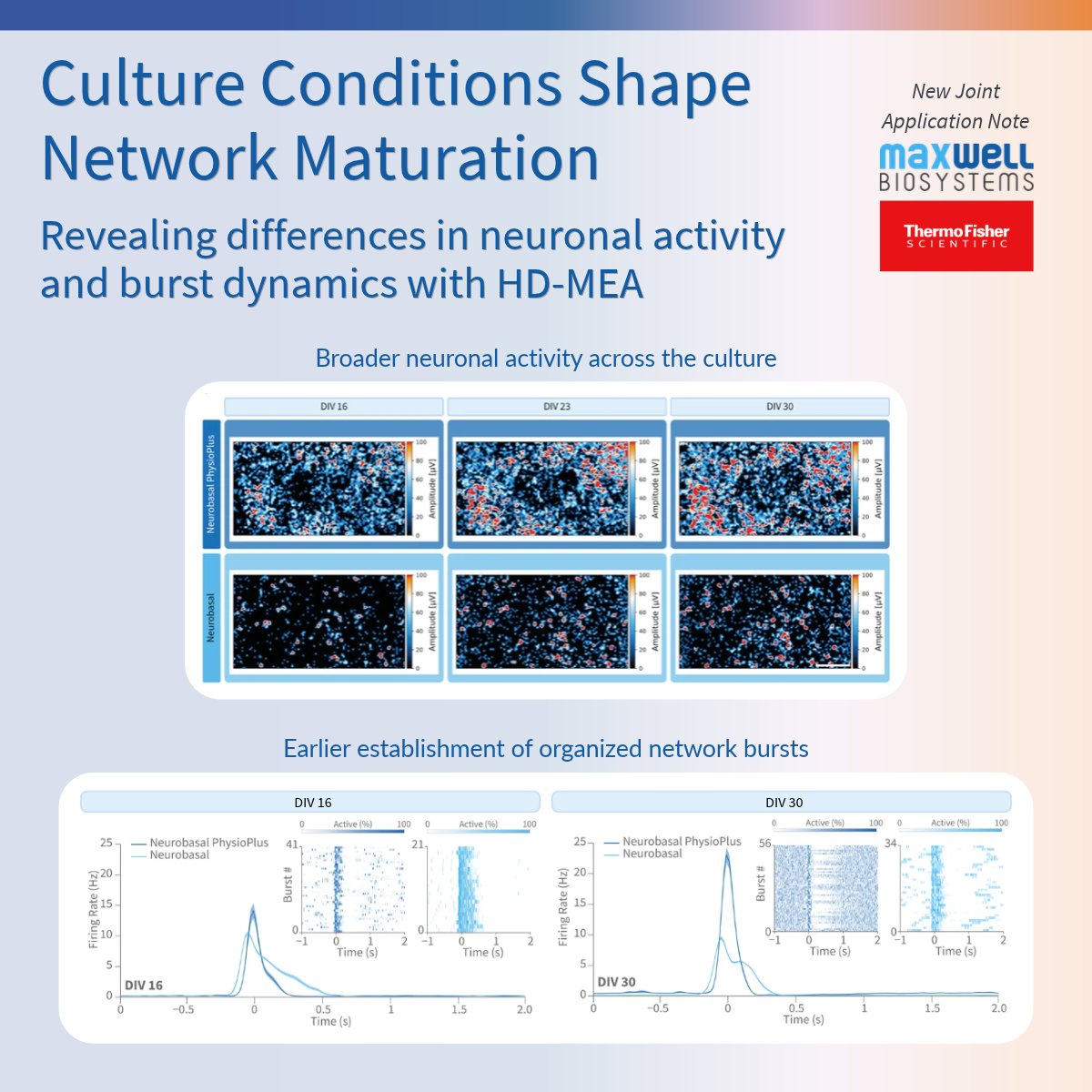
Using the MaxTwo 24-Well Plate, we compared primary cortical neurons grown in two different media. Cultures maintained in Gibco™ Neurobasal™ PhysioPlus Medium showed broader neuronal activity and developed regular, synchronized network bursts earlier.
But network maturation is more than firing rates and burst rhythm.
With the new burst-shape analysis in MaxLab Live 26.1, we can investigate network dynamics in greater detail, from burst shape and duration to how rapidly neurons are recruited into synchronized activity and how they disengage as each burst ends.
🔬 What did we observe?
Neurons cultured in Neurobasal™ PhysioPlus Medium established a stable burst shape early on, while the kinetics of network activation and termination continued to evolve as the cultures matured.
These findings highlight how combining optimized culture conditions with high-resolution functional recordings and advanced analysis can provide a more comprehensive understanding of neuronal network maturation and establish more reliable baselines for future experiments.
👉 Explore the findings in our new application note: https://t.co/glLJjY4iIz
🔬 Attending #SfN26? Visit us at booth #636 to learn more about these results and discover what you can now measure with MaxLab Live!
#Neuroscience #Electrophysiology #HDMEA #SfN26
Go beyond network burst frequency and duration with MaxLab Live 26.1!
More than 50 years ago, microelectrode arrays revealed that neuronal networks burst. Today, we can go a step further and characterize how they burst.
Network bursting is a hallmark of neuronal activity in vitro. But measuring how often bursts occur and how long they last tells only part of the story. Two networks might exhibit similar burst rates and durations while displaying very different activity patterns.
With the new Burst Shape Metrics in MaxLab Live 26.1, researchers can characterize how network activity rises, peaks, and decays, capturing subtle functional differences beyond conventional burst measurements.
What does this mean for your research?
Our latest blog explores experimental examples, showing how burst shape analysis can provide deeper insights into neuronal network maturation across different culture conditions and reveal distinct responses to compounds.
👉 Discover what the shape of a burst can reveal: https://t.co/DONjf1D2F9
#Neuroscience #Electrophysiology #HDMEA #NeuronalNetworks #DrugDiscovery
💙 MaxLab Live 26.1 is here! Bringing more biological insights to every recording.
Our latest software release strengthens our analysis capabilities, helping researchers go beyond individual measurements and gain a more comprehensive understanding of their experimental models.
First in focus: the new Cardiac Analysis.
When investigating the effects of a compound on cardiac activity, beat rate tells only part of the story. Changes in rhythm, electrical propagation, and field potential waveforms provide complementary insights into cardiac function and compound response.
With the new Cardiac Analysis in MaxLab Live 26.1, researchers can explore these readouts together through a dedicated, automated workflow, making it easier to characterize cardiac models, compare experimental conditions and identify compound-induced changes.
What does this look like in practice?
Our new blog explores real experimental examples, from slower conduction following disopyramide treatment to changes in cardiac rhythm and repolarization timing.
👉 Discover how to go beyond beat rate: https://t.co/Tp5pOe3wEw
And there is more to come! Next, we will take a closer look at the new neuronal network metrics in MaxLab Live 26.1.
#CardiacResearch #Cardiotoxicity #DrugDiscovery #Electrophysiology #HDMEA
💧 What if water looks clean according to conventional measurements, but still disrupts neuronal communication?
In a new Environmental Science & Technology (@ACSPublications) paper, Chen et al. show how conventional water-quality indicators and morphological measurements can miss subtle neurotoxic effects of contaminant mixtures.
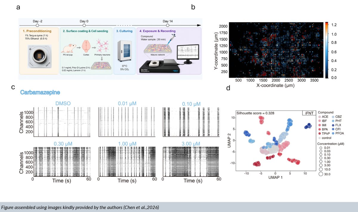
Using MaxWell Biosystems' MaxOne High-Density Microelectrode Array (HD-MEA) System, the researchers developed in vitro Functional Neurotoxicity Testing (iFNT) to assess primary cortical neurons exposed to increasing concentrations of individual chemicals, a contaminant mixture, and real wastewater samples without preconcentration.
They combined 14 electrophysiological metrics from MaxLab Live recordings, spanning neuronal firing, bursting, and network synchronization, into multidimensional dose–response profiles.
The results? HD-MEA functional profiling detected neurotoxic effects at 5 to over 500-fold lower concentrations than conventional neurite-outgrowth assays for individual compounds, with higher sensitivity also observed for chemical mixtures and wastewater samples.
This work highlights how rich HD-MEA data can reveal functional neurotoxicity beyond conventional morphological measurements, opening new opportunities for environmental monitoring.
👉 Read the full publication here: https://t.co/WYT4QvJCGt
👏Congratulations to Jiayu Chen, Kewei Liao, Ruixin Li, Hongqiang Ren, and Haidong Hu from the Water Quality Risk Assessment and Intelligent Monitoring Laboratory, Nanjing University @NJU1902 , China! 🇨🇳
🧬 From a groundbreaking discovery to two decades of scientific transformation.
Some scientific gatherings are more than just conferences. They are moments to reflect on how far a field has come, celebrate the people who made it possible, and imagine what lies ahead.
This is exactly how we felt at the iPSC 20th Anniversary Symposium in Kyoto.
From the beautiful orchestral performance that opened the symposium to the unique opportunity to hear directly from Prof. Shinya Yamanaka, the atmosphere was truly special. Throughout the three days, we could feel what this milestone meant to the researchers who have dedicated years, and in many cases entire careers, to advancing iPSC research.
It was inspiring to reflect on two decades of discoveries that have transformed our understanding of human biology and disease, surrounded by a community that continues to push the boundaries of what is possible.
🔬 For our team, being part of this remarkable gathering was also an opportunity to contribute to the scientific exchange and share new approaches to studying human neural models.
During our Innovation Showcase, Dr. Marie Obien @engeleneo explored how HD-MEA technology enables multiscale functional phenotyping of human iPSC-derived neural models, while Dr. Yi Sun shared insights into isogenic ALS disease-in-a-dish models, highlighting the value of longitudinal axon tracking in patient-derived motor neurons.
Our two poster presentations further demonstrated how HD-MEA technology can help characterize neuronal maturation in midbrain organoids and identify early functional alterations in neuronal networks and axons in FTD models.
💡 We left Kyoto feeling inspired by how far the field has come and excited about the discoveries still ahead. Being part of this scientific journey reinforces our commitment to supporting researchers with new technologies, meaningful collaborations, and deeper functional insights into human neural models.
🙏 A heartfelt thank you to the @ISSCR organizers for creating such a memorable celebration, to Dr. Yi Sun for her contribution to our Innovation Showcase, and to everyone who joined our session, visited our posters, or stopped by our booth to exchange ideas.
#ISSCR #HDMEA #Neuroscience #ALS #iPSC #MxWConnect
🧠Attending Neuroscience 2026 in Washington, DC? Let's continue the scientific conversations beyond the conference floor!
Join us, @AnatomicTech and @NanionTech for an evening of science and networking at our SfN Mixer.
Bringing together researchers from academia and industry, this gathering is an opportunity to connect with the neuroscience and drug discovery community in a relaxed and informal setting.
🔬 Whether you are working with hiPSC-derived neuronal models, investigating ion channels, exploring automated patch clamp and HD-MEA technology, or developing functional assays for drug discovery, we look forward to exchanging ideas, discussing experimental approaches, and sharing experiences.
🍸 And of course, our signature cocktails will be waiting for you!
📍 Where: Washington, DC
📅 When: November 16th, 2026
👉 Space is limited! Contact [email protected] or reach out to a member of our team to secure your spot.
See you in Washington, DC!
#Neuroscience2026 #SfN2026 #Neuroscience #HDMEA #MxWConnect
ISSCR 2026 Kyoto may have wrapped up, but our commitment to Amyotrophic lateral sclerosis (ALS) research continues.
While the conference brought together researchers to celebrate advances in iPSC science and explore new opportunities for disease modeling and regenerative medicine, one of our most meaningful moments happened beyond the scientific sessions.
🏃♂️On the crisp morning of Day 3, three members of our team @u_frey ,@engeleneo and @m_hruskaplochan took to the streets of Kyoto for a sunrise Run for ALS. We were delighted to be joined by other runners including Prof. Jun TAKAHASHI @juntakajun (@CiRA_KU_E Center for iPS Cell Research and Application (CiRA)) and Keith Alm (@ISSCR International Society for Stem Cell Research), sharing those early morning miles to raise awareness and show our support for people living with ALS.
ALS is a progressive neurodegenerative disease that affects motor neurons, gradually impairing the ability to move, speak, swallow, and breathe. Behind every diagnosis are people and families facing profound challenges, reminding us why continued scientific progress and collective action matter.
But our commitment extends beyond technology and the lab. Through initiatives like Run for ALS, we come together as colleagues and as part of a broader community united by the hope of making a difference.
💙 Every contribution counts. Support our initiative here: https://t.co/WcYwNzhGKl
🙏🏻 A heartfelt thank you to everyone who joined us, cheered us on, or contributed to our fundraiser. What started in Zurich during NeuMoS continued through the streets of Kyoto, and our journey is far from over. Stay tuned for our next Run for ALS as we keep moving together for a cause that matters! 💙
#ALS #RunForALS #ALSResearch #ALSAwareness #ISSCR #MxWConnect
🫂One organoid? Two? Three? Which network can learn to discern one input from another? The more, the merrier.
In a new @CommsBio (@NaturePortfolio) paper, Chow et al. used MaxWell Biosystems’ MaxOne High-Density Microelectrode Array (#HDMEA) System to build and longitudinally stimulate solo, duo, and trio human cerebral #organoid networks, asking whether repeated electrical input could refine their ability to discriminate two different signals.
Using MaxOne+, the team could monitor all interconnected modules simultaneously while keeping their activity spatially resolved, enabling spike sorting and functional connectivity analysis across the network. These analyses revealed that, after two weeks of repeated input (aka training), only the three-organoid networks consistently improved stimulus discrimination, with the output organoid responding differently depending on which upstream organoid was activated. This functional refinement was also reflected in spontaneous activity, where network propagation became increasingly biased from the input organoids toward the output.
This work highlights how modular network architecture together with repeated external cues can drive stimulus-specific functional refinement in human brain organoid networks.
👉 Read the full publication here: https://t.co/jSODlM2ydD
👏 Congratulations to Siu Yu A. Chow, Huaruo Hu LinkedIn, @Tomoyadu , Takuya Asakura, Sota Sugimura, and @YoshihoIkeuchi from @UTokyo_News_en, CNRS, and @SoftBank on their new publication!
#Connectoid #Organoids #FunctionalConnectivity #OrganoidIntelligence #Neuroengineering #Neurocomputing #ElectricalStimulation #iPSC #Neuroplasticity
💤 Can a human brain #assembloid reproduce the electrical signatures of #anaesthesia?
In a new British Journal of Anaesthesia paper, Toker et al. asked whether a minimal human cortical circuit, without the thalamus or other subcortical structures, could reproduce propofol-induced neural dynamics. Their cortical–ganglionic eminence assembloids provided this minimal circuit, combining excitatory and inhibitory neurons with propofol-sensitive GABAA receptors.
The team captured three hallmark electrophysiological features of anaesthetic states. Using MaxWell Biosystems’ MaxOne HD-MEA System and spike sorting, they observed an abrupt and sustained suppression of single-unit firing after propofol exposure, while complementary LFP recordings revealed a shift toward prominent slow/delta activity together with reduced signal complexity.
By reproducing key anaesthesia-like electrophysiological dynamics in a human in vitro model, this work opens new possibilities for mechanistic studies of anaesthetic action and future drug screening.
👉 Read the full publication: https://t.co/dSZV4JBM3d
👏 Congratulations to Daniel Toker and all co-authors
#BrainOrganoids #HDMEA #MaxOne #iPSC #Neuroscience #GABA #Pharmacology
🧠 Two human dopaminergic neurospheroids. One axonal bridge. Can HD-MEAs reveal whether the two networks are actually interacting?
In a new Biofabrication (@IOPPublishing) paper, Ausilio et al. developed a microfluidic microphysiological system (MPS) in which two human iPSC-derived dopaminergic neurospheroids are kept spatially separated while axons grow through a defined microchannel, forming a stable inter-spheroid fascicle.
But does this morphological connection also translate into a functional one? In this case, yes.
Using MaxWell Biosystems’ MaxOne+High-Density Microelectrode Array (HD-MEA) System, the team followed the two neurospheroids longitudinally and could spatially resolve activity from each network while monitoring their maturation over time.
Going one step further, the authors separated local interactions within each spheroid from long-range interactions between them, revealing bidirectional lag-consistent activity across the connecting axonal tract, compatible with functional connectivitybetween the two neuronal modules.
This work brings 3D human dopaminergic circuitry models, microfluidic MPS, and HD-MEA electrophysiology together in one platform to investigate how spatially separated neuronal networks connect and communicate.
👉 Read the full publication here – https://t.co/PGICwEzkjf
👏 Congratulations to Chiara Ausilio, Annachiara Scalzone, Valentina Mollo, and Paolo Antonio Netti from Istituto Italiano di Tecnologia (IIT) and Università degli Studi di Napoli Federico II on their new publication in Biofabrication / IOP Publishing.
#Connectoid #MicrophysiologicalSystems #MPS #HDMEA #Neurospheroids #Microfluidics #BrainOnChip #Neuroengineering #iPSC
It was a pleasure to welcome our partners to MaxWell Biosystems in Zurich, Switzerland, for our second annual IUK–Innosuisse consortium meeting!
A big thank you to Zameel Cader, Satyan Chintawar, Galbha Duggal, and Anna Underhill from Oxford StemTech, and Joseph Bateman and Harry Mayes from King’s College London, for the insightful discussions, productive exchanges, and great time together.
Together, we are developing a high-throughput drug testing platform combining patient-specific brain assembloids with high-density microelectrode array (HD-MEA) recordings, with the goal of advancing precision medicine for epilepsy.
It is exciting to see this collaboration progressing and to bring together complementary expertise in stem cell models, electrophysiology, and drug discovery to develop new approaches for patient-specific therapeutic testing.
We are grateful to Innosuisse and Innovate UK (IUK) for supporting this collaborative project.
🔗 Want to learn more about the project? Find more info here: https://t.co/wTC1zGUVwp
#Epilepsy #PrecisionMedicine #Organoids #NAMs #HighThroughput #HDMEA
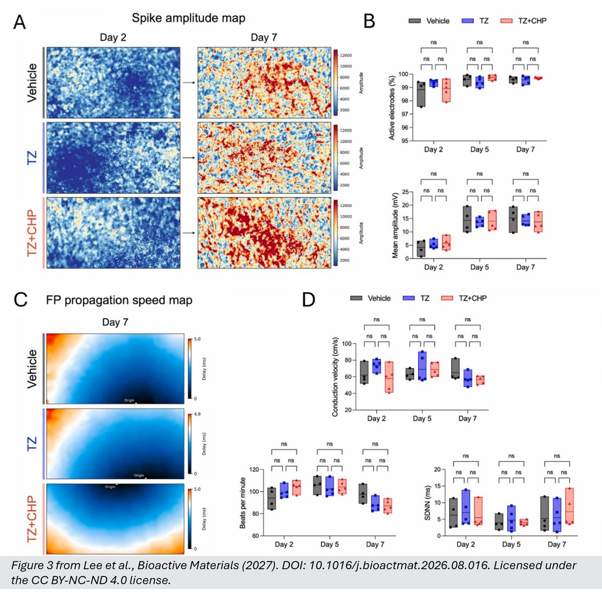
🫀 Can engineered cardiomyocytes improve cardiac repair after myocardial infarction?
In a new Bioactive Materials paper published by KeAi Publishing, Lee et al. tackle one of the major challenges of cardiac cell therapy: the poor retention and survival of transplanted human pluripotent stem cell-derived cardiomyocytes (hPSC-CMs) in the injured heart.
The team developed a rapid surface-engineering strategy using lipid insertion and click chemistry to attach a collagen hybridizing peptide (CHP) to hPSC-CMs. By targeting denatured collagen in injured myocardium, CHP-modified cells showed improved adhesion and engraftment, supporting better cardiac repair after transplantation in a rat ischemia-reperfusion model.
But what about beating function?
Using the MaxOne High-Density Microelectrode Array (HD-MEA) System, the team followed the cultures on days 2, 5, and 7 after modification. Active beating area, field-potential amplitude, beating rate, rhythm variability, and conduction velocity remained comparable across conditions, with propagation maps further confirming coordinated electrical conduction across the network — showing that the surface-engineered cardiomyocytes retained their electrophysiological phenotype.
Together, the study highlights how HD-MEA functional phenotyping can provide a quality-control step for engineered cell therapies, connecting cell-surface bioengineering with cardiac repair in vivo.
👉 Read the sull publication here:
https://t.co/6h2XZqOf15
👏Congratulations to Donghyun Lee, Woo-Sup Sim, Jae-Hyun Park, Dong-Yeon Han, Soo-Jeong Kim, Jin Ryeol An, Mi Seon Seo, Jin-Ju Kim, Seo Young Cheon, Hyejin Hong, Seong Woo Choi, Kiwon Ban, Heebeom Koo, and Hun-Jun Park, from The Catholic University of Korea, @donggukuniv, and @CityUHKDG.
#CardiacResearch #Cardiomyocytes #StemCells #RegenerativeMedicine #HDMEA #Electrophysiology #CellTherapy #Bioengineering
In 3D pharmacology, a robust baseline is only the starting point. The next question is: how does a compound reshape network function?
In our new joint application note with @InSphero, 3D InSight™ Human Neural Microtissues recorded on the MaxTwo 24-Well Plate showed reproducible spontaneous activity and synchronized network bursting across independent microtissues, establishing a consistent functional baseline for compound testing.
Following pharmacological modulation, HD-MEA recordings revealed distinct network responses to several neuroactive compounds. But the analysis goes beyond conventional network metrics. With the new burst-shape analysis in MaxLab Live, changes in the population bursts dynamics can be characterized in greater detail, from burst onset and peak activity to decay.
Together, reproducible 3D neural models, scalable HD-MEA recordings, and advanced network analysis bring greater granularity to the functional characterization of compound responses.
👉 Read the full application note: https://t.co/bl9o2mfzNN
#Pharmacology #Spheroids #Neuroscience #Electrophysiology #HDMEA
🧠 Advancing functional characterization of iPSC-based models of motor neuron disease
As the field advances from individual iPSC-derived neuronal populations toward increasingly sophisticated 2D and 3D models, new opportunities are emerging to capture the cellular complexity of diseases such as ALS, SMA, and inherited peripheral neuropathies. At the same time, questions around functional characterization, reproducibility, model quality, and translational relevance are becoming increasingly important.
⚡ How can electrophysiology contribute to this next generation of disease models?
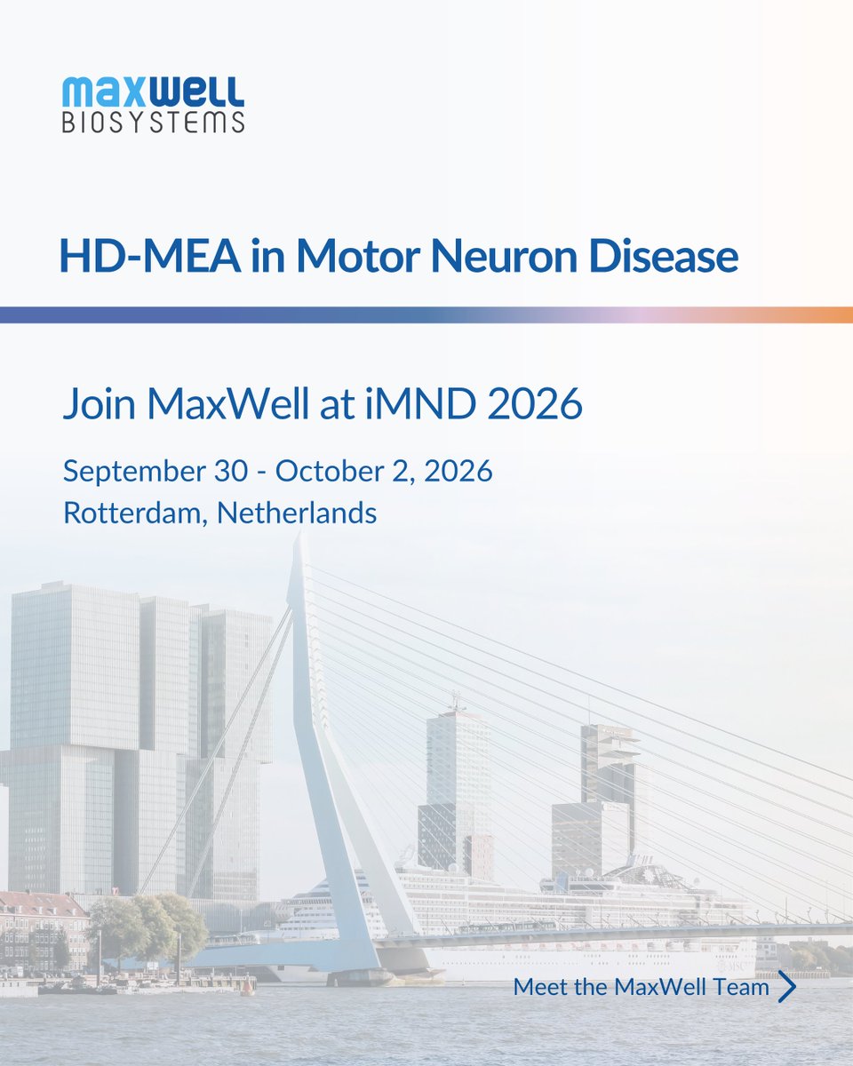
At the iPSC Modeling in Neuromuscular Disorders (iMND) Meeting in Rotterdam, meet Dr. Marilina Douloudi and Dr. Tom Dufor, Field Application Scientists at MaxWell Biosystems, and discuss your models, functional phenotyping, and how HD-MEA technology can complement your iPSC workflow.
Do not miss their poster presentation:
📌 “Electrophysiological Profiling and Phenotypic Characterization in ALS and FTD Models Using High-Density Microelectrode Arrays”
Discover how HD-MEA can add a functional dimension to iPSC-based disease modeling by complementing molecular and cellular readouts with high-resolution measurements of neuronal activity, helping uncover functional alterations associated with neurodegenerative disease.
📍 Rotterdam, The Netherlands
📅 September 30 – October 2, 2026
#iMND2026 #DiseaseModeling #Electrophysiology #HDMEA #MxWConnect
🧫 From thalamic input to human cortical development
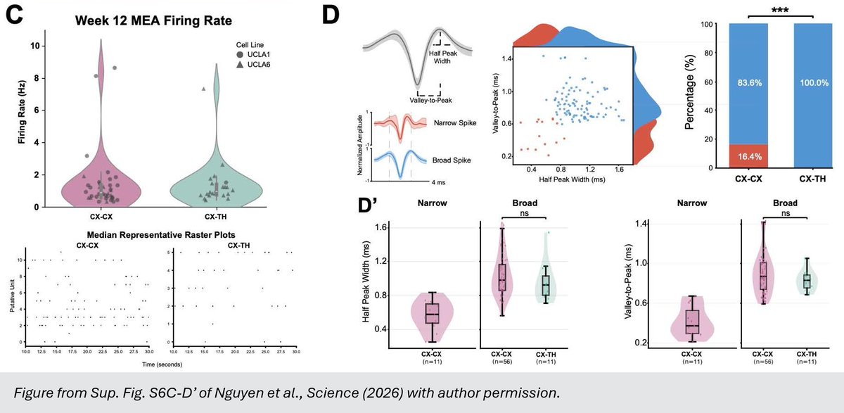
In a new paper published in @ScienceMagazine, Claudia V. Nguyen and colleagues use human thalamocortical assembloids, to investigate how signals from one brain region shape the development of another.
What makes this especially interesting is how early thalamic input influences the production of cortical excitatory neurons. The team identified NRXN1-mediated contacts between thalamic axons and cortical progenitor cells as a mechanism contributing to this process.
Using the MaxTwo HD-MEA system, the researchers characterized electrical activity in 12-week-old assembloids. Spike-waveform analysis revealed a shift towards broad-spiking waveforms in thalamocortical assembloids compared with cortex–cortex controls, adding an electrophysiological perspective to the study of how thalamic input shapes human cortical development (Sup. Fig. S6).
This creates an exciting experimental platform for studying how interactions between brain regions shape human cortical development, and how disease-associated NRXN1 mutations may disrupt this process.
Read the full publication here: https://t.co/WCPCGhZpe6
🎉 Congratulations to Claudia V. Nguyen, Antoni Martija, Daniel C. Jaklic, Rista White, Marty G. Yang, Patricia R. Nano, Jose A. Soto, Jessenya Mil, Dakshesh Rana, Jacqueline M. Martin, Hunter E. Schweiger, Sebastian Hernandez, Elisa Fazzari, Yu Liu, Jack M. Parent, Mohammed A. Mostajo-Radji, Daniel H. Geschwind and Aparna Bhaduri, from @UCLA, @ucsc, @UMich and the VA Ann Arbor Healthcare System!
#HDMEA #Organoids #Assembloids #Neurodevelopment #Electrophysiology
👁️ Before the eyes even open, the retina is already “talking” to the brain. But what happens in neurodevelopmental disorders with visual impairment?
In a new Frontiers in Neuroscience paper (@FrontNeurosci), Gupta et al. investigate how loss of Ube3a, the gene underlying Angelman syndrome, affects spontaneous retinal activity during early development, a particularly interesting question given the visual abnormalities commonly associated with the disorder.
Before vision is fully established, developing retinas generate retinal waves, coordinated bursts of neuronal activity that travel across the tissue and help guide the wiring of the visual system.
Using the MaxOne High-Density Microelectrode Array (HD-MEA) System, the team recorded 32 ex vivo retinas from wild-type (WT) and Angelman-model mouse pups. Angelman-model retinas showed reduced retinal wave frequency and lower overall RGC activity, indicating less patterned activity during this critical developmental period.
But what about the waves that did occur? They were largely normal: wave propagation speed, duration, area, shape, local synchrony, and distance-dependent functional connectivity were comparable to WT controls. This suggests that Ube3a deficiency primarily disrupts the initiation of retinal waves rather than their ability to propagate across the retinal network.
This is a nice example of how the spatial and temporal resolution of MaxOne can move beyond detecting “more or less activity” and reveal which specific property of a developing neural circuit is altered in disease.
👉 Read the full publication here –
https://t.co/I8tjPx3fUu
👏 Congratulations to Rishikesh Kumar Gupta, Alexander J. Fisch, Fernanda S. Orsi, and Alexandre Tiriac from Vanderbilt University (@VanderbiltU) and Vanderbilt University Medical Center (@VUMCDiscoveries) 🇺🇸
#HDMEA #Retina #Neurodevelopment #DiseaseModeling #Neuroscience
Supplementary Video S1 from Gupta et al., Frontiers in Neuroscience (2026). DOI: 10.3389/fnins.2026.1895084. Licensed under CC BY 4.0.