Huzzah for @stevenmeisler.com, whose preprint on our new fully-processed ABCD dMRI data resource is now out. The super-clear results from this monumental effort will undoubtably change the way we approach studies of white matter development going forward.
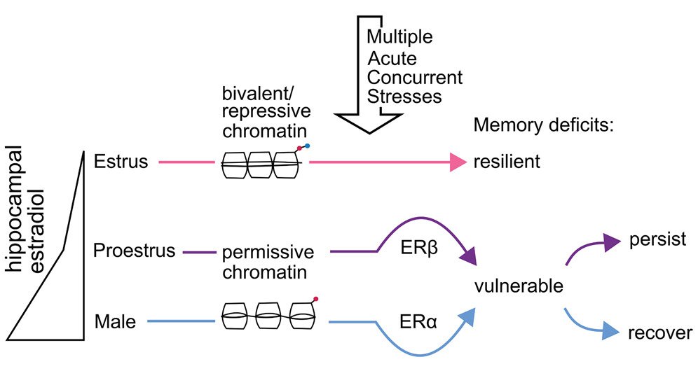
New research out in @NeuroCellPress finds that hippocampal estrogen levels, receptor types & epigenetics contribute to sex-specific memory vulnerabilities to concurrent acute stresses ft. @kiararodz_, @DrJuliaW & Elizabeth Heller (@PennSysPharmATT)
https://t.co/lDKFEPDUU1
David Kleinfeld’s Sprague Lecture at @PennMINS deeply thought-provoking. Volitional control of behavior highlights that touch isn’t passive—the brain actively samples the world. Essential thinking for somatosensory neuroscience. #ActiveSensing#Neuroscience#Touch
@ShorterLab@PennNGG@PennMINS Thank you @MadayLab for the invitation and a wonderful visit! Had a great time discussing science with the students and faculty.
The MRI Neuroanatomy Post 🧠
Can you identify the gyri between the Sylvian fissure and hippocampal formation?
1. Superior temporal gyrus, STG
2. Middle temporal gyrus, MTG
3. Inferior temporal gyrus, ITG
4. Fusiform gyrus, FG
5. Parahippocampal gyrus, PHG
Note the intradural segment of the vertebral artery, V4 (red)
Semicircular canals of the vestibular apparatus are also seen (asterisk)
Trace these structures on the opposite side!
#anatomy #mri #brain #neuroanatomy #Neuroscience #radiology #neurosurgery #Neurociencia #MedEd #education #match2025 #AI #Technologynews #MedTwitter #futureradres #research #ArtificialInteligence @futureradres
The MRI Neuroanatomy Post 🗞️
The parieto-occipital fissure (yellow) divides the occipital lobe from the parietal lobe and intersects the calcarine sulcus (pink) - resembling a tilted "Y".
The primary visual cortex lies along the calcarine sulcus.
Above the calcarine sulcus lies the cuneus (green) - the site of basic visual processing.
Below the calcarine sulcus lies the lingual gyrus (blue) - needed for face & landmark perception and recognition, and visual memory.
#MRI #Neuroanatomy #neuroscience #radiology #anatomy #neurotwitter #neurosurgery #MedEd #education #MedTwitter #neuro #ArtificialInteligence @futureradres
MRI Neuroanatomy 101 🧠
The red nuclei (red), mammillary bodies (pink), optic tracts (yellow), & cerebral peduncles (dark blue) are marked here.
Fibers of the cerebral peduncle are organized from anteromedial to posterolateral as:
- frontopontine (FP)
- corticobulbar (CB)
- corticospinal (CS)
- temporo-parieto-occipito-pontine (TPP)
Notice the ICA branching into the first segments of the anterior cerebral artery (A1) and middle cerebral artery (M1).
Try to trace these structures on the opposite side!
#radiology #neurotwitter #Neuroscience #Anatomy #Neurosurgery #education #MedTwitter #AI #MRI #brain #vascular #KnowledgeIsPower #AI #ArtificialIntelligence #algorithm
There is just over a week left to apply to the MindCORE Postdoctoral Fellowship @Penn. Applications close Sunday, December 1.
https://t.co/niuXIS8uT0
@pennbgs@UPennGAPSA@LingSocAm@PennMINS
A human DCC variant causing mirror movement disorder reveals that the WAVE regulatory complex mediates axon guidance by netrin-1–DCC ft. @kchau91, Yixin Zang, Sarah Gagnon & @BashawLab (@PennMINS) @PennNGG@scisignal https://t.co/y61o6b83Dg
I won't be at @SfNtweets this year but spread the word for me friends! The Giovanniello Lab @CSARtemplemed is looking for a lab technician for a January 2025 start date. Winter grads 🎓, I'm looking at you 👀
Interested in stress, drugs, and habits? Feel free to DM or email.