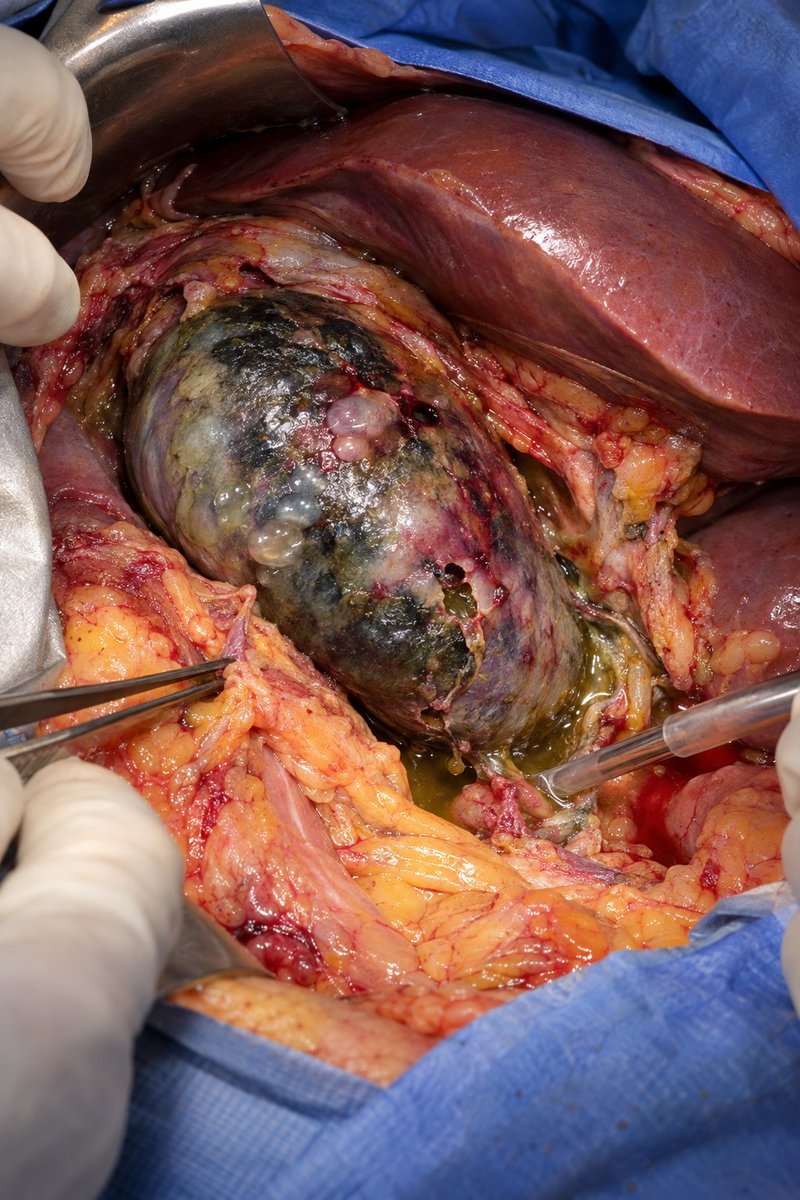
The laparoscopic cholecystectomy begins routinely. As the surgeon lifts the gallbladder, the camera reveals a liver covered in coarse, irregular nodules.
The surgeon pauses. “This was not in the preoperative report.”
What unexpected liver condition has likely been discovered?
During laparoscopic cholecystectomy, severe inflammation has completely distorted Calot’s triangle—and the surgeon is no longer certain which structure is the cystic duct.
What must be done before applying any clip, and what is the safest bailout strategy if the anatomy remains unclear?
Echo From Scratch | Post #24
PSAX at the Mitral Valve Level
Drop the parasternal short-axis plane from the aortic valve level to the mitral valve, and you get a completely different view of the mitral apparatus.
What you see
In diastole, the thin anterior and posterior mitral leaflets open widely, approaching the LV wall.
In systole, they close and form the coaptation zone.
The posterior leaflet has 3 major scallops:
🟧P1: lateral
🟨P2: central
🟩P3: medial
The two mitral commissures are located at the lateral and medial ends of the valve.
The papillary muscle connection
There is an important anatomical pattern:
The medial portions of both leaflets are supported by the medial, or posteromedial, papillary muscle.
The lateral portions are supported by the lateral, or anterolateral, papillary muscle.
This arrangement helps maintain leaflet position throughout the cardiac cycle.
The chordae
Chordae tendineae form a branching network between the papillary muscles and mitral leaflets.
They branch into:
⚪Primary chordae
🔵Secondary chordae
⚫Tertiary chordae
Most attach near the free edge of the leaflets and are called marginal chordae.
Others attach to the ventricular surface and are called basal or strut chordae.
Occasionally, abnormal-looking chordal attachments can be seen in otherwise normal hearts, so anatomy must always be interpreted in context.
Understanding this 3D arrangement makes mitral valve pathology much easier to localize.
#Echofirst
Dural arteriovenous fistulas are known for their high bleeding risk, but their venous hypertension and venous congestion can also cause profound deficits, like in this patient with a superior petrosal sinus DAVF who became “locked in” from medullary ischemia and edema. Vascular findings on CTA led to diagnosis and avoided a brainstem biopsy…
PICA vs AICA vs MCA
TWO MOST IMPORTANT DIFFERENTIATING POINTS ARE:
PICA: Patients have Dysphagia and Hoarseness
AICA: Patientz have hearing loss / Tinnitus.
USMLE Series.
A man in his late 50s, a former competitive bodybuilder w/ extensive tanning bed exposure, presented w/ a 25 × 25 cm nonhealing ulcerated plaque on the upper back.
O/E: a large pink ulcerated plaque w/ rolled borders involving most of the upper back, along w/ multiple scattered waxy plaques w/ telangiectasias on the torso.
He was afebrile & nontoxic, w/out palmoplantar pits, milia, skeletal abnormalities, lymphadenopathy, or hx of odontogenic cysts. CBC, CMP, & CK were w/in normal limits.
What’s the diagnosis?
A child was brought to the clinic because the parents noticed continuous yellow fluid leaking from the belly button since birth.
What is the diagnosis?