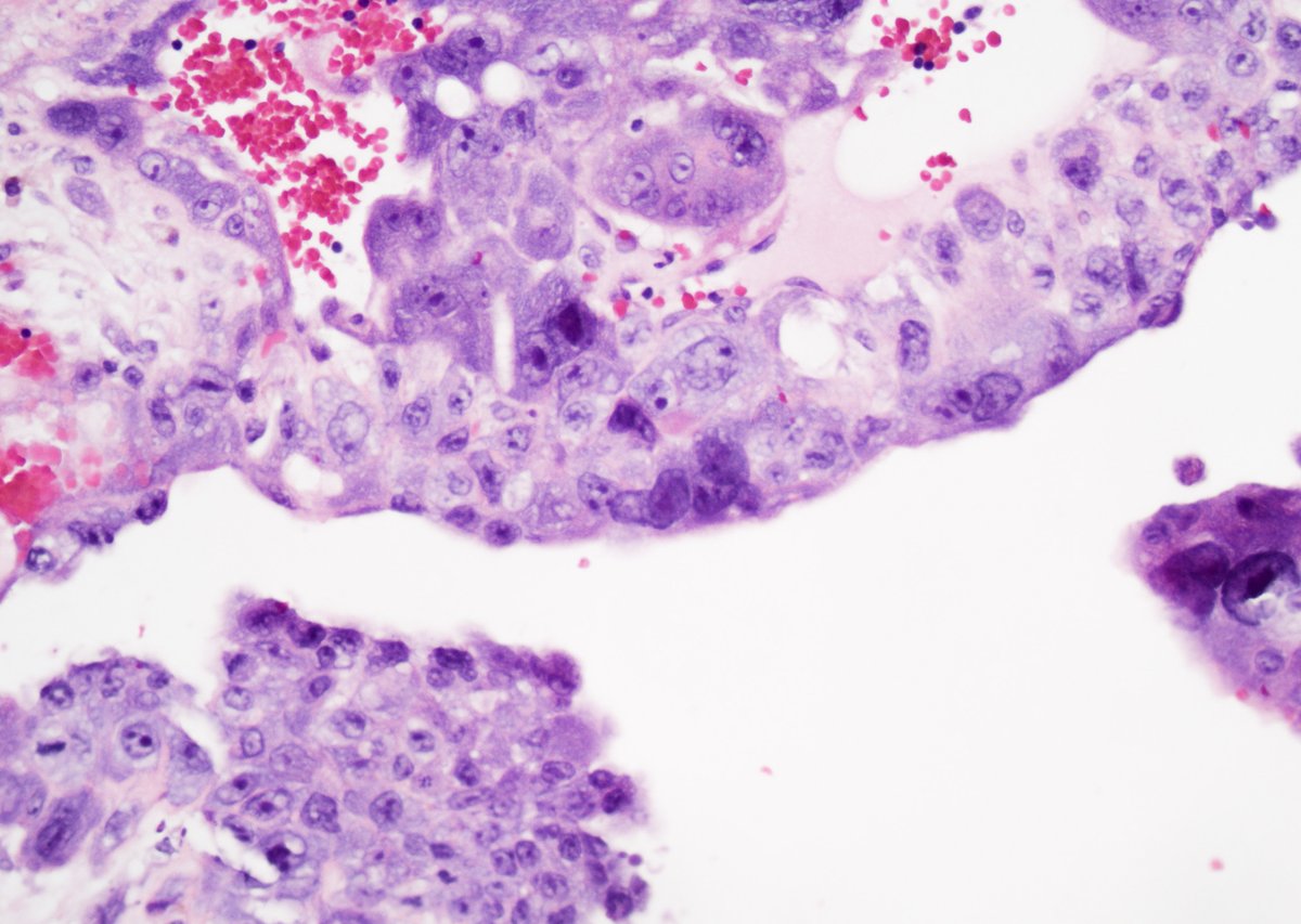
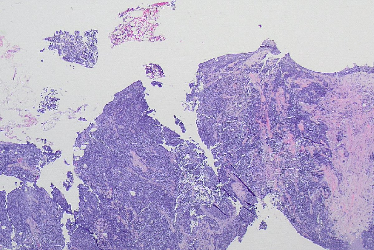
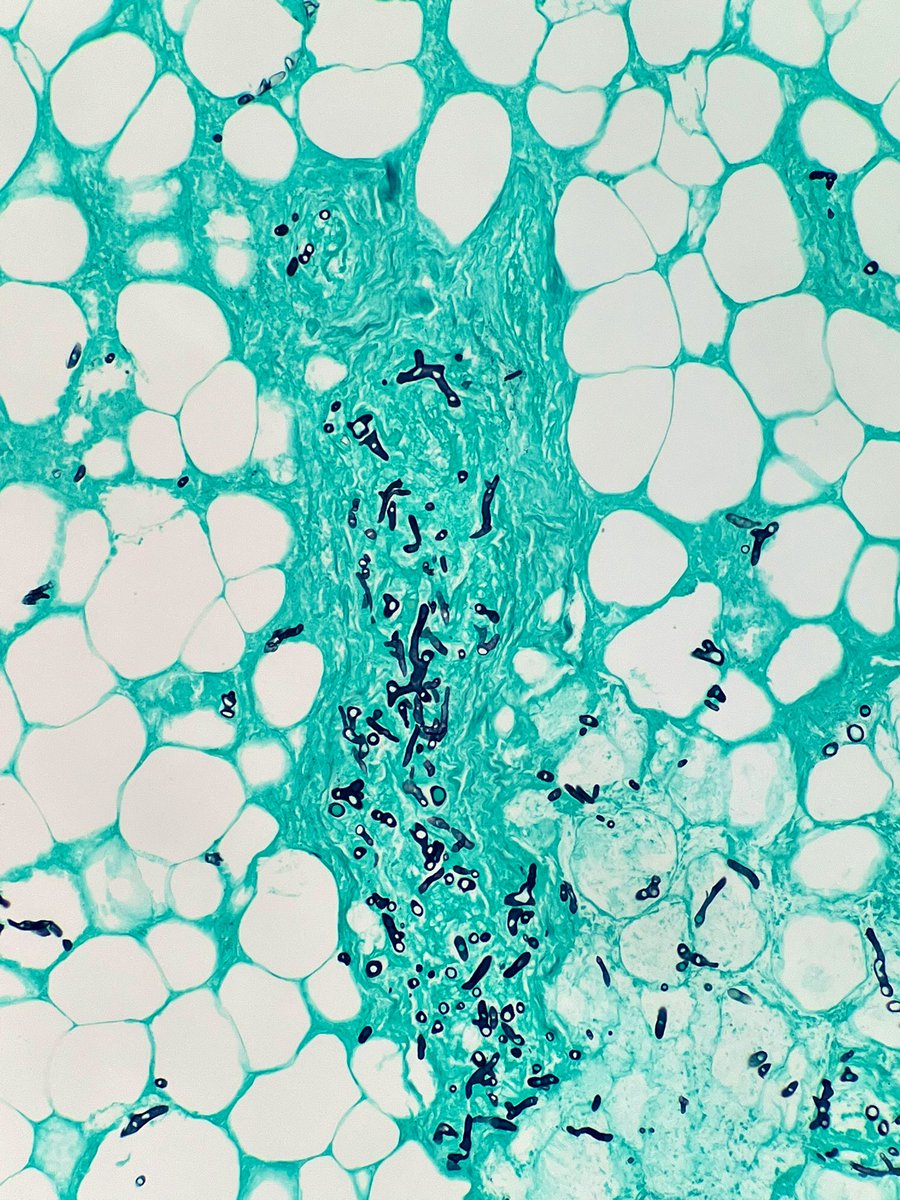
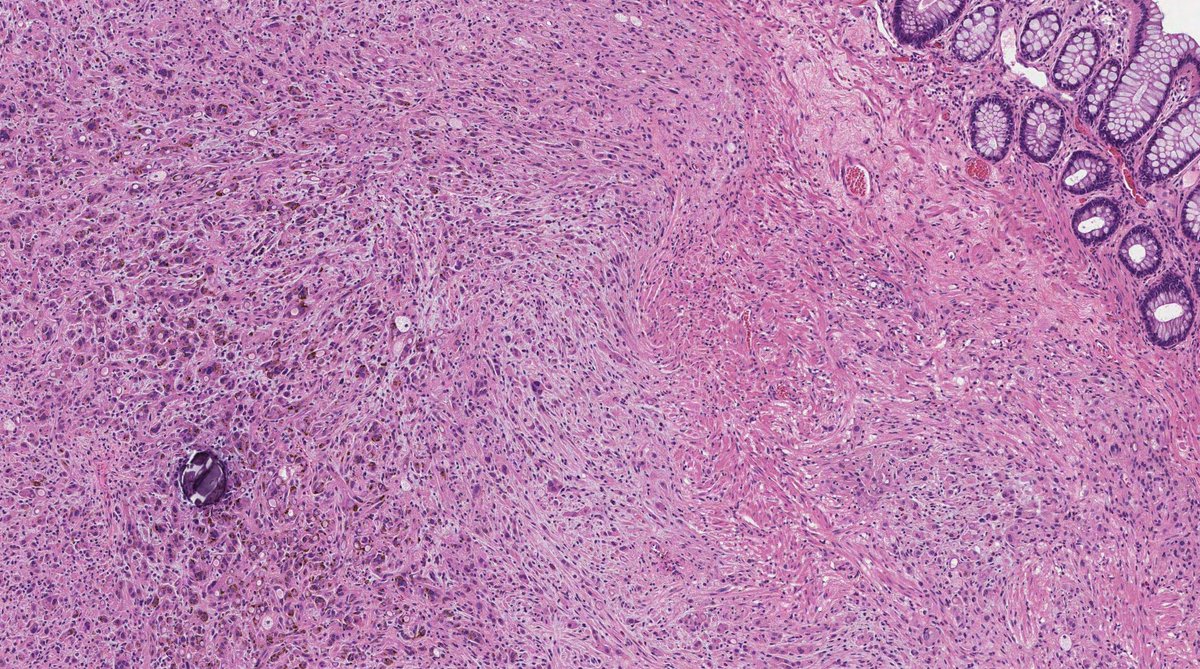
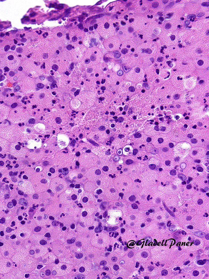
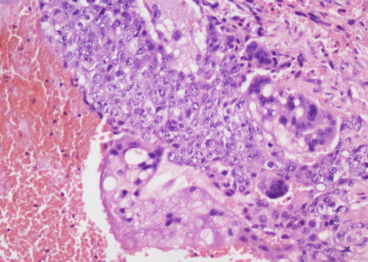
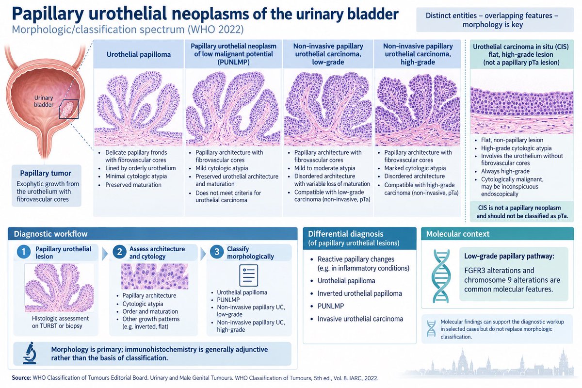
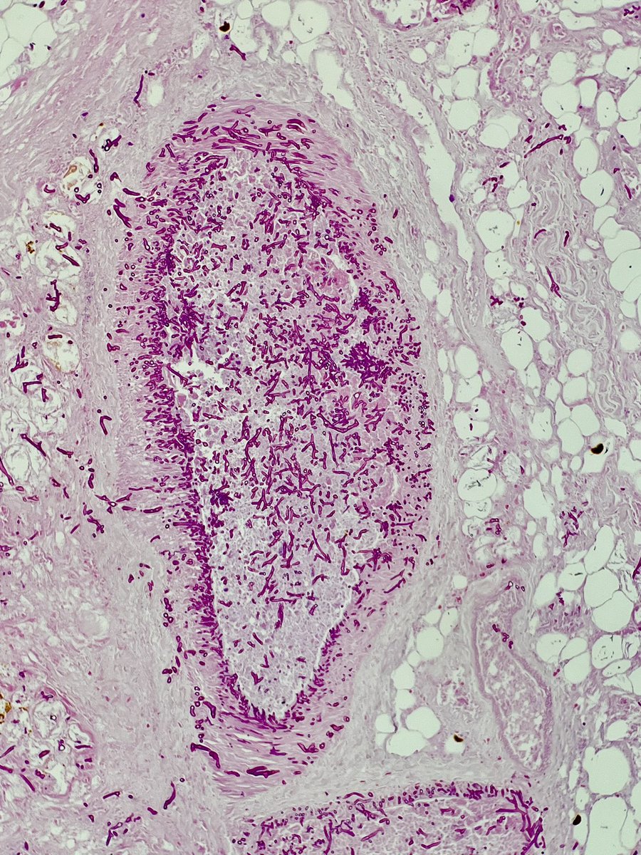
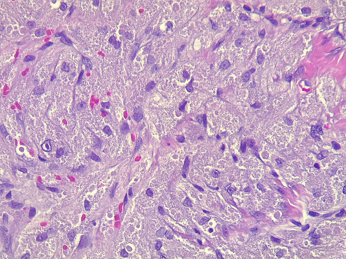
This triple negative breast cancer with neuroendocrine morphology with Ki-67 of 90% and no known lung mass. How the biopsy should be signed out? #breastpath
Immunohistochemical Approach to the Classification and Diagnosis of Ovarian Tumors
Primary ovarian tumors are broadly classified into three major categories: epithelial tumors, germ cell tumors, and sex cord-stromal tumors.
The selection of immunohistochemical markers for the classification of ovarian tumors may include:
Epithelial tumor markers: CK, EMA, PAX8, CK7, CK20, etc.
Germ cell tumor markers: SALL4, PLAP, OCT4, etc.
Sex cord-stromal tumor markers: α-inhibin, calretinin, SF-1, etc.
Hormone receptor markers: ER and PR.
🧠 Neuropathology quiz:
Brain mass — from intraoperative consultation to final workup.
1️⃣ Touch prep
2️⃣ Frozen section
3️⃣ Permanent H&E
4️⃣ IHC
What’s your diagnosis, and what stain is shown in image 4?
Pathologic Diagnostic Algorithm for Trophoblastic Lesions
Based on clinical and morphologic features, trophoblastic tumors and tumor-like lesions can generally be readily distinguished. However, selected cases may pose diagnostic challenges, particularly in limited biopsy specimens and endometrial curettage samples. The following diagnostic algorithm may be used as a reference.
Patient presents with a slow growing subcutaneous knee mass (< 2 cm) present for years. Diagnosis? What is the cell of origin for this entity? Answer and pearls in the comments! #PathTwitter#MedEd#BSTPath
WHO 6th ed Gyn path update:
➡️SBT with microinvasion (<5 mm):no longer recommended,describe descriptively.
➡️The term “microinvasive ca” for LGSC like areas <5 mm in SBT is also no longer recommended.Just call it LGSC in SBT😮
This will take some time to get used to!
#gynpath
Immunohistochemical Marker Selection in the Diagnosis and Classification of Lymphomas
When lymphohematopoietic neoplasms (primarily lymphomas) cannot be readily distinguished from other undifferentiated small-cell tumors of non-lymphohematopoietic origin, an initial antibody panel that includes leukocyte common antigen (LCA/CD45), S-100, and cytokeratin (CK) should be applied to establish the tissue of origin.
When the histologic findings are already sufficient to support a diagnosis of lymphoma, lineage-associated markers should be used to determine the neoplastic cell type. A practical first-line panel consists of two pan-T-cell markers (CD3 and CD43) and two pan-B-cell markers (CD20 and PAX5), together with CD15 and CD30. This combination usually permits a straightforward separation of Hodgkin lymphoma from non-Hodgkin lymphoma and, within the latter group, of B-cell from T-cell neoplasms.
For lesions that display a follicular or nodular growth pattern, markers such as CD10, BCL6, CD21, CD23, BCL2, and Ki-67 are useful for delineating the relationship between the nodules and residual lymphoid follicles.
When Hodgkin lymphoma is favored morphologically, the recommended panel includes CD30, CD15, LCA, PAX5, MUM1, and EBV-encoded RNA (EBER) in situ hybridization. If anaplastic large-cell lymphoma enters the differential diagnosis, EMA, ALK, and CD43 should be added. If T-cell/histiocyte-rich large B-cell lymphoma must be excluded, BOB.1, OCT2, CD20, and CD3 should be included.
Small B-cell lymphomas require simultaneous consideration of small lymphocytic lymphoma/chronic lymphocytic leukemia (SLL/CLL), marginal-zone lymphoma (MZL), follicular lymphoma (FL), and mantle-cell lymphoma (MCL). An appropriate panel comprises CD20, PAX5, CD19, CD5, CD23, CD10, BCL2, cyclin D1, SOX11, LEF1, and MNDA. For neoplasms showing clear-cut immature (blastic) differentiation, the differential diagnosis includes acute lymphoblastic leukemia/lymphoblastic lymphoma (ALL/LBL) and acute myeloid leukemia (AML). In addition to lymphoblastic markers (TdT, CD99, CD10, CD34), myeloid antigens such as myeloperoxidase (MPO) and CD117 should be assessed to exclude lymphoblastic lymphoma with myeloid or bilineage differentiation. Judicious selection of these markers greatly facilitates accurate diagnosis and subclassification.
In plasma-cell-rich lesions, useful markers include CD38, CD138, immunoglobulin light chains (κ and λ), MUM1, and cyclin D1. Demonstration of light-chain restriction (κ or λ) helps distinguish neoplastic from reactive plasma-cell proliferations.
For lesions suspected to represent highly aggressive mature B-cell neoplasms—including the majority of diffuse large B-cell lymphomas, Burkitt lymphoma, B-cell lymphomas with features intermediate between these two entities, high-grade B-cell lymphoma, and high-grade follicular lymphoma—the recommended panel includes CD10, BCL6, BCL2, MUM1, MYC, CD5, and p53, supplemented by EBER in situ hybridization.
In diagnostically difficult cases, additional markers, sometimes applied in successive rounds, may be required; however, restraint in panel size remains important. Because of known antigenic cross-reactivity, two independent T-cell markers and two independent B-cell markers are advisable to provide more reliable evidence of lineage. Combined morphologic evaluation and immunophenotyping allow accurate diagnosis and classification of the great majority of lymphomas.
Molecular studies. When the above investigations still leave the diagnosis unresolved, ancillary molecular techniques—including antigen-receptor gene-rearrangement analysis, fluorescence in situ hybridization (FISH), and gene-expression profiling—should be employed.
Everyday appendix
Fibrous obliteration of the appendiceal tip
Lumen replaced by spindle cells in loose stroma ± eosinophils, hypertrophied nerves & fat
Mucosa, crypts & lymphoid follicles lost.
Incidental; almost always tip-limited
#pathology#everydayGI#PathX
Atypical ductal hyperplasia (ADH) vs low-grade DCIS
Distinguishing them remains challenging, with significant interobserver variability among pathologists.
WHO recommends a conservative approach for lesions of limited extent, especially on core biopsy
The preferred quantitative criteria described by Dr. Page:
Low-grade DCIS requires complete involvement of at least two separate duct spaces by a proliferation showing both the cytologic and architectural features of low-grade DCIS.
ADH is typically small/focal (often <2–3 mm). Larger foci may still be accepted as ADH when associated with a radial scar/complex sclerosing lesion or papilloma.
• ADH → no need to report margins
• DCIS → margins required
Pic 2: Core biopsy, signed after consensus conference as multifocal ADH, approaching low grade DCIS.
Pic 3: Excisional biopsy/partial mastectomy: DCIS grade 1
Dr. Turner. Breast Pathology CAP on Demand https://t.co/flTFNvANfS
#pathology #CAPondemand #PathX #Breastpath #PathTwitter
High-grade endometrial cancer: morphology is only the beginning
MMR → p53 → POLE
± targeted IHC for unusual morphology
A p53-abnormal tumor can wear many faces—endometrioid, serous, mixed or dedifferentiated
Don’t just ask: “What does it look like?”
Ask: What is driving it?
40F, last menstrual period 15 days ago, history of abnormal pap.
What is your interpretation?
A) Benign endometrial cells
B) AGC, endometrial
C) AGC, endocervical
D) HSIL
#CytoPath#GynPath#PathTwitter#Pathology
🖼️https://t.co/3bQiVMmna6