Kidney biopsy: Great teaching case
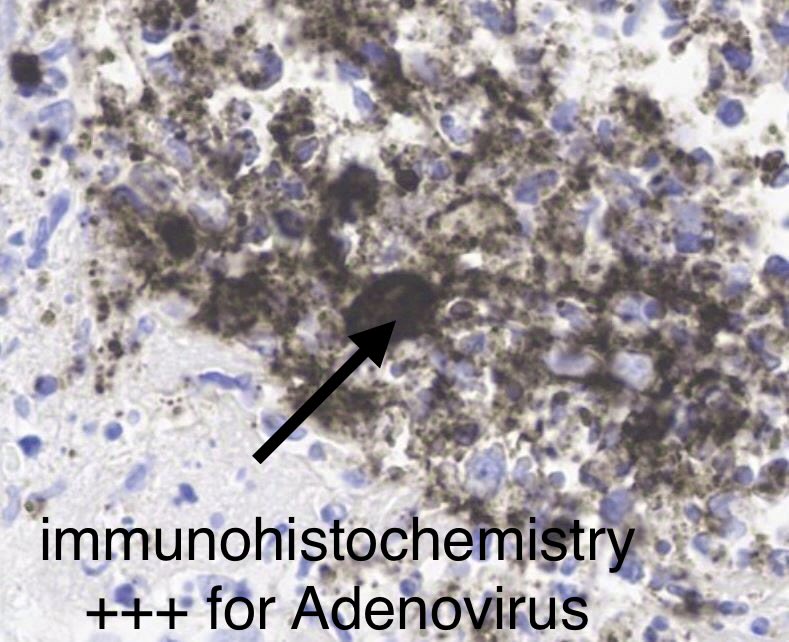
Severe inflammation and necrosis with abscess formation.
Look carefully=you see cells with large nuclei in the mix of inflammatory cells.
IHC= Positive staining for ADENOVIRUS.
DX: Adenovirus-associated interstitial nephritis.
55-yr old with kidney &
heart transplant, fevers, rise in serum creatinine.
This prospective study in #ASNJASN of 976 adults measured the association between subclinical primary aldosteronism and kidney outcomes. Read more: https://t.co/7d7isydJVr
Can fish oil supplementation help lower risk of cardiovascular events among individuals receiving hemodialysis? In this #ASNKidneyNews article, current evidence is reviewed and explained: https://t.co/1RLb0VIuyE
I scored > 300 lupus nephritis biopsies for chronicity indices with Maria Gutierrez/Chile.
Bx scoring was simple: minimal, mild, moderate, severe.
The chronicity score correlates very well with outcomes/prognosis.
Score should be included in Bx report.
https://t.co/IIC763OFOn
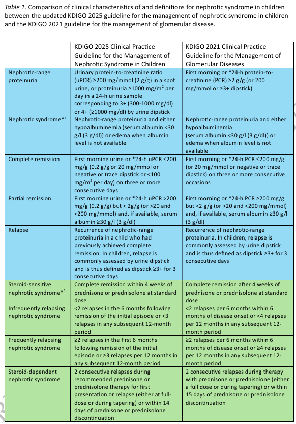
The 2025 KDIGO guideline on the management of nephrotic syndrome in children:
a comment of the European Renal Association Immunonephrology Working Group
https://t.co/cVrRJ19aMR
@ERAkidney
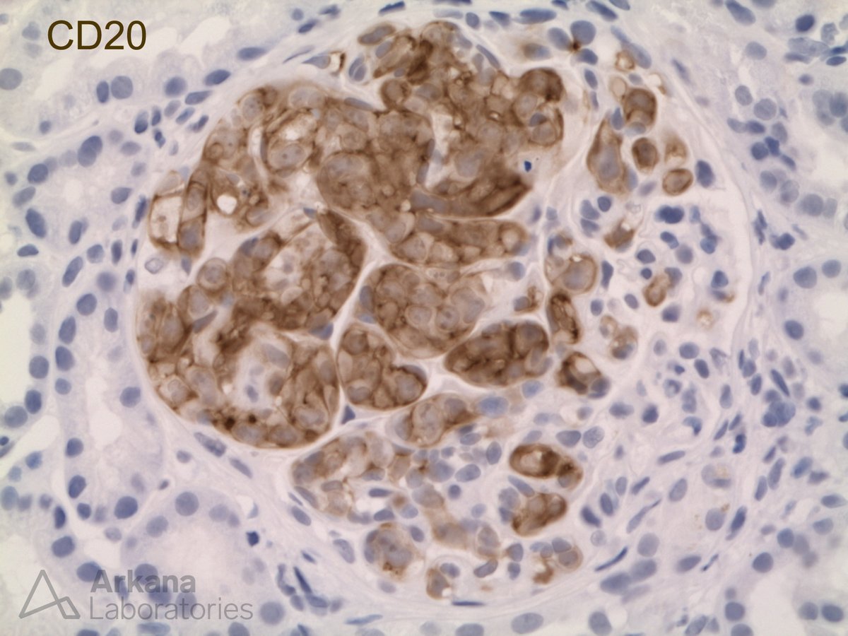
#TeachingPoints A 75 year old woman presented for her annual checkup with no complaints. She had a past medical history of hypertension and her blood pressure at this visit was 160/80. Laboratory findings were significant for an elevated serum creatinine at 2.1 mg/dL (baseline 1.0 mg/dL) as well as proteinuria of 855 mg/24 hr. All serologies, including proteinase-3, myeloperoxidase, and anti-nuclear antibodies were negative. A kidney biopsy was performed to evaluate the decreased kidney function and proteinuria. It revealed a total of five glomeruli were present, three of which showed a global endocapillary proliferation of large atypical cells with frequent mitotic figures (Photomicrographs A and B). These atypical cells were isolated to the glomeruli and stained positive for CD20 . The remaining glomeruli did not contain any proliferative changes. Electron microscopy and immunofluorescence were both negative for immune deposits.
This is a rare subtype of extranodal diffuse large B cell lymphoma that has been known in the past by several different names and was originally thought to be a proliferation of endothelial cells; however, it is currently classified by the WHO as “intravascular large B-cell lymphoma” (IVBCL). IVLBCL most frequently affects adults over 60 and is typically widely disseminated in the small vessels of many organs at presentation. It most often presents without tumor masses or lymphadenopathy and lymphoma cells are usually not seen in peripheral blood smears. Clinical symptoms are highly variable and usually result from the occlusion of small vessels. The vast majority of cases are diagnosed in the bone marrow, liver, spleen, or skin. However, there are case reports of the diagnosis being made by kidney biopsy. In these cases, it usually manifests with slowly progressive renal failure and proteinuria which is frequently in the nephrotic range.
#RenalPath
Very excited to share that our DELiVR method is now open access published @NatureMethods. We created a simple, brain-wide cell analysis deep learning tool, no coding needed! Fiji Plugin makes it accessible to all. https://t.co/qjFk3p5aba
by @Dorie00@Rami96614090 @moritz_negwer
Free access through May 12: Quantitative assessment of glomerular basement membrane collagen IV α chains in paraffin sections from patients with focal segmental glomerulosclerosis and #AlportSyndrome gene variants. @pongpratch https://t.co/QjLzEl13RM
Just out today, #WorldKidneyDay2024 and start of the #AlportSyndrome Workshop in Cyprus, Open Access article: Alport syndrome and Alport kidney diseases – elucidating... : Current Opinion in Nephrology and Hypertension https://t.co/Etu4rsFwci @pongpratch