Associate Professor of Medicine & Cell Biology, Washington University in St Louis. We study the cell cytoskeleton, centrosomes, cilia, kidney and lung diseases.
Our latest study is now published @JCI_insight and OMG WE GOT THE COVER! 😱😱😱
Congrats to my amazing postdoc @ChengTao_PKD for this herculean effort (~5 yrs), and to @LangnerEP for the beautiful image that made the cover 😍
Lemme tell you about our study real quick👇
This is really cool (and wild):
Scientists simulated a complete living cell for the first time. Every molecule, every reaction, from DNA replication to cell division.
The paper (Luthey-Schulten et al., Cell 2026, https://t.co/PXxXWKC8yp), just out today, used JCVI-Syn3A — a synthetic minimal bacterium with fewer than 500 genes. A 3D+time simulation of the full 105-minute cell cycle: DNA replication, protein translation, metabolism, division. Every gene, protein, RNA, and chemical reaction tracked through physical space.
It took years to build. Multiple GPUs. Six days of compute time per run.
And this is the simplest possible cell.
A human cell has ~20,000 genes. It lives in tissue. It interacts with neighbors. It differentiates. It responds to drugs in ways that depend on context we haven't fully measured.
Mechanistic simulation of the minimal cell costs 6 GPU-days for 105 minutes of biology. You cannot scale that to human cells. The complexity isn't 40x harder. It's exponentially harder.
This is why the field pivoted to data-driven models. You can't hand-encode the regulatory wiring of a human hepatocyte. But you can learn it — if you have the right perturbation data collected across enough diverse biological contexts.
The two approaches aren't competing. Papers like this generate the ground truth that future ML models need for validation. But the path to a clinically useful virtual cell runs through foundation models, not through scaling up mechanistic simulation.
Amazing work!
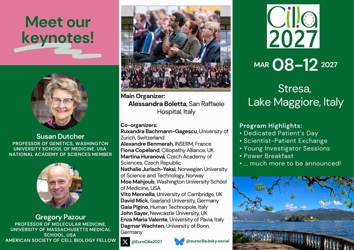
𝗠𝗲𝗲𝘁 𝗼𝘂𝗿 𝗸𝗲𝘆𝗻𝗼𝘁𝗲 𝘀𝗽𝗲𝗮𝗸𝗲𝗿𝘀 𝗳𝗼𝗿 #𝗖𝗶𝗹𝗶𝗮𝟮𝟬𝟮𝟳!
We are grateful for their participation and look forward to welcoming the cilia community on 8-12, March 2027.
Wishing you all a happy and inspiring 2026.
#Cilia#Milan
Proud moment for WashU Nephrology! Dr. Moe Mahjoub @MahjoubLab, with Drs. Susan Dutcher and Steven Brody, are advancing lung disease research with two #NIH R01 awards, one renewal and one newly funded project! #LungResearch#NIH#WashU See More:
https://t.co/q1Wd5uibKA
Flashback #FluorescenceFriday to some of the first light-sheet imaging we were doing. Video shows the collecting duct system of the developing kidney labeled with a pan-cytokeratin antibody. Courtesy of former talented technician Deanna Hardesty.
🔊New preprint from our lab!
Zygotene cilia regulates meiosis, germ cell development, and fertility in zebrafish, mice, and humans
https://t.co/qg1v5Mjku5
It's another NOA day!
So happy (relieved) to finally get this one. What started out as a crazy idea pitched to the WashU Cilia group a couple of years ago is now a fully funded project.
Major thanks to @NIH@NHLBI for continuing to fund our work on motile ciliopathies 🙏
Meet the PKD Foundation Board of Directors committed to helping us chart a bold path forward, one that advances science, empowers patients, and brings us closer to a future without PKD.
Meet the full Board ⬇️
https://t.co/zRw99g3kl5
Applications are due soon! If you're working to move the field of PKD research forward, we invite you to apply for a PKD Foundation Research or Fellowship Grant. Learn more ⬇️
Exciting new preprint alert! Our latest research is a tour de force exploring the biology of centriolar satellites, membrane-less organelles essential for development and physiology. Below tweets summarize our findings; full paper here 👇
https://t.co/qedbHEcay4