Effects of resistance exercise dose on pain intensity and disease impact among individuals with fibromyalgia: a systematic review with meta-analysis
https://t.co/r8EJ9z8nfI
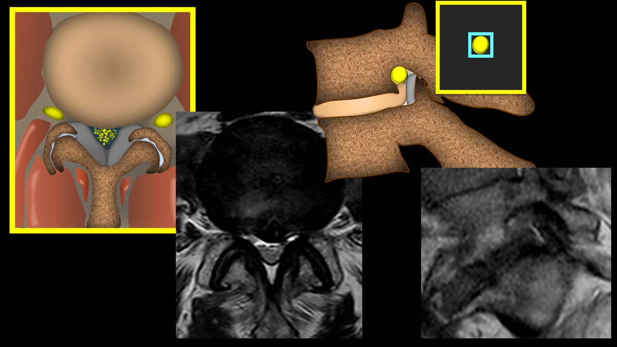
Deep gluteal syndrome: anatomy, imaging, and management of sciatic nerve entrapments in the subgluteal space
doi:10.1007/s00256-015-2124-6
Deep gluteal syndrome (DGS) is an often-missed condition causing buttock, hip, and posterior thigh pain, sometimes with radicular pain. It's characterized by sciatic nerve entrapment in the subgluteal space, a region encompassing various anatomical structures. The term "piriformis syndrome" is considered a subset of DGS, not encompassing the broader range of pathologies included within DGS.
The subgluteal space's anatomy is complex, including the piriformis muscle and other external rotators (superior and inferior gemelli, obturator internus, and quadratus femoris muscles), along with neurovascular bundles. MR neurography (MRN) is the imaging modality of choice for DGS assessment, providing crucial visualization of this space and its components. The article emphasizes the importance of understanding the complex anatomical relationships within the subgluteal space to accurately diagnose and manage DGS.
Several etiologies contribute to DGS, including: fibrous and fibrovascular bands (compressive or adhesive, affecting sciatic nerve mobility); piriformis syndrome (muscle hypertrophy, anomalous attachments, or dynamic entrapment); obturator internus/gemelli syndrome; quadratus femoris and ischiofemoral pathology; hamstring conditions; gluteal disorders; and orthopedic issues. Fibrous bands are a primary cause of DGS and are classified by location and mechanism (compressive, adhesive, or undefined).
Clinical presentation of DGS is varied, potentially mimicking other conditions. Symptoms include buttock or hip pain, sciatica, difficulty sitting, and antalgic posture. Diagnostic tests include physical examination (Lasègue, Pace's, Freiberg's, Beatty, FAIR tests) and the crucial infiltration test (diagnostic and therapeutic). MRN imaging is essential to pinpoint the precise location and nature of nerve entrapment.
Treatment is typically initiated with conservative measures (rest, anti-inflammatories, physical therapy). Injections (anesthetic/corticosteroids or botox) are used for diagnosis and pain relief. Endoscopic procedures (neurolysis, tenotomy, or resection) are reserved for cases unresponsive to conservative management. The authors highlight the classification of fibrous bands as crucial information for surgeons, informing appropriate instruments, portal placement, and surgical approach.
The article concludes by emphasizing the importance of recognizing the multifactorial etiology of DGS, the critical role of MRN, the utility of the infiltration test, and the value of endoscopic techniques in diagnosis and treatment. Radiologists play a crucial role in guiding the diagnosis and treatment strategy.
Let's chat about frozen shoulders... A 🧵
There are a few ortho conditions that can make a mess of things due to its natural history, initial presentation, and progression.
Frozen shoulders are one of those diagnoses.
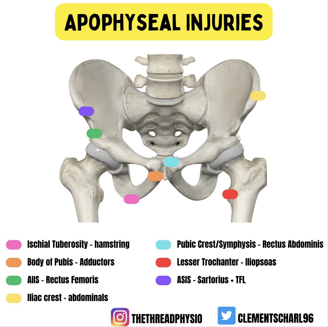
1) A brief 🧵on Avulsion Fractures
Always keep on the radar 🚨 in any adolescent presenting with acute pelvic pain OR recalcitrant Sx which are not responding to conservative efforts
Hot off the press - DCM SR on clinical signs :)
https://t.co/5saFSLY5RU
* Tromner and hyper-reflexia most sensitive
* Babinski, clonus, Tromner, inverted supinator most specific
* Consider imaging if +ve tests
* Don’t be reassured by normal tests if strong DCM history
Stress fractures - a detailed thread 🦴🦴🦴
I thought I'd put together an overview & some clinical nuggets from the 'coal-face'
First up, I prefer to call them 'bone stress injuries' - majority don't have a # line on imaging & the language can be scary / nocebic for some
It's always the same story... Felt a pop, and had some pain, the pain got much worse a few days later.
Now they're miserable.
Mild/mod OA--->Degen root---> subchondral fx/intense edema.
You know the diagnosis in one minute if you listen.
https://t.co/XMWmiUwHkw
Prognostic Factors of Axial Spondyloarthritis (AxSpA)
Thanks to @DerekGriffin86@pdkirwan and @DrJN_SportsMed for pushing for this thread.
Remember AxSpA can vary greatly from person to person, and several factors can influence outcomes
Read on for details!
1/🧵
Pre-print: Getting out of neutral: the risks and rewards of lumbar spine flexion during lifting exercises
Here is a link to our article in the latest @UKSCA journal.
@GregLehman
https://t.co/908DnyY0OP
Thread alert 🚨🚨🚨
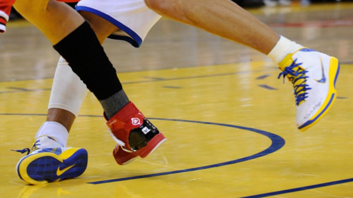
Ankle inversion injuries don't just result in lateral ligament sprains....
...there are plenty of other injuries to bear in mind when you assess, both acutely & further down the line
Not many know, but I had pretty bad back pain from the age of 13-21. Stopped playing sport, fear avoidant, many chiro/physio sessions etc etc
I was asked recently, "How did I get out of pain?"
Since threads are what the cool kids do, here is my response 🧵
Thread alert - posterior ankle & heel pain 🚨🚨🚨
Most cases are ‘common garden’ achilles tendinopathy
However, here is a selection of conditions I've seen over time in clinic which can masquerade & are worth considering if the picture is atypical or patient not progressing
Thread alert 🚨🚨🚨 - Medial knee pain!
What else should we be considering beyond OA & MCL sprains?
Here are some differentials to consider & their clinical presentation 🧐