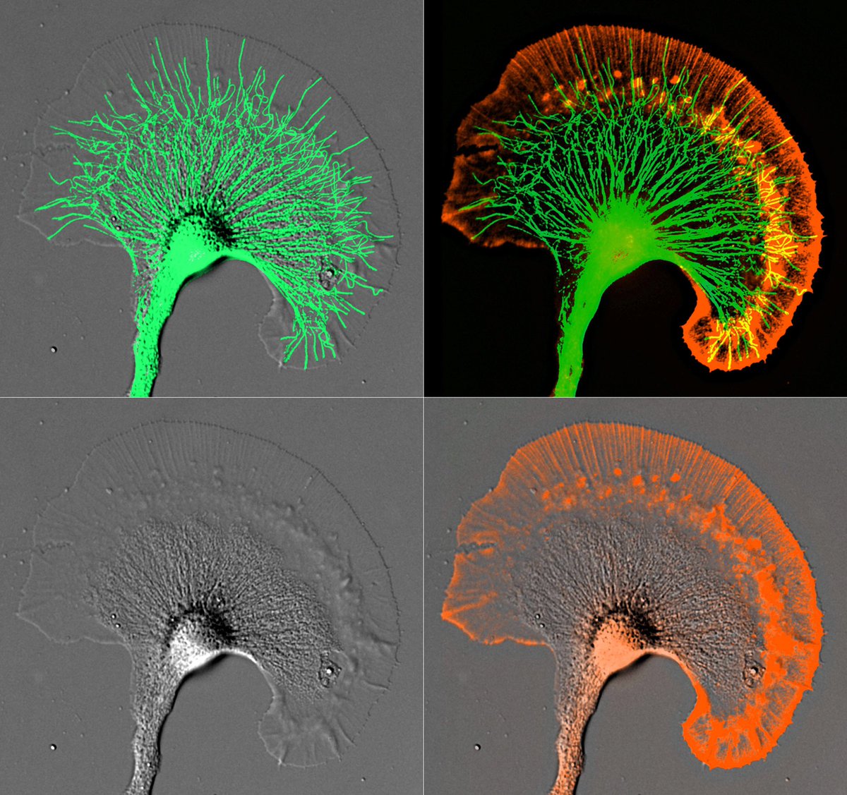
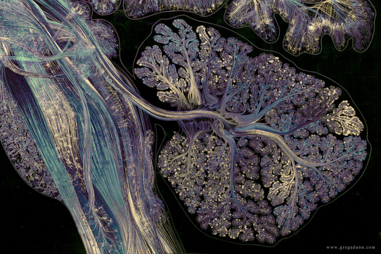
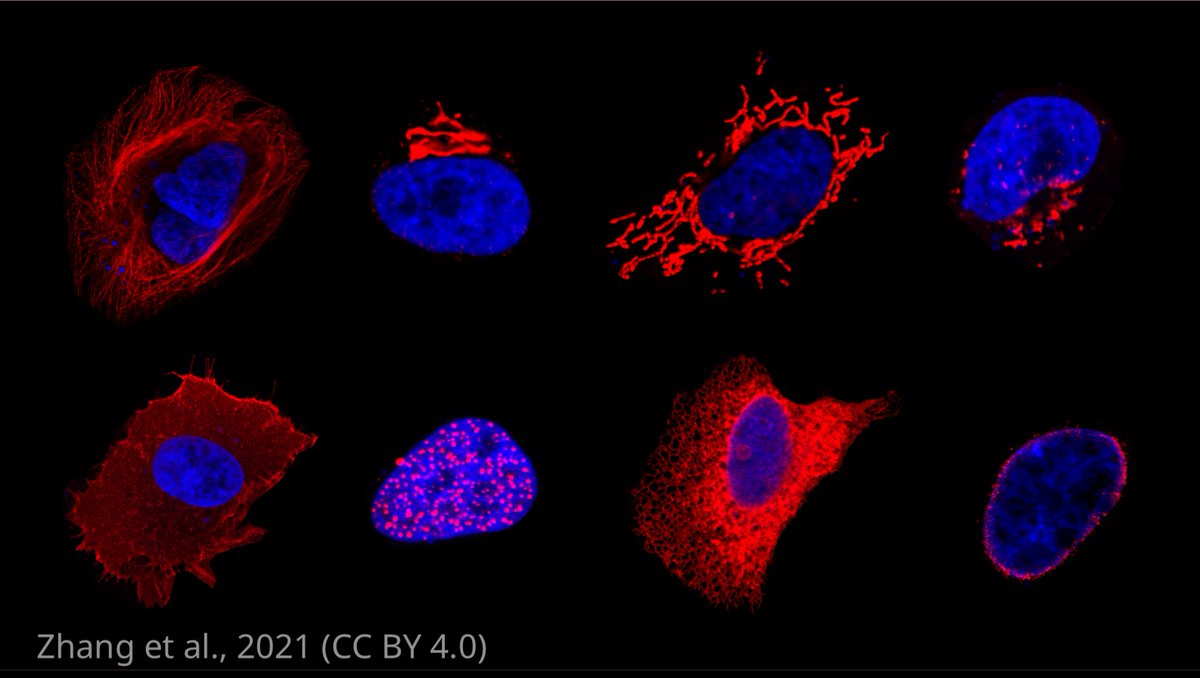
A neuronal growth cone photographed through a microscope. DIC, actin filaments and microtubules are shown. One of my many @NikonSmallWorld rejects from the 2000’s. #NSWRejects
This data made my day ! ... I am exploring autonomic implications of the "motor structure" of the century... the cerebellum. @VoicesScience@UHouston@UHBiomedEng
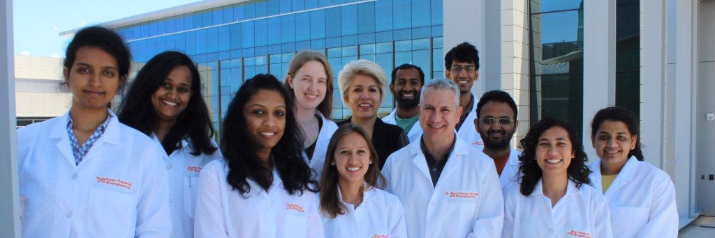
We welcome our STEM Research Inquiry Summer Enrichment scholars,MD students and high schools performing hands-on research at our lab, our internal scholastics director @AleGnzlzzz will be leading this effort together with our grad students. Go research! @UHouston@UHEngineering
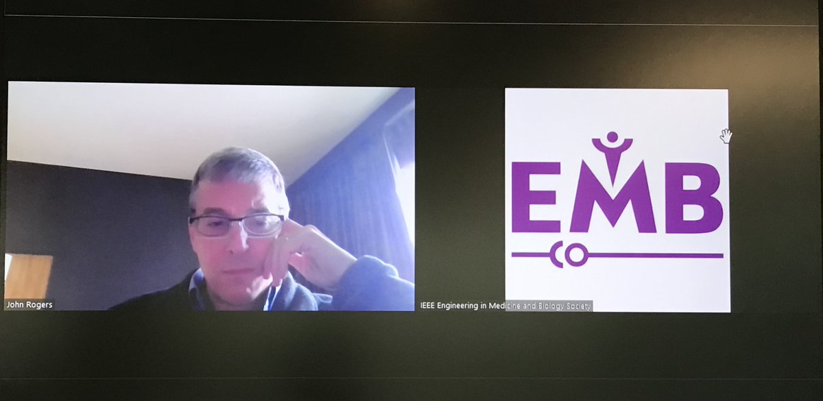
Today, we had an honor to host Dr. John Rogers of @NorthwesternU as a keynote speaker at the @IEEEembs Healthcare Innovations in Point Care of Technology Conference in Houston.
We tremendously enjoyed his highly inspiring talk this morning!
Just now
I am very proud of the @UHBiomedEng students and postdocs at the @IEEEembs Healthcare Innovations in Point of Care Technologies 2022. in 2013. Wonderful presentation by David. Many thanks to Dr Mario Romero-Ortega for the program and leadership.
Visualization of nerves (light blue) and endothelial cells (e.g. blood vessels, dark blue) in a 13.5-day old mouse embryo:
#neuroscience by Nathan Burns
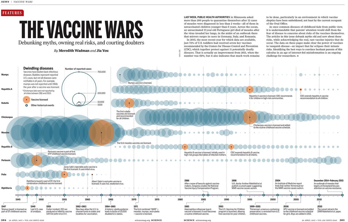
As #vaccines for #COVID19 are distributed all across the globe, we look back at this data visualization from 2017 that illustrates the power of vaccines to vanquish disease—an impact that far eclipses their minute risks. ➡ https://t.co/Mr2QfEDvi3
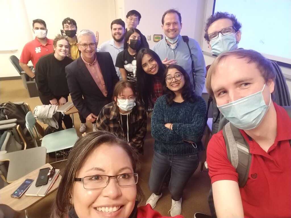
Had a great time chatting with @KipLudwig about his experiences translating bioelectronic medicine into clinical practice, and potential career pathways post-PhD. Thank you @RomeroLabUH for hosting, and to you Dr. Ludwig, for that amazing talk!
Two participants using a new neural-machine interface for #prosthetic arms demonstrated increased physical ownership and agency of their #bionic limb, a new @SciRobotics study shows.
Read more: https://t.co/zv6Wk5Gnfn