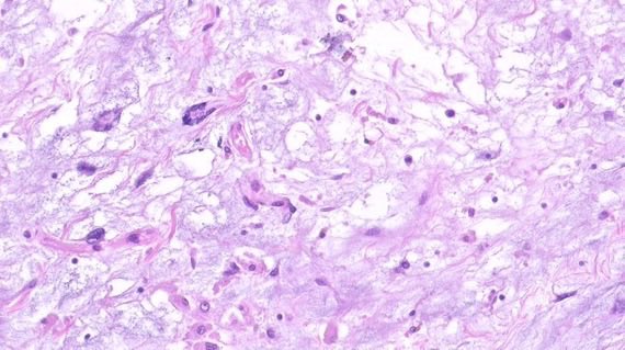
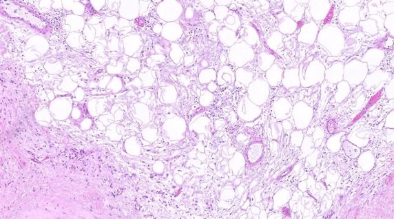
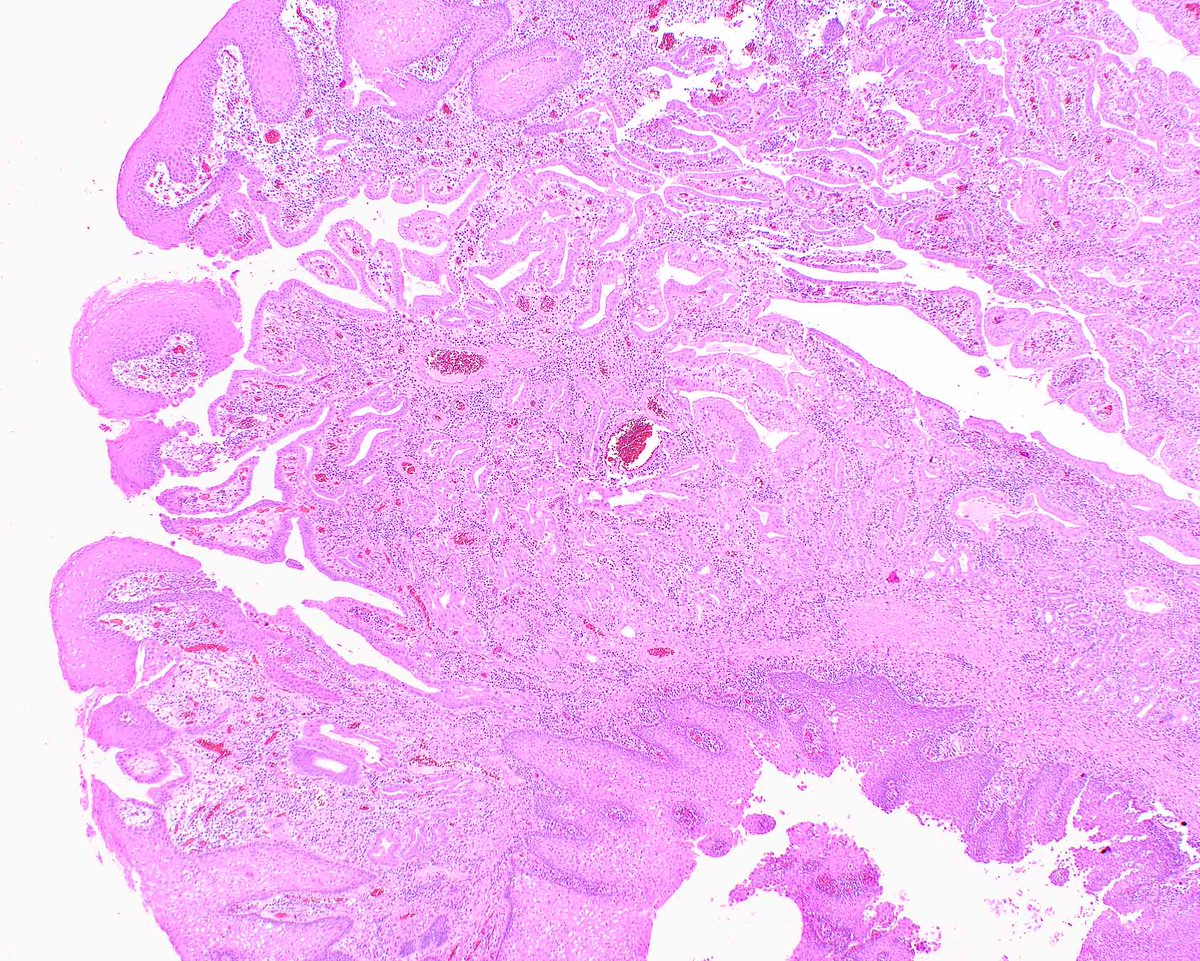
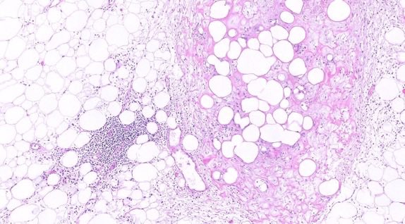
🟪Uterine myxoid leiomyosarcoma🟪
Macroscopic:
➡️WHO: myxoid tumours may be gelatinous and friable
➡️May look deceptively circumscribed. Still sample the interface
Diagnostic criteria 🔬
WHO Female Genital 5e, essential criteria box, myxoid:
One or more of:
➡️Moderate to severe cytological atypia (2+/3+)
➡️Tumour cell necrosis
➡️> 0.4 mitoses/mm² (equating to > 1 mitosis/10 HPF of 0.55 mm diameter and 0.24 mm² area)
➡️Infiltrative borders / irregular margins
For contrast, conventional spindle LMS (same box): two or more of marked atypia (2+/3+), tumour cell necrosis, or ≥ 4 mitoses/mm² (equating to ≥ 10 mitoses/10 HPF of 0.55 mm diameter). That mitotic number does not apply here.
Myxoid leiomyoma (WHO leiomyoma box):
➡️Circumscribed, hypocellular and myxoid, lacking cytological atypia or mitoses
STUMP (WHO): exceeds the leiomyoma box, below the LMS box.
Tips:
☑️Extensive sampling may be required to find the diagnostic areas (WHO)
☑️Confirm myxoid stroma (Alcian blue–positive acid mucin), not hydropic oedema (WHO leiomyoma chapter)
☑️Desmin, h-caldesmon, and SMA may be weak or patchy in myxoid tumours (WHO)
☑️CD10 is often positive (WHO). It does not separate this from a stromal tumour
☑️If it sits between the two boxes, that is STUMP. WHO: try to land leiomyoma or LMS first, with generous sampling
#PathTwitter
WHAT'S NEW IN GASTROESOPHAGEAL TUMORS (WHO 6TH EDITION 2026)
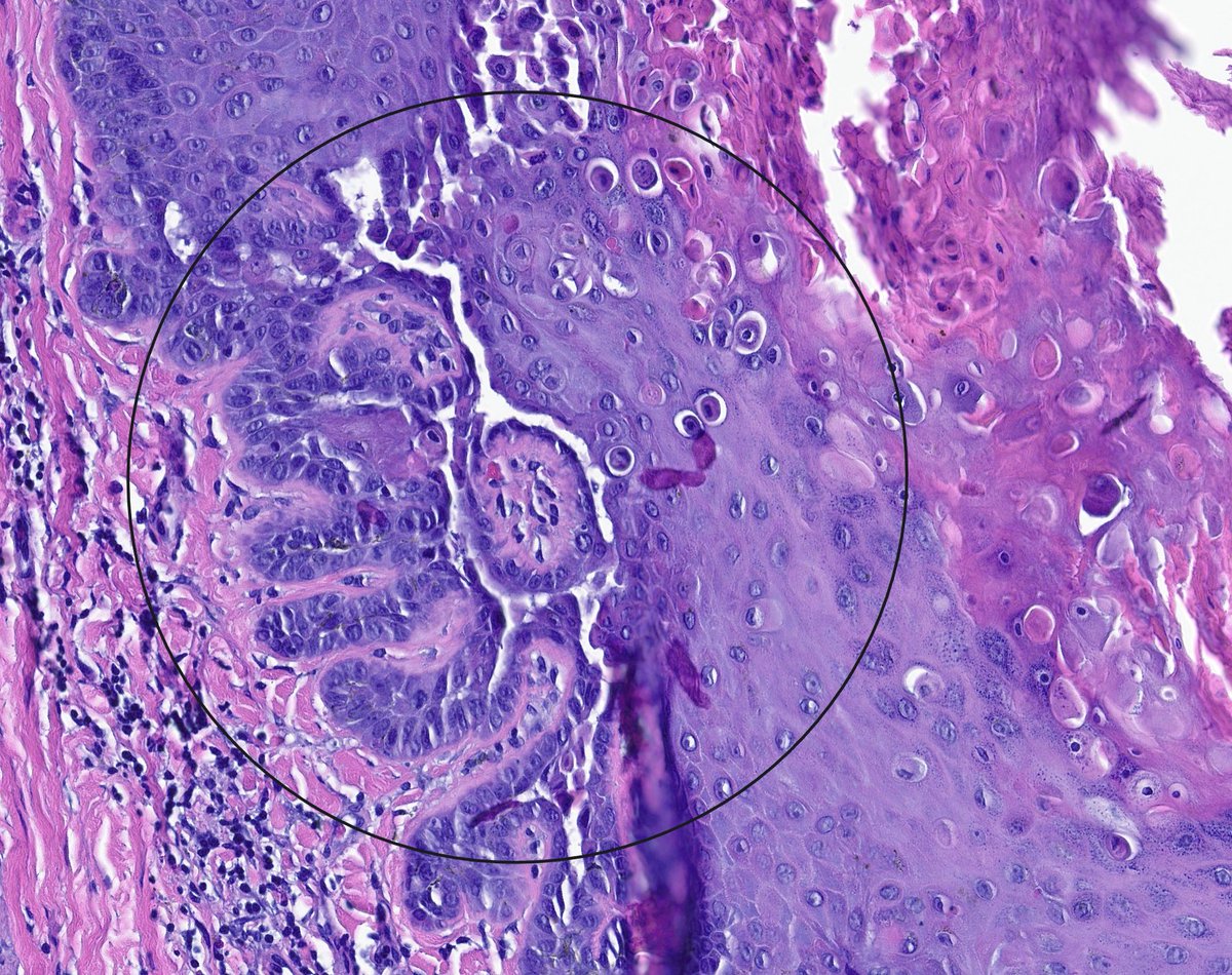
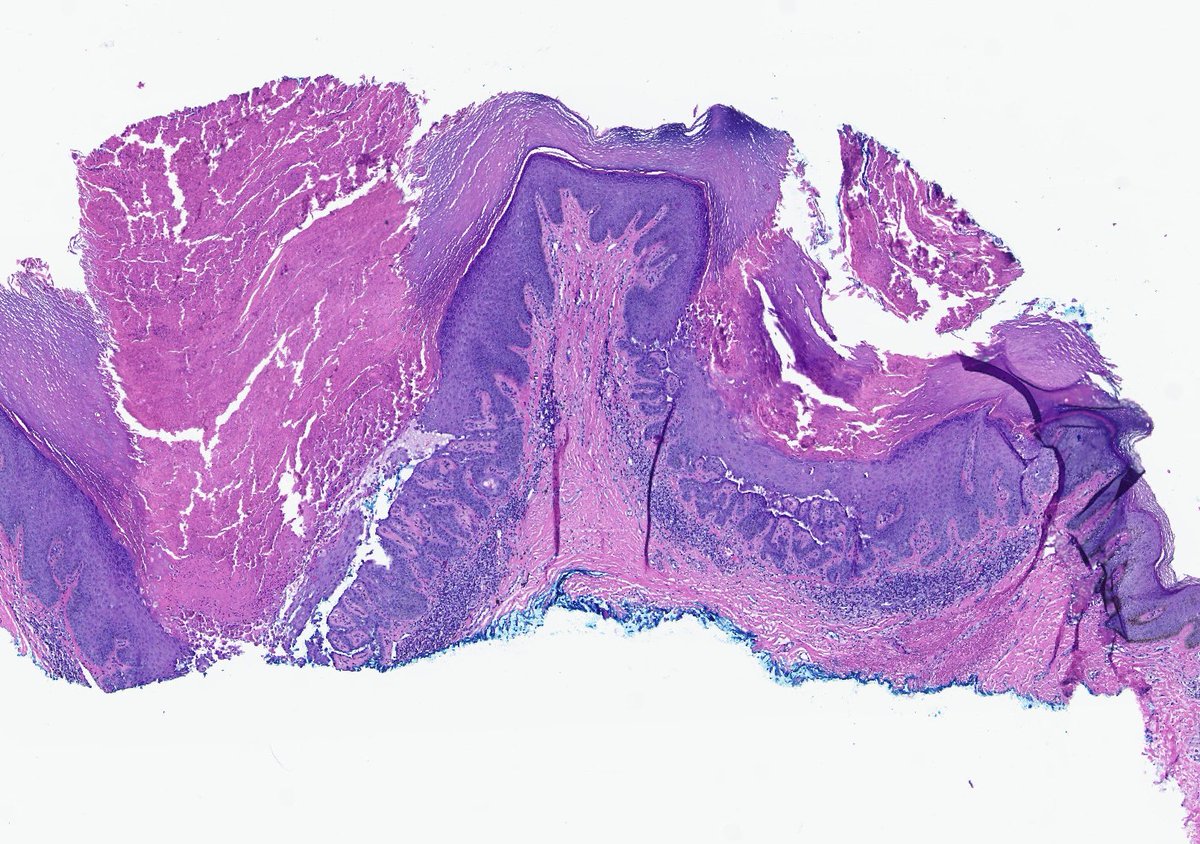
Epidermoid metaplasia was added as a precursor lesion (new to WHO)
Dr. Montgomery PathCast GIPS
https://t.co/cLz2yW20FH #PathX#pathology
A great research paper is more than good results.
It tells a clear story:
1. Introduction → Why the study matters
2. Methods → How the study was conducted
3. Results → What was found
4. Discussion → What the findings mean
Strong research isn't just about discovering something new. It's about communicating it clearly so others can understand, evaluate, and build on your work.
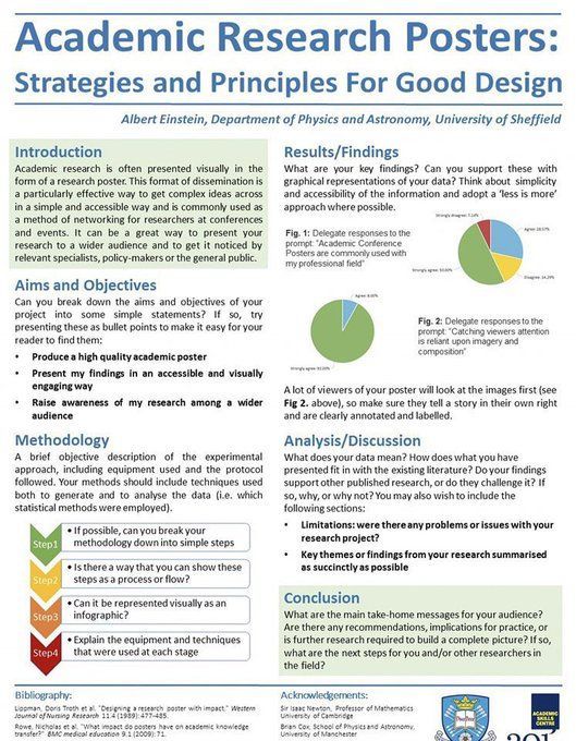
طريقة كتابة وتصميم وعرض الـ Research Poster بشكل احترافي 🎓📌
هذا الدليل مفيد جدًا للمبتدئين، خصوصًا إذا كانت هذه أول مشاركة لك في مؤتمر علمي أو أول مرة تعرض فيها بحثك 👇🏼
SYP+ adenocarcinoma has been previously reported, and occasionally these cases display focal CgA+, but this didn't feel right. So I threw in the 2nd gen NE marker INSM1...
To be able to discern between these entities, allow me to introduce this fabulous algorithm from Dr Rindi and colleagues in @EndoPath!
https://t.co/sq8jZcxeI5
Incidental polyp in the terminal ileum of a colon cancer right hemicolectomy. IHC negative apart from patchy CD34. This is a benign calcifying fibrous tumour. Can be found anywhere in the GI tract and do not have any syndromic associations (IgG4 IHC was less than 40%) #GIpath
#CytoQuiz 🧐🔬
A) Solitary Fibrous Tumor
B) Inflammatory Myofibroblastic Tumor
C) Schwannoma
D) Dermatofibrosarcoma Protuberans
#Pathology#Cytology
Clue in caption 🧐
How to differentiate between adenoid cystic carcinoma and basaloid squamous cell carcinoma?
p40 stains basal/myoepithelial cells only in ACC and is diffuse in SCC.
Pitfall ⚠️: SOX10 and MYB IHC can be positive in both.
Dr. Nishino - 2025 Diagnostic Pathologic Update #USCAP #pathology #PathX