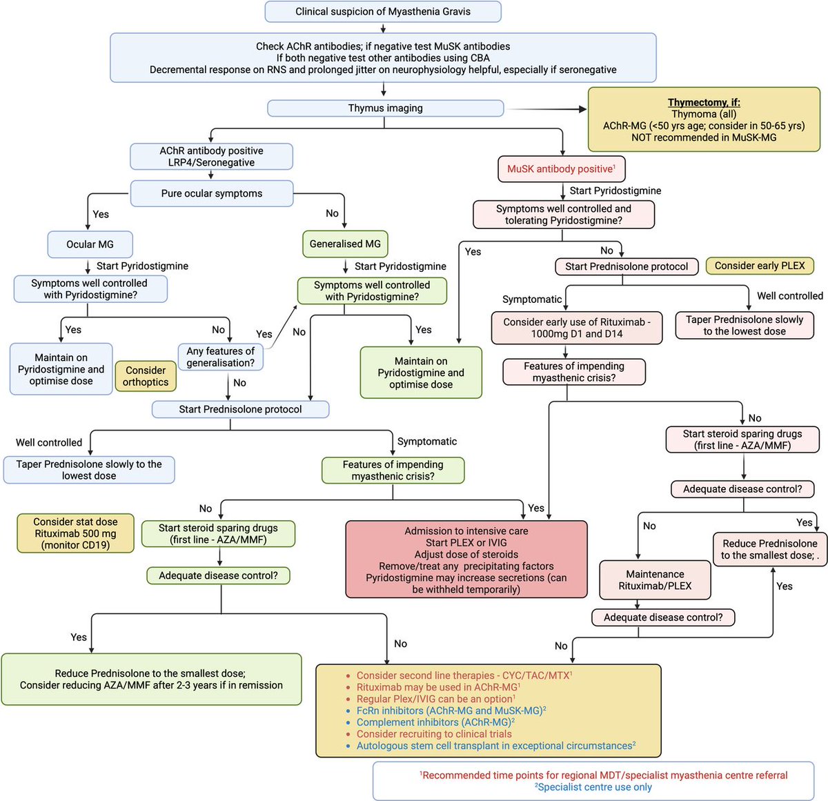
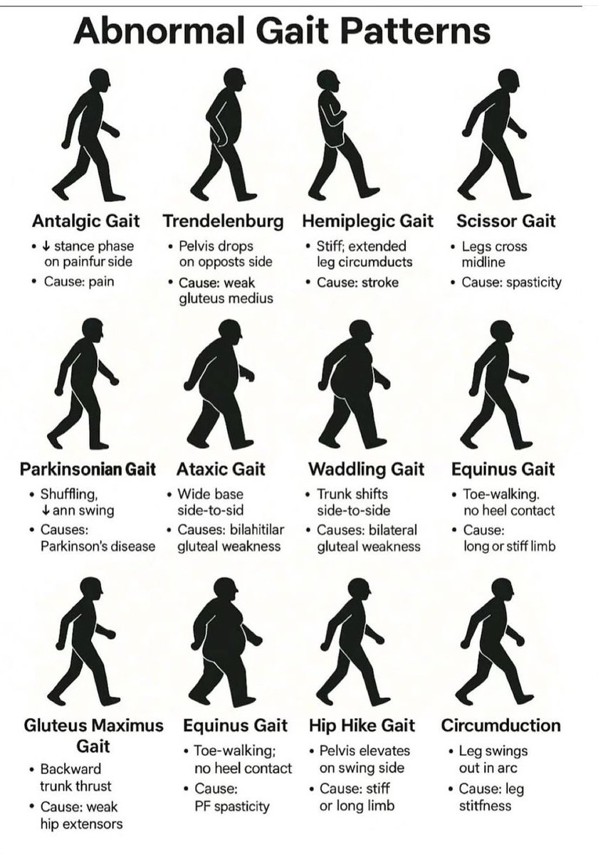
I’ll have some time coming up, so I’ll be returning to this topic as a free webinar. Date dropping in the next few weeks. Keep an eye out👁
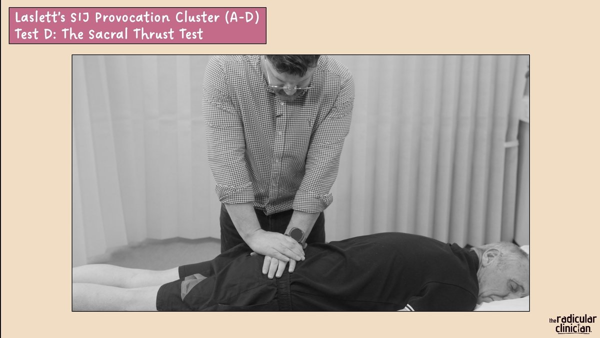
The SIJ in a World of Non-Specific Low Back Pain! 🤓
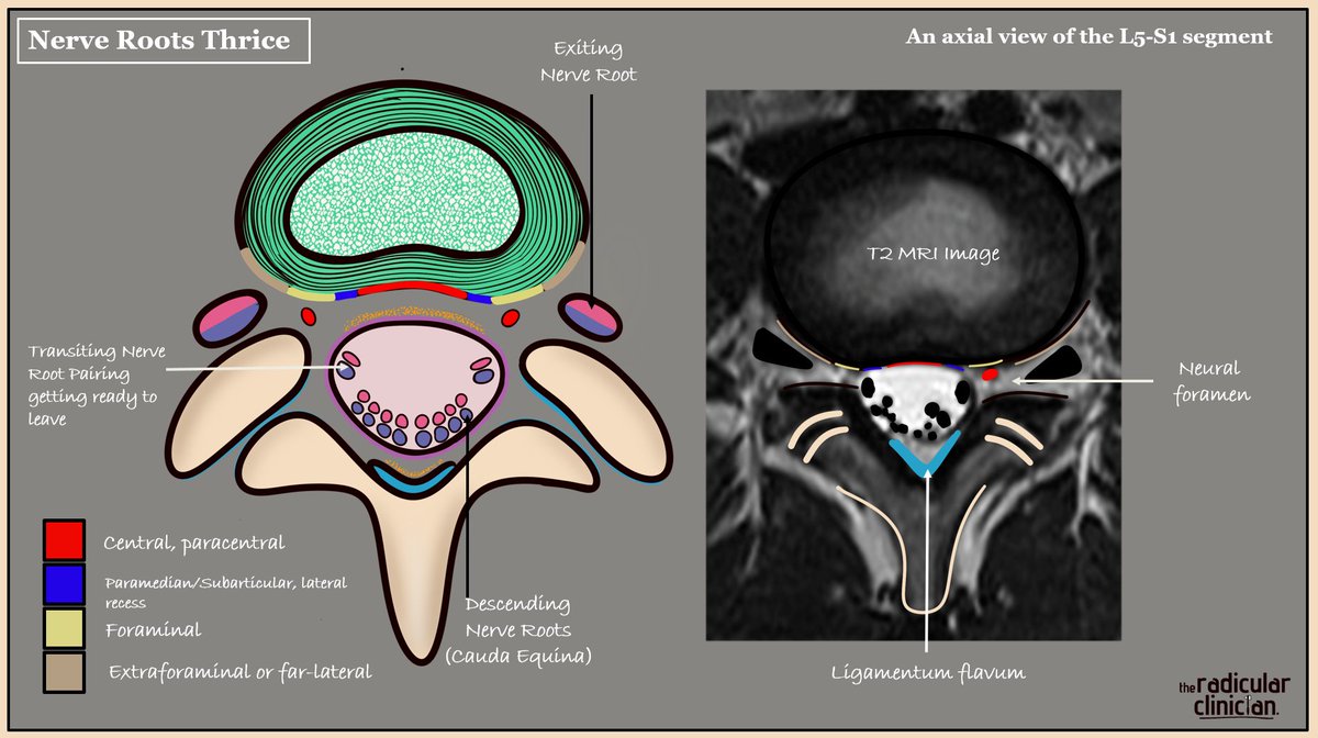
Nerve Roots 'Thrice'
Clinical anatomy applied
Lumbosacral nerve roots can be described in three geographical locations on axial T2 MRI, each with important clinical implications if they are compressed or inflamed. The picture illustrates the L5-S1 level, so we are looking at the L5 disc. Focus on the colour codes in order to appreciate disc location in relation to each geographical nerve root location.
Descending (bilateral S2, S3, S4 and S5 roots as part of the cauda equina).
An acute central or paracentral disc herniation here is rare but can cause cauda equina syndrome if compressed. Also relevant with crowding as seen with LSS.
Transiting/Traversing (here bilateral S1 roots).
As the bilateral transiting nerve roots leave the thecal sac centrally, they remain enveloped by its outer connective tissue layer, referred to as the nerve root sleeve. The lateral recess is the most common site of disc related compression because of its anatomical position and the root being fixed in place. This is why radicular pain is more commonly noted in one leg. Note that the nerve roots do not lie flat when you do.
Exiting (here L5).
As the nerve root enters its respective neural foramen. This is the second most common site of compression, typically from a far lateral disc herniation or from crowding within the foramen.
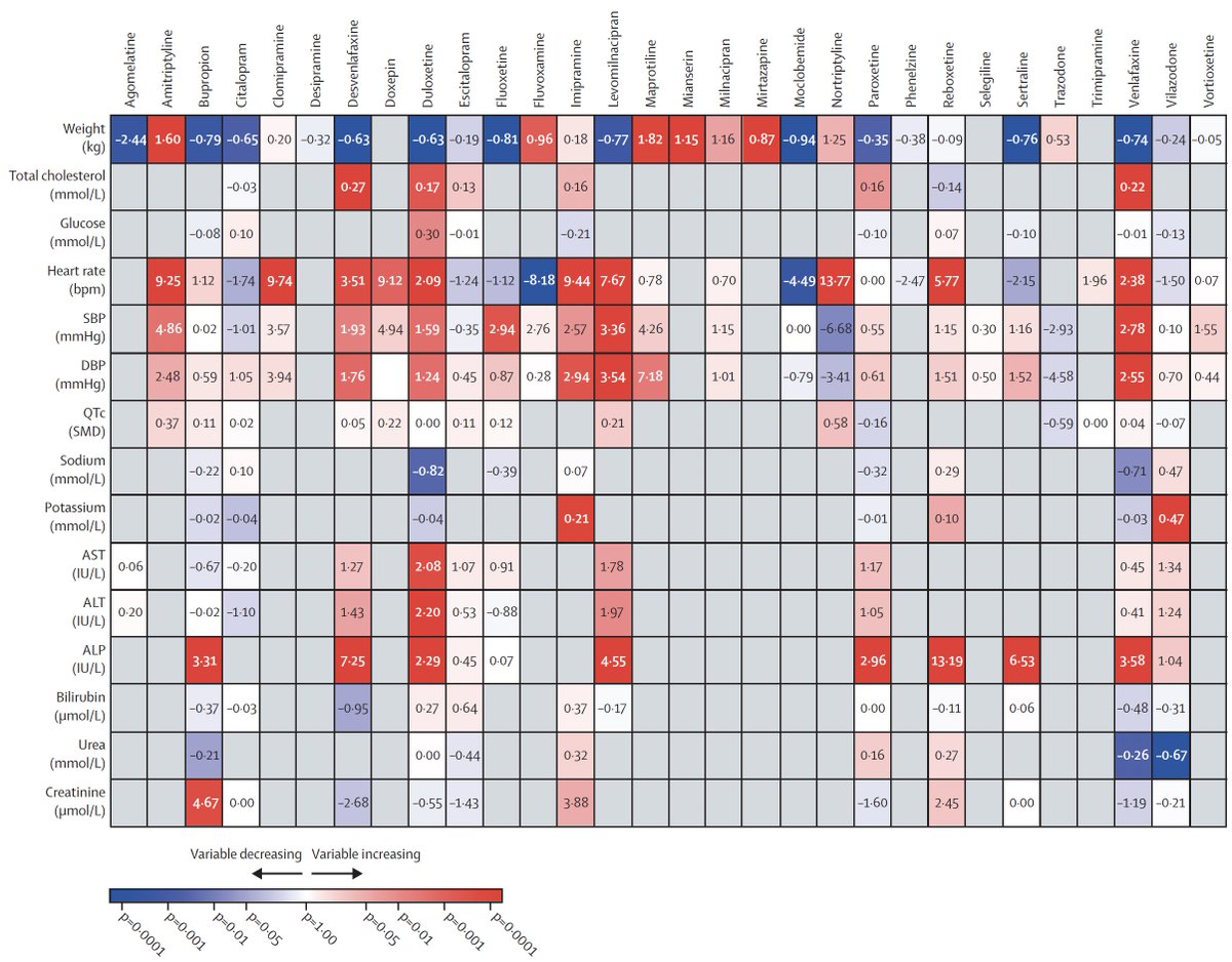
Antidepressants have a range of adverse cardiometabolic and physiologic side effects, as seen in a new systematic review of 30 such drugs
https://t.co/8VxIzW8JwQ












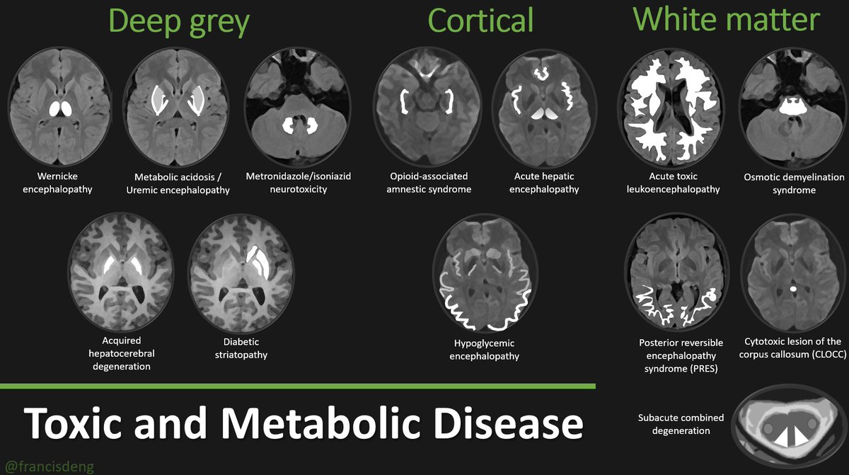
![MarlonVFZR's tweet photo. 🧬𝗘𝗻𝗰𝗲𝗳𝗮𝗹𝗼𝗽𝗮𝘁𝗶́𝗮𝘀 𝘁𝗼́𝘅𝗶𝗰𝗮𝘀 𝘆 𝗺𝗲𝘁𝗮𝗯𝗼́𝗹𝗶𝗰𝗮𝘀 – 𝗥𝗠 𝗰𝗲𝗿𝗲𝗯𝗿𝗮𝗹🧠
⬇️⬇️⬇️⬇️
1. 𝙎𝙪𝙨𝙩𝙖𝙣𝙘𝙞𝙖 𝙜𝙧𝙞𝙨 𝙥𝙧𝙤𝙛𝙪𝙣𝙙𝙖 (𝘿𝙚𝙚𝙥 𝙜𝙧𝙚𝙮)
📌𝙒𝙚𝙧𝙣𝙞𝙘𝙠𝙚: afectación del tálamo y cuerpos mamilares.
📌𝘼𝙘𝙞𝙙𝙤𝙨𝙞𝙨/𝙪𝙧𝙚́𝙢𝙞𝙘𝙖: lesiones en ganglios basales.
📌𝙈𝙚𝙩𝙧𝙤𝙣𝙞𝙙𝙖𝙯𝙤𝙡/𝙞𝙨𝙤𝙣𝙞𝙖𝙯𝙞𝙙𝙖: lesiones del tronco encefálico y cerebelo.
📌𝙃𝙚𝙥𝙖𝙩𝙤𝙘𝙚𝙧𝙚𝙗𝙧𝙖𝙡 𝙖𝙙𝙦𝙪𝙞𝙧𝙞𝙙𝙖 / 𝘿𝙞𝙖𝙗𝙚́𝙩𝙞𝙘𝙖: simetría en ganglios basales.
2. 𝘾𝙤𝙧𝙩𝙞𝙘𝙖𝙡
📌𝙊𝙥𝙞𝙤𝙞𝙙𝙚𝙨 / 𝘼𝙢𝙣𝙚𝙨𝙞𝙖: afectación del hipocampo.
📌𝙃𝙚𝙥𝙖́𝙩𝙞𝙘𝙖 𝙖𝙜𝙪𝙙𝙖: edema cortical difuso.
📌𝙃𝙞𝙥𝙤𝙜𝙡𝙪𝙘𝙚́𝙢𝙞𝙘𝙖: lesiones occipito-frontales simétricas.
📌𝙋𝙍𝙀𝙎: edema parieto-occipital reversible.
𝟯. 𝗦𝘂𝘀𝘁𝗮𝗻𝗰𝗶𝗮 𝗯𝗹𝗮𝗻𝗰𝗮
📌𝙇𝙚𝙪𝙘𝙤𝙚𝙣𝙘𝙚𝙛𝙖𝙡𝙤𝙥𝙖𝙩𝙞́𝙖 𝙩𝙤́𝙭𝙞𝙘𝙖: necrosis difusa de la sustancia blanca.
📌𝘿𝙚𝙨𝙢𝙞𝙚𝙡𝙞𝙣𝙞𝙯𝙖𝙘𝙞𝙤́𝙣 𝙤𝙨𝙢𝙤́𝙩𝙞𝙘𝙖: protuberancia y ganglios basales.
📌𝘾𝙇𝙊𝘾𝘾: lesión citotóxica del cuerpo calloso (splenium).
📌𝘿𝙚𝙜𝙚𝙣𝙚𝙧𝙖𝙘𝙞𝙤́𝙣 𝙘𝙤𝙢𝙗𝙞𝙣𝙖𝙙𝙖 𝙨𝙪𝙗𝙖𝙜𝙪𝙙𝙖: médula dorsal por déficit de B12.
👇🏼👇🏼👇🏼👇🏼
📚📖 Más en el blog #ClubCrit 👉 [https://t.co/zqkNIKC5Wd]
#Encefalopatia #MRI #RMN #ClubCrit #MedTwitter #CritCare #UCI #ICU #CriticalCare #Diagnóstico #icu #intensivecare #MedTwitter #diagnosis #management #Riesgo #ICU #POCUS #UCI #Tratamiento #MedicinaBasadaEnEvidencia #Emergencia #Terapia #MedEd #Medicina #Emergencias #FOAMed #FOAMcc #MedTwitter #CriticalCare #MedEd #CuidadoCrítico #MedX #IntensiveCare #EducaciónMédica #MedIntensiva #MedXCommunity #MedicinaCrítica #MedED #CritCare #ICUManagement #MustRead #LecturaRecomendada](https://pbs.twimg.com/media/G3D9O_bWYAAwtUG.jpg)